
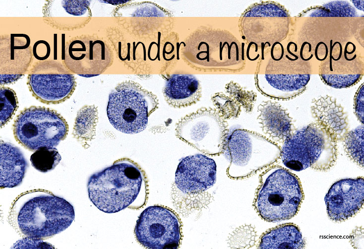
Pollen under a Microscope
Pollen grains are the male gametophytes, and they are formed in anthers, the male parts…
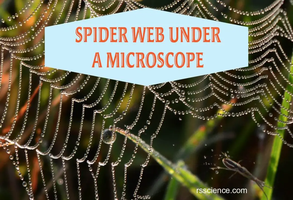
Spider Web Under a Microscope
Spider silk is one of the strongest natural materials on Earth. Despite common belief, a…
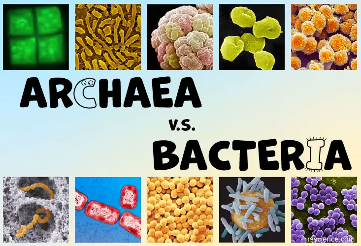
Archaea vs Bacteria – What are the Similarities, Differences, and Examples
Bacteria and archaea are single cell prokaryote. They are similar in sizes, shapes, reproduction, and…
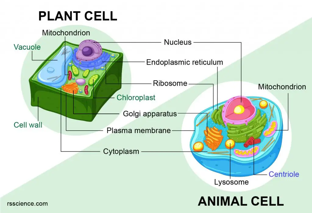
Animal vs. Plant cells – Similarities, Differences, Chart, and Examples
Animal and plant cells are eukaryotic cells. They all have nuclei, cell membranes, and organelles…
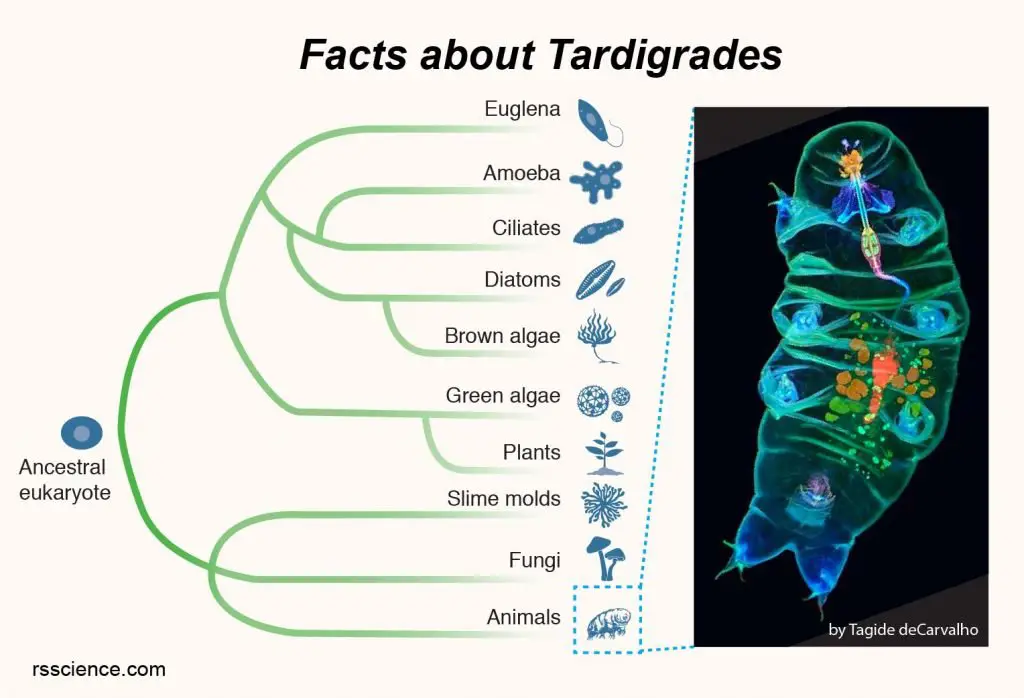
What are Tardigrades (Water Bears) – extreme conditions survivor
Tardigrades, also known as water bears, look like chubby, microscopic bears walking slowly with eight…
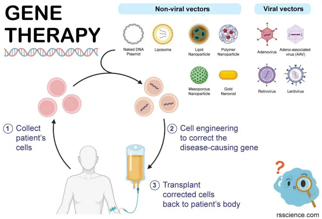
Gene Therapy: A Promising Biotechnology for the Treatment of Genetic Diseases and Cancers – Basic Introduction
Gene therapy is a medical treatment that involves introducing or altering genes within a person’s…
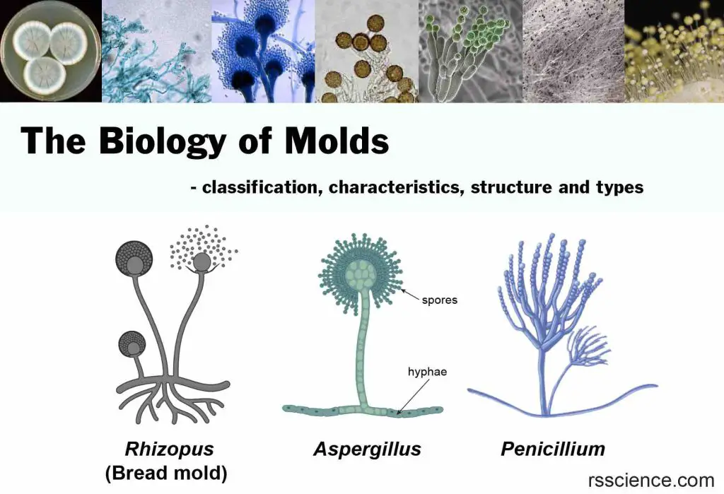
The Biology of Molds (Moulds) – classification, characteristics, structure and types
Mold is a type of fungus that grows in multicellular fiber-liked structures called hyphae and…
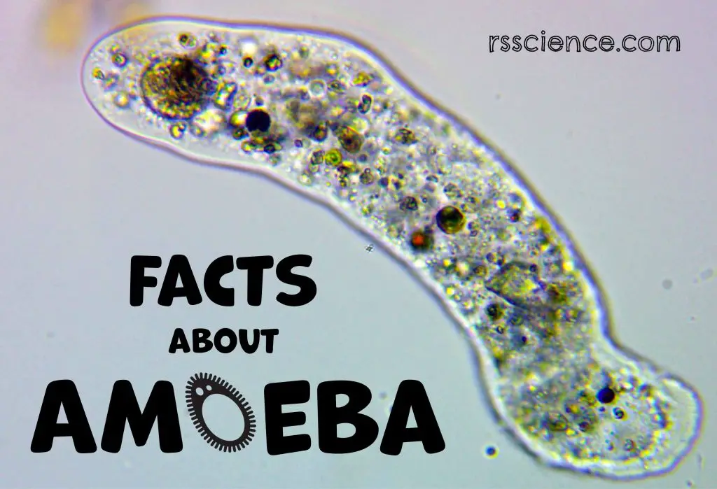
Facts about Amoeba, structure, behavior and reproduction
Amoeba is a group of primitive protists. They move through extension and retraction of their…
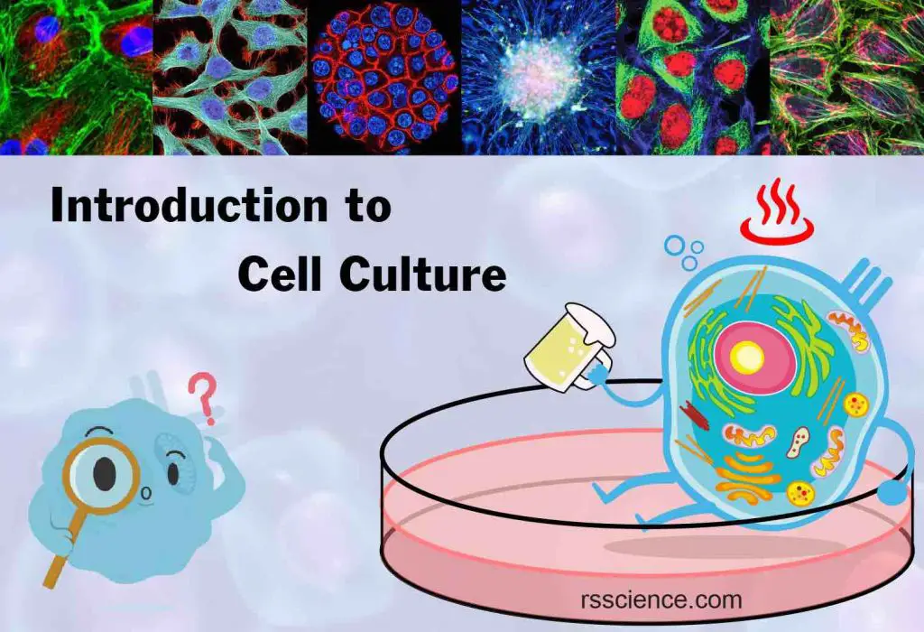
Cell Culture Basics – The Foundation of Biotechnology
Cell culture is a technique to grow and maintain cells in a Petri dish or…

The Secret of Bird Feathers – What’s a Feather Look Like Under a Microscope?
A feather is a light, strong structure that grows on the skin of birds. Their…

6 Science Humor Images That Make You Smile
A picture is worth a thousand words. I want to share with you 6 science…
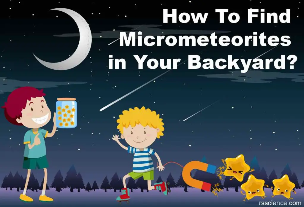
Shooting stars! How To Find Micrometeorites in Your Backyard?
Thousands of tons of star debris fall on Earth annually. You can use a strong…
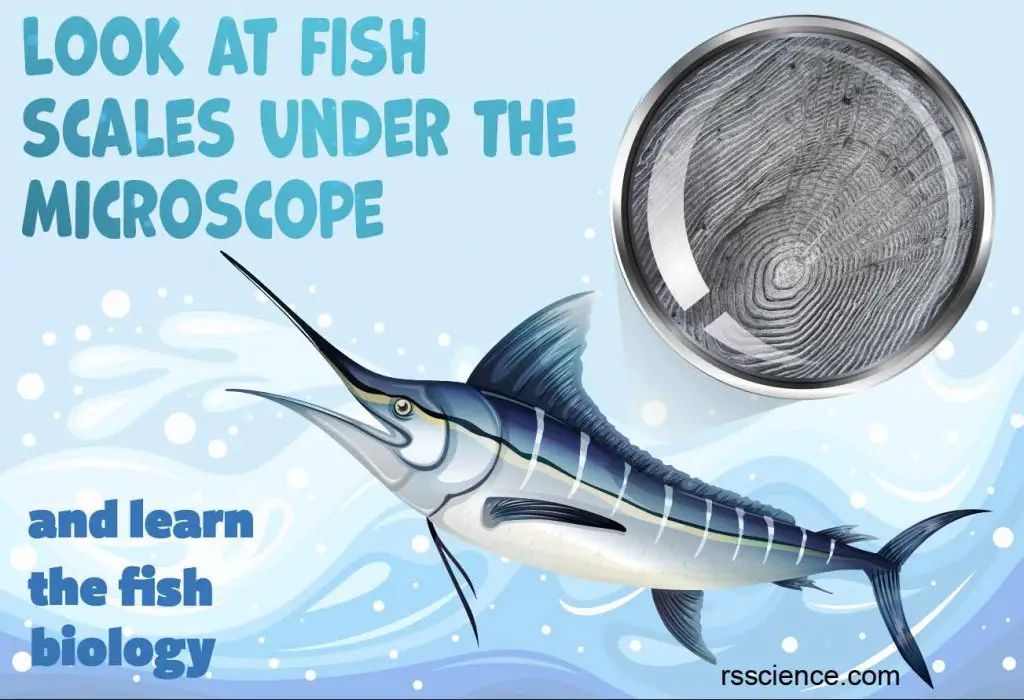
Fish Biology and Fish Scales – Look at fish scales under the microscope
Fish scales are produced from the inner layer of fish’s skin, and their function includes…
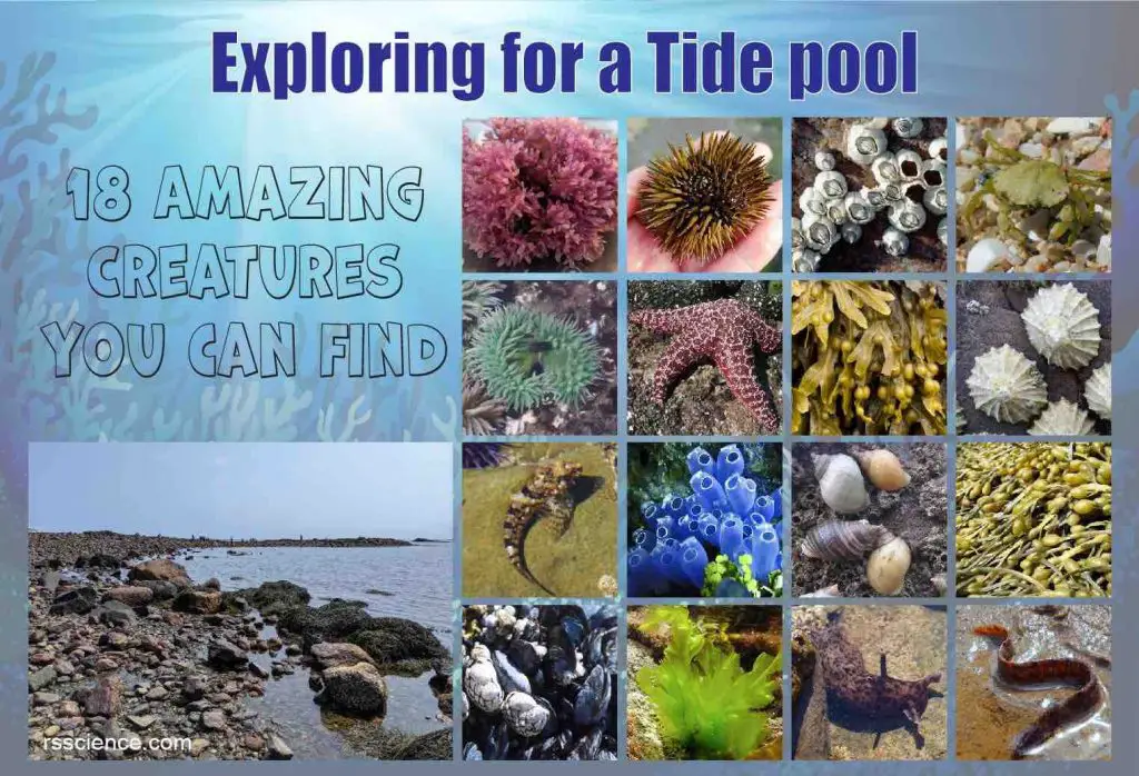
Exploring a tide pool – 18 Amazing Creatures You can find
A tide pool is an isolated pocket of seawater found in the ocean’s intertidal zone…
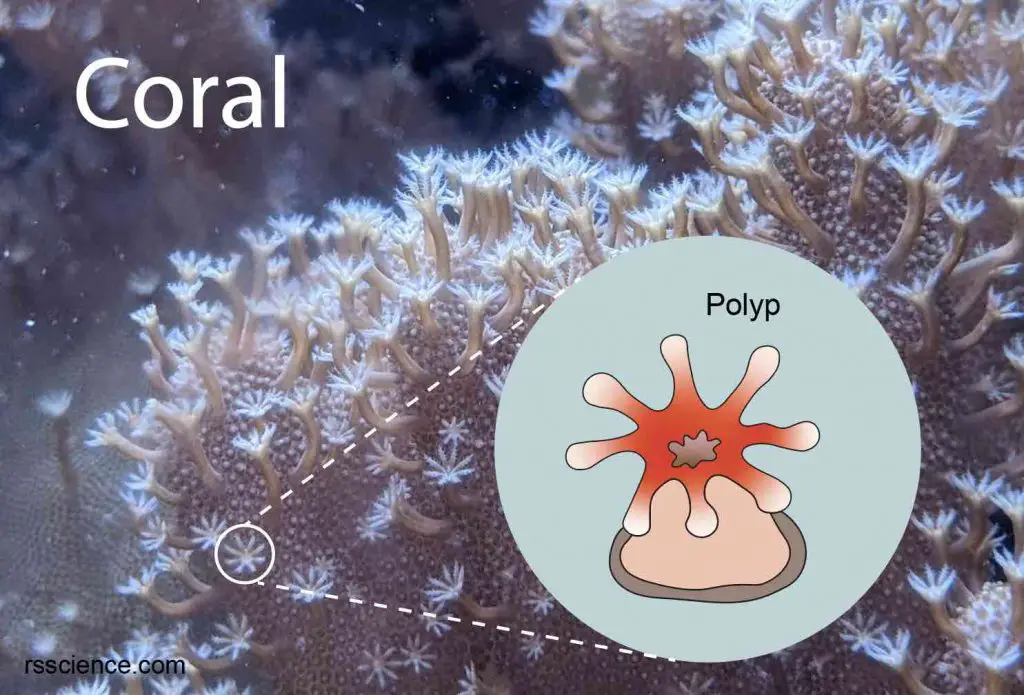
Coral – classification, characteristics, structure and types
Corals are marine animals that resemble miniature sea anemones. The soft, jelly-like body of an…
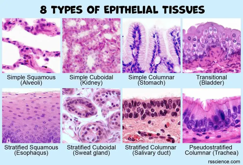
Classification and Types of Epithelial Tissues
Epithelial tissues are classified by the number of cell layers (simple, stratified, pseudostratified), the cell…
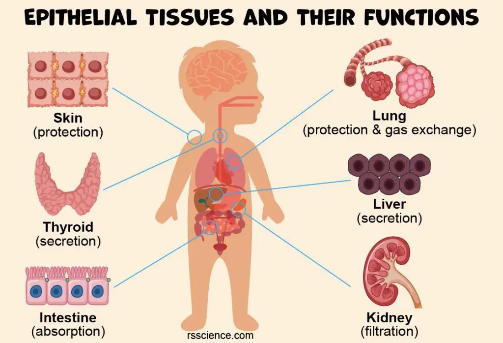
Epithelium – Definition, Characteristics, Cell Structures, Types, and Functions
Epithelium (plural = epithelia) is a protective, continuous sheet of compactly packed cells. It covers…
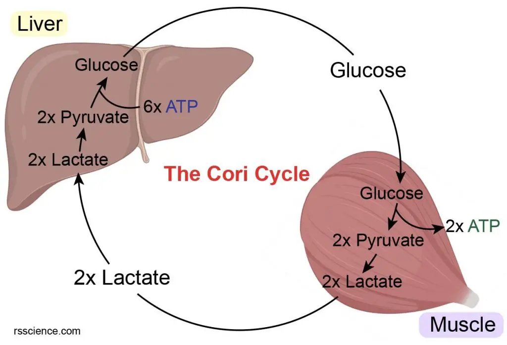
What does the Cori cycle do? – definition, steps, and importance
Cori cycle is to get rid of the lactate, which is generated by muscle contracting…
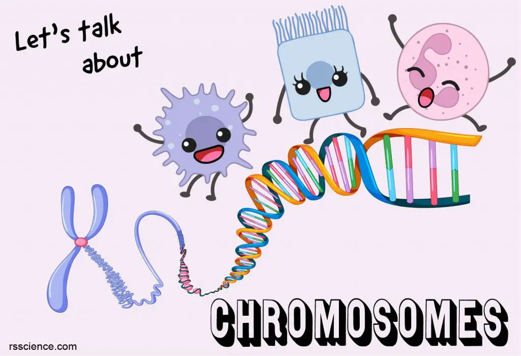
What is a Chromosome? – Function and structure
Chromosomes are made up of bundles of tightly coiled DNA molecules and proteins called histones….
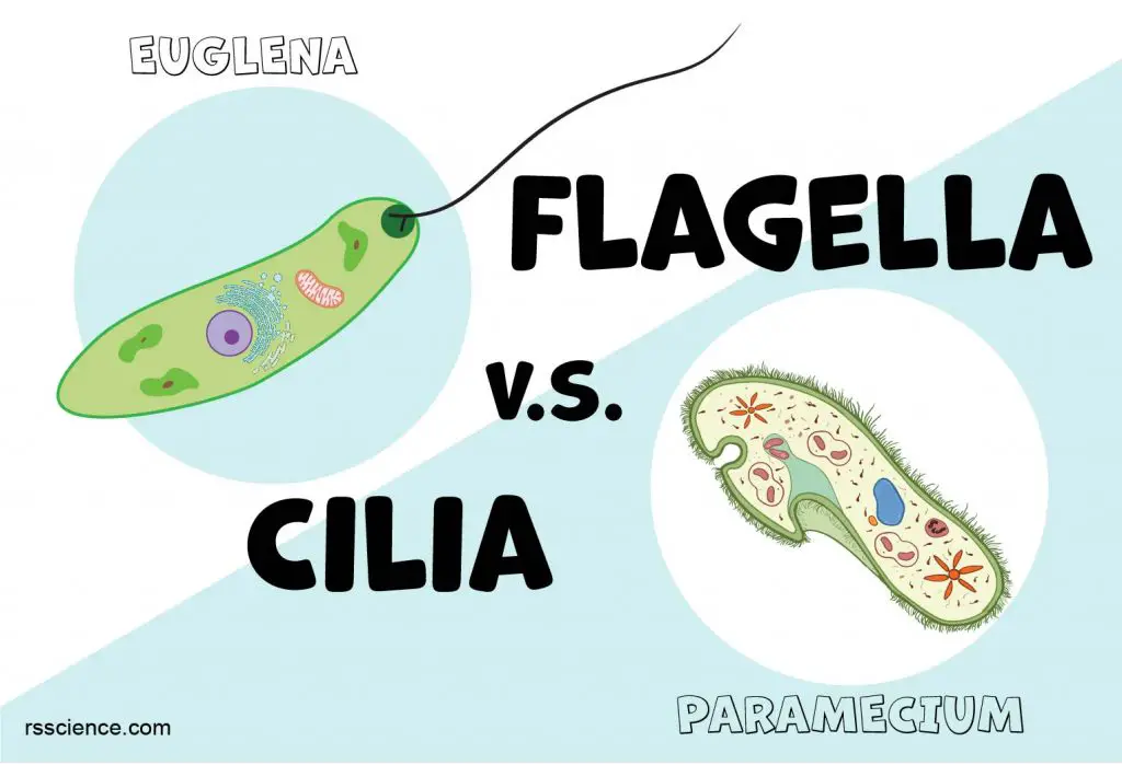
Flagella and Cilia – Definition, Structure, and Functions
Flagella and cilia are two types of cellular structures that allow movement in most microorganisms…
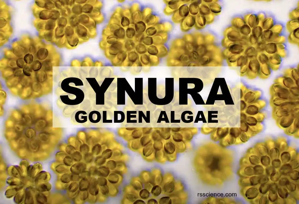
Synura – Biology, Classification, Characteristics, and Reproduction
Synura is a small group of golden-brown algae found mostly in freshwater. They are covered…
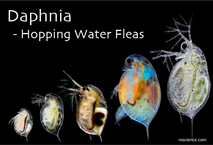
Daphnia – Biology, Classification, Characteristics, and Reproduction
Daphnias, also called water fleas, are small planktonic crustaceans. Its body is usually 0.2–6.0 mm…
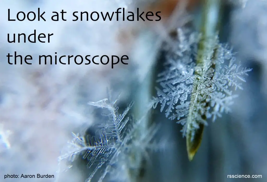
Look at snowflakes under the microscope
Wilson A. Bentley took the first photograph of a snowflake under a microscope. His collection…
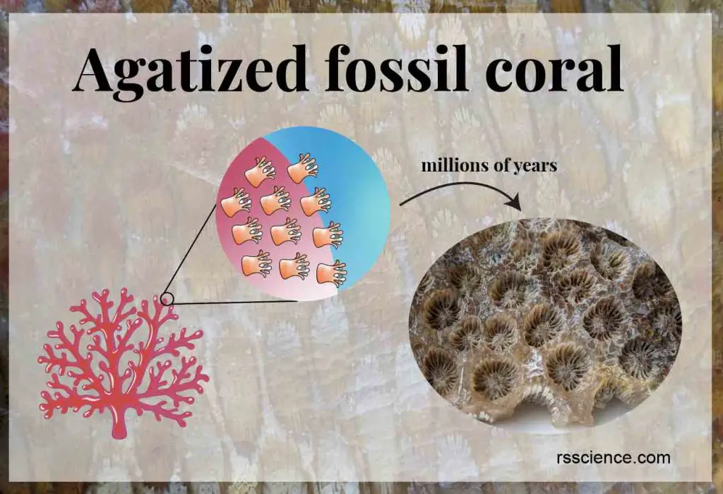
Agatized fossil coral
Agatized fossil coral is a natural gemstone with a beautiful flower pattern that resembles the…
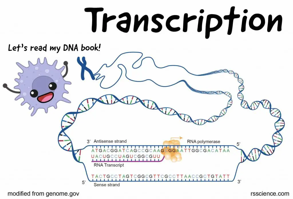
What is Transcription? – From DNA to RNA
Transcription is the process of making an RNA copy of a DNA segment. This copy…
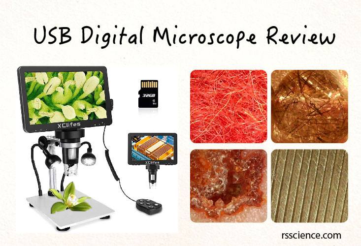
USB Digital Microscope Review
Functionally, USB digital microscopes are similar to stereo microscopes, ideal to view 3D objects. This…
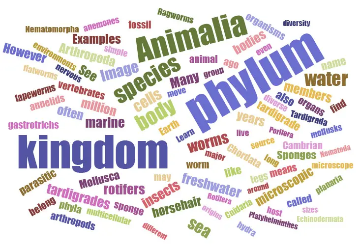
Kingdom Animalia – Different Phylum and their examples
All animals are members of the Kingdom Animalia. Examples of Phylum include Chordata, Arthropoda, Mollusca, Cnidaria,…
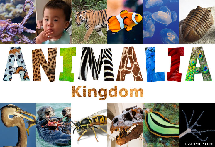
Kingdom Animalia – Classification, Characteristics, and Evolution
All animals are members of the Kingdom Animalia and It is estimated that around 9 or…
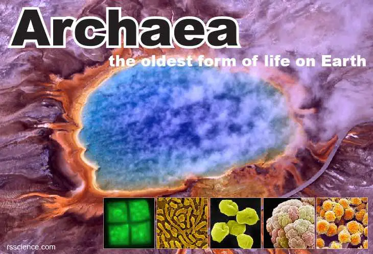
Archaea – Definition, Structure, Types & Extremophile Habitats
Archaea are single cell organisms without a nucleus and other membrane-bounded organelles. Archaea are not…
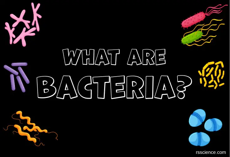
Bacteria – Definition, Structure, Types & Infections
Bacteria are single-celled microorganisms. Because they don’t have nuclei or membrane-bound organelles, they are classified…

Planarian, the Master of Regeneration – the Science of Stem Cells and Genes
A fragment of planarian can regrow into a new animal in two weeks. Genes such…
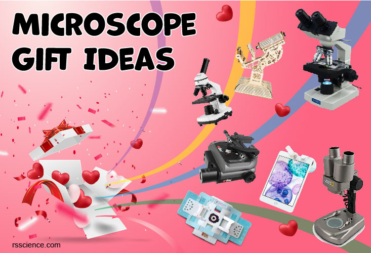
Unique Gift Ideas for Microscopes
Microscopes are wonderful gifts to inspire young kids to fall in love with science. It…
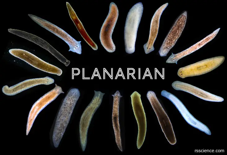
Planarian – Biology, Classification, Characteristics, and Regeneration
Planarians are a group of free-living flatworms. They have a remarkable ability to regenerate an…

Hydra – Biology, Classification, Characteristics, and Reproduction
Hydras are classified under the phylum Cnidaria; some of its relatives are jellyfish and sea…
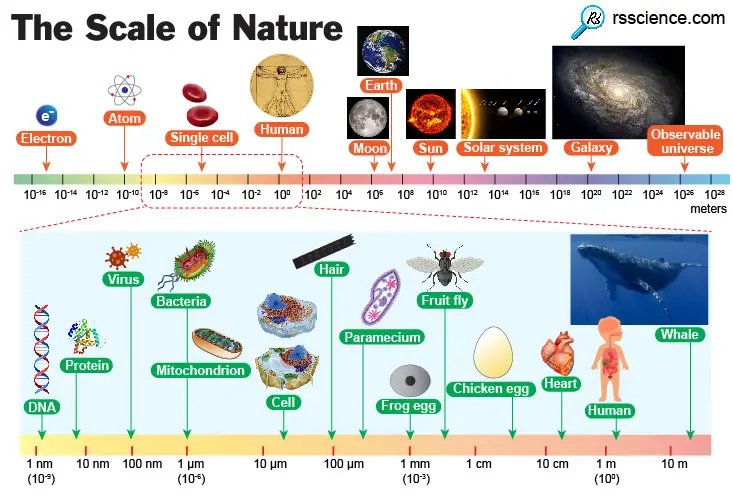
6 Reasons Why the Biological Scale is Important
Why the Biological Scale is Important? Knowing what you can see and what you cannot…
Size Matters – The Scale of Biology – Examples and Fun Facts
From the diameter of DNA molecules (2 nanometers) to the height of an adult (1.75…
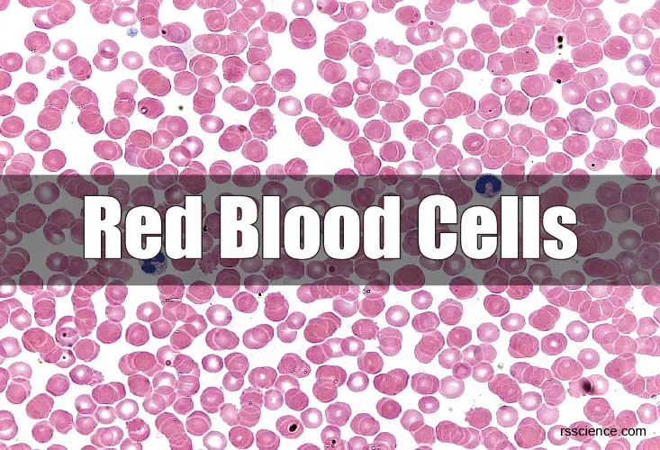
Red Blood Cells – Definition, Biology, and Observation under the Microscope
What are red blood cells – an overview Red blood cells (RBCs, also called erythrocytes)…
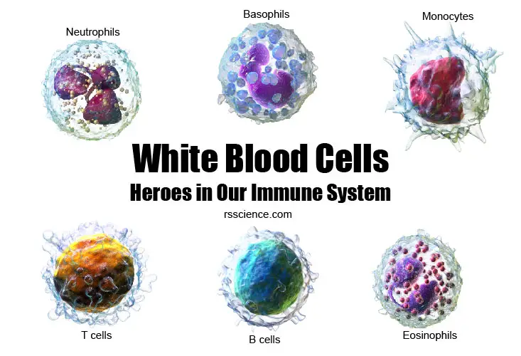
White blood cells – Types, Biology, and Observation under the Microscope
White blood cells are a critical part of our body’s immune system. Types of white…
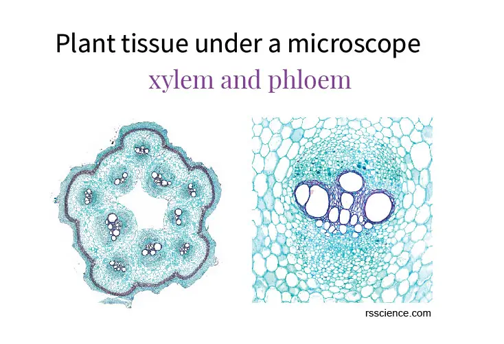
Plant tissue under a microscope – xylem and phloem
Guide to plant anatomy and biology using microscope premade slide sets. We can observed roots,…
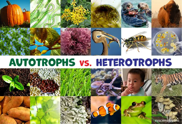
Autotrophs vs. Heterotrophs – Definition and Examples
Autotrophs and heterotrophs are two nutritional groups in ecosystems. Autotrophs produce their own food whereas…

How to Read the Amino Acids Codon Chart? – Genetic Code and mRNA Translation
Cells need proteins to perform their functions. Amino acids codon chart (codon table) is used…
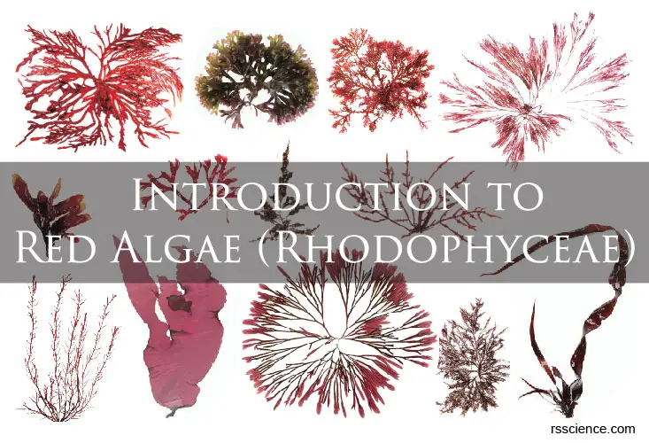
Red algae – characteristics, structure, reproduction, and examples
Red algae, also called Rhodophyta, are a large group of eukaryotic organisms that can photosynthesis….
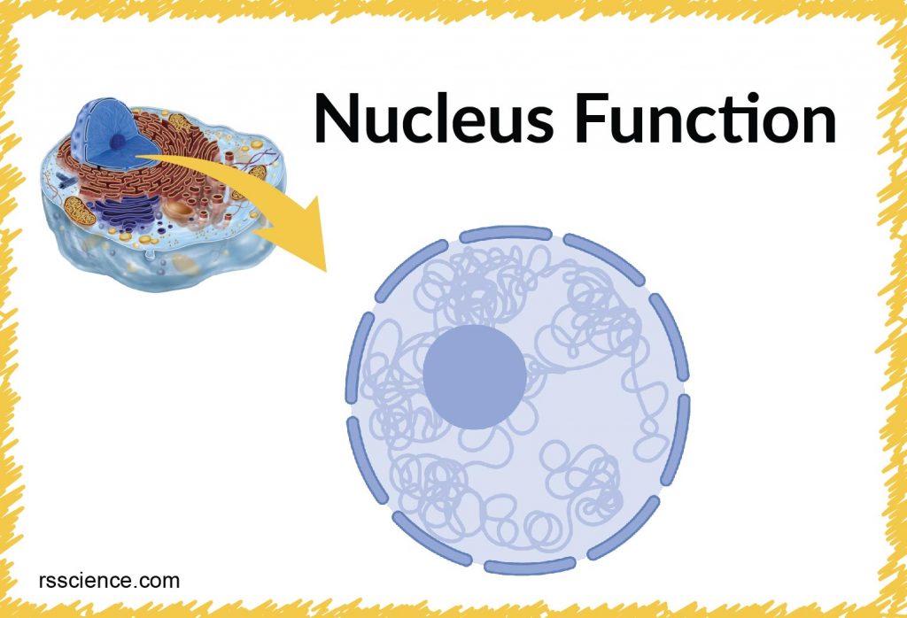
Cell Nucleus – function, structure, and under a microscope
Nucleus is a double-membrane organelle, and it controls cell activities and carries genetic information to…
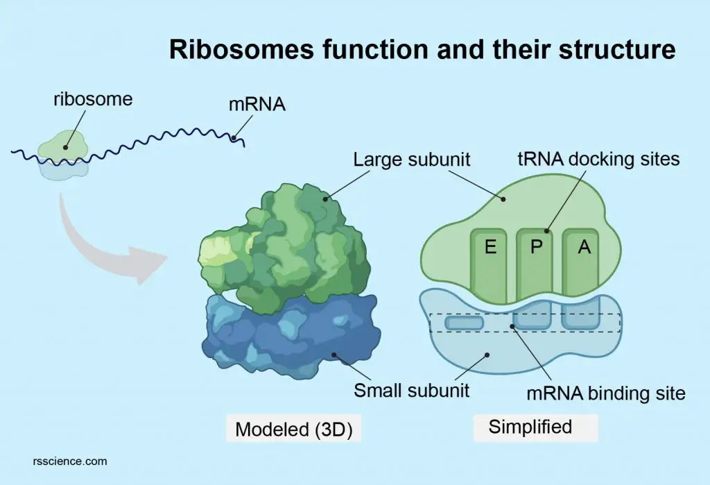
Ribosome – protein factory – definition, function, structure, and biology
Ribosomes are complex molecular machines that make proteins inside cells. Ribosomes consist of two subunits…
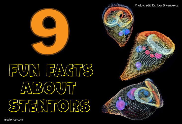
9 Fun Facts About Stentors
Stentors are one of the biggest single-cell microorganisms. They also have fascinating avoidance behaviors,…
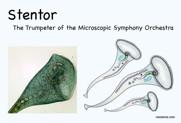
Stentor – The Trumpeter of the Microscopic Symphony Orchestra
Stentor, also called Trumpet Animalcule, is a single cell protist. They have a horn-shaped body…
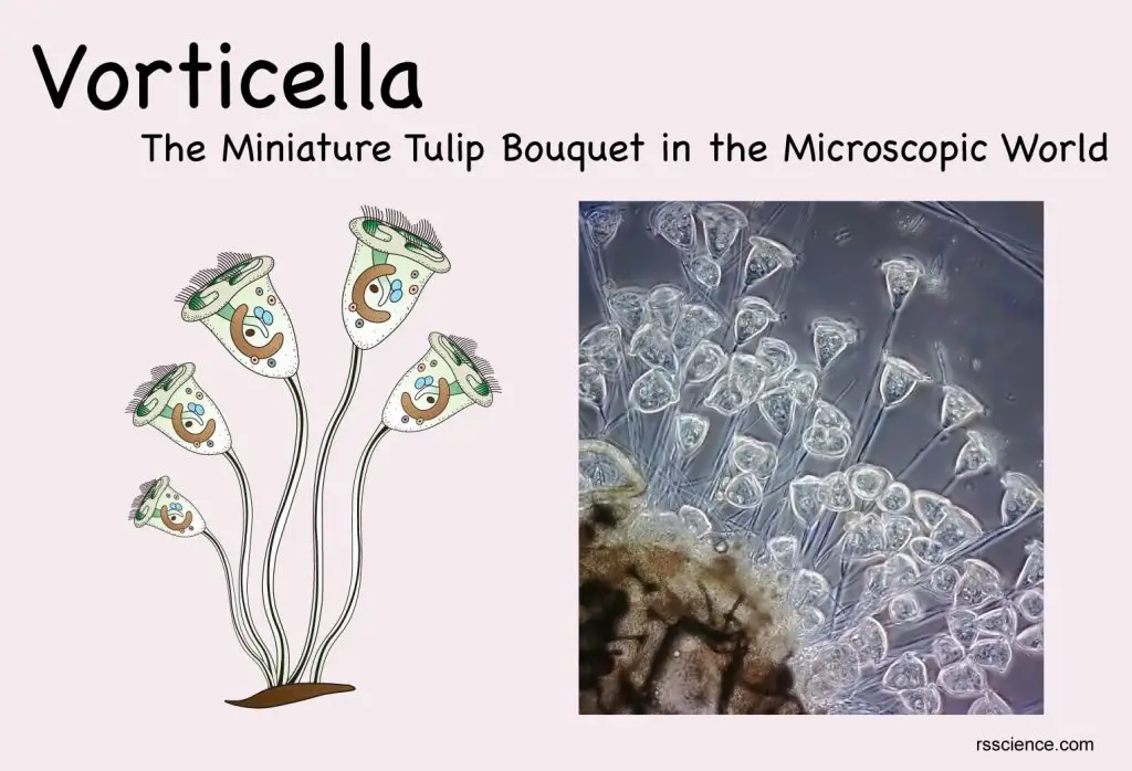
Vorticella – The Miniature Tulip Bouquet in the Microscopic World
Vorticella is a type of ciliate protozoans. They are tiny, single-cellular, and animal-like microorganisms with…
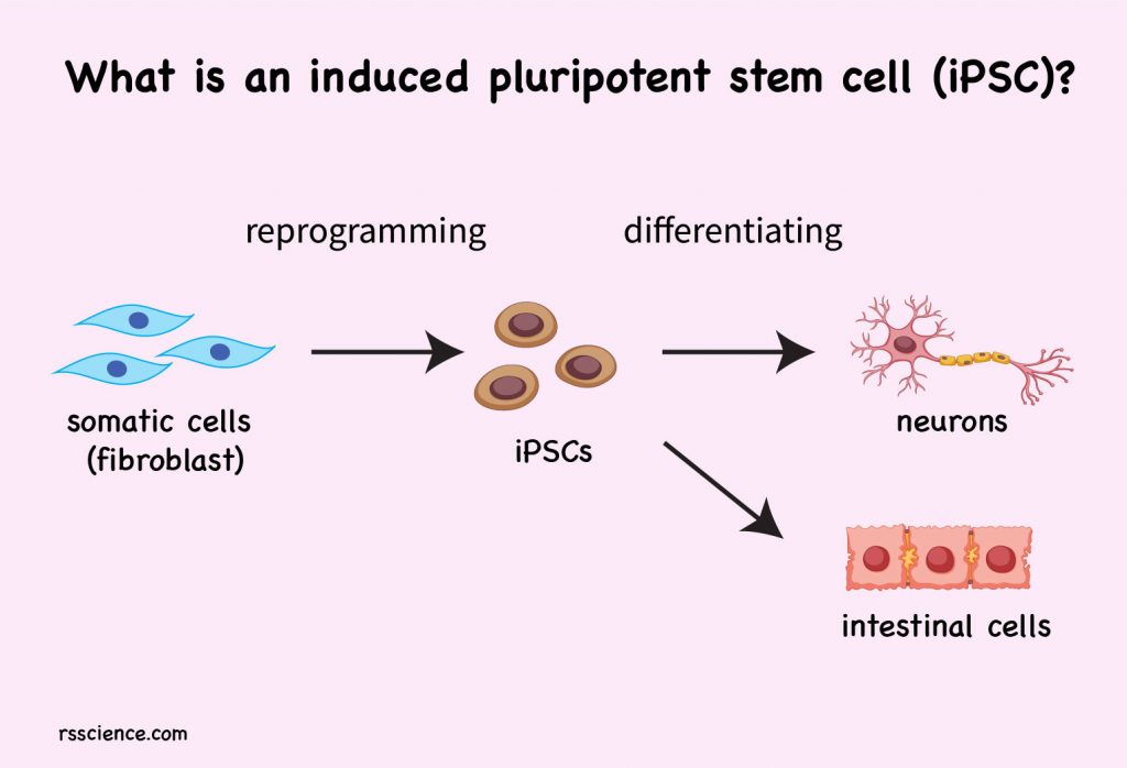
What is an induced pluripotent stem cell (iPSC)?
Induced pluripotent stem cells (iPSCs) are pluripotent stem cells that can be generated and reprogrammed…
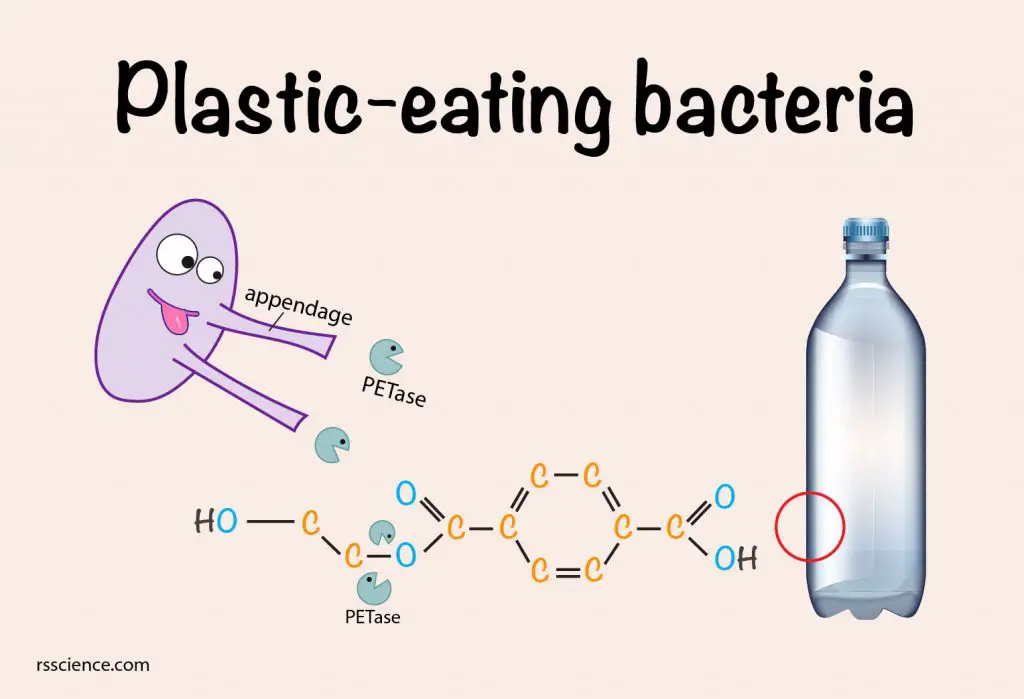
Plastic Eating Bacteria – how they work
The plastic-eating bacteria, called Ideonella sakaiensis 201-F6, can secreate enzymes (PETase and MHETase) to break…
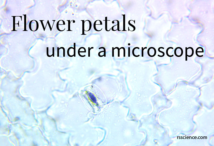
Flower Petals under a Microscope
Petals are modified leaves that surround the reproductive parts of flowers. They are often brightly…

3 Science Projects You Can Do in the Spring
1. Identify lives belonging to each of the Five Kingdoms; 2. See how lives survive…
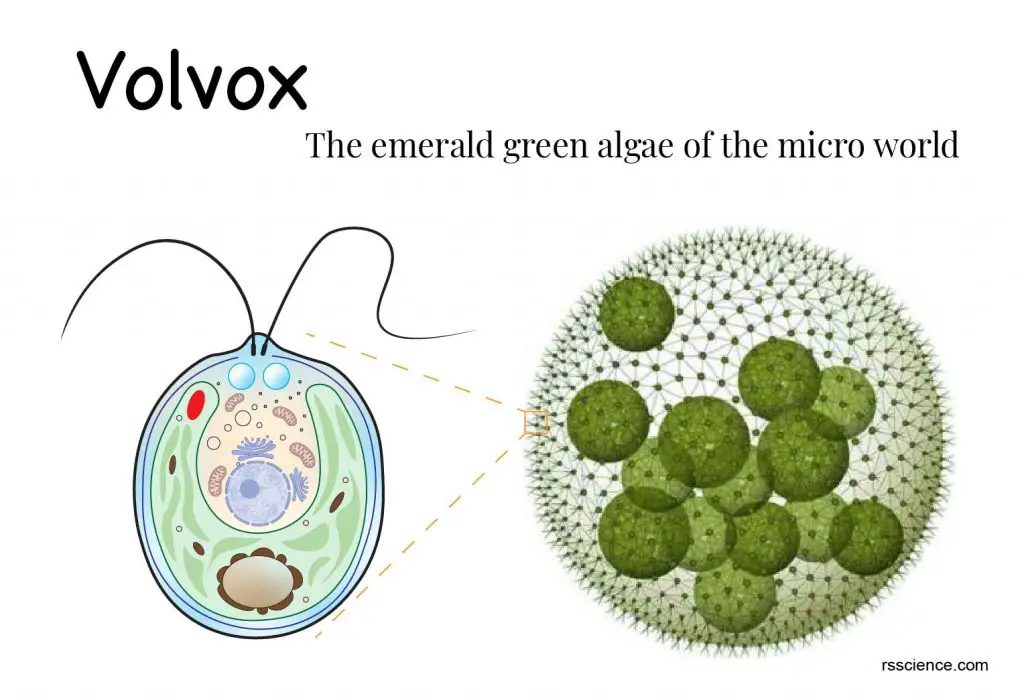
Volvox – The emerald green algae of the micro world
What is a Volvox? A quick overview Volvox is a genus of green algae. Volvoxes…
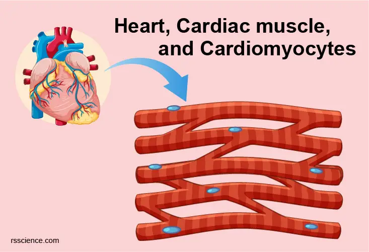
Cardiomyocytes (Cardiac Muscle Cells) – Structure, Function, Cell Biology and Histology
Cardiomyocytes are the muscle cells that make up the heart muscle. Cardiomyocytes go through a…
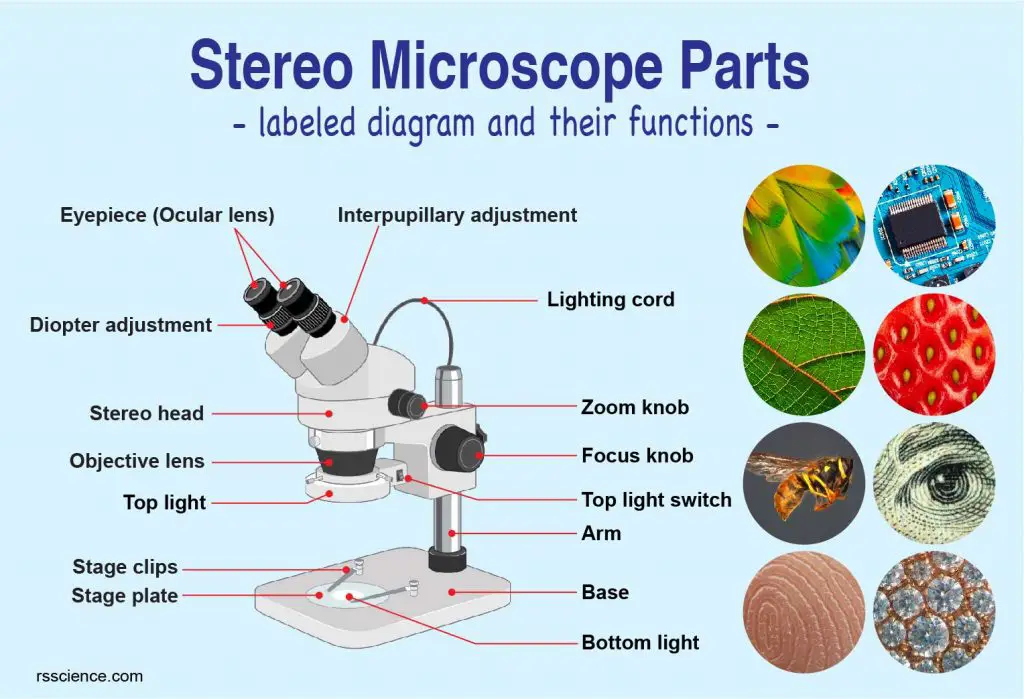
Parts of Stereo Microscope (Dissecting microscope) – labeled diagram, functions, and how to use it
A Stereo microscope is like a powerful magnifying glass, good for thick and solid specimens…

How to Identify Mono vs. Dicot Plants in Your Garden
Common examples of monocots are corn, rice, wheat, orchids, bamboos, and bananas. Dicots are Roses,…
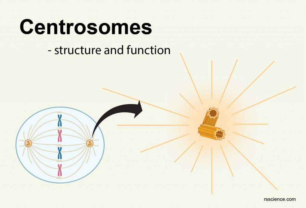
Centrosomes – the engine of cell division – definition, structure, function, and biology
A centrosome comprises two centrioles that serve as microtubule organizing center (MTOC). Its function is…
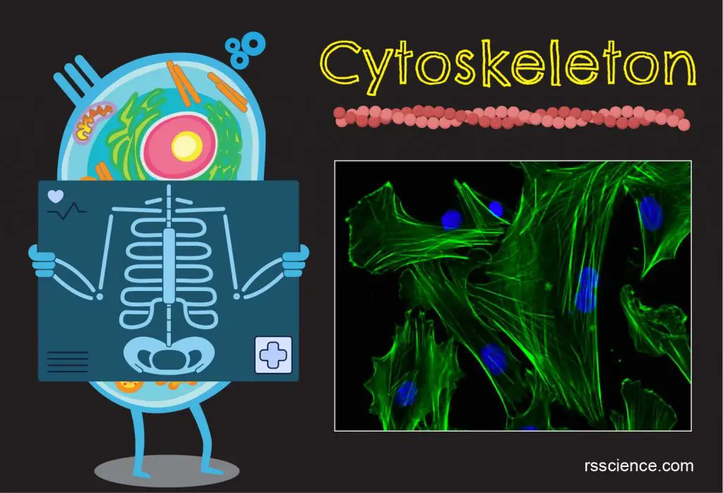
Cytoskeleton – the muscle and the bone of a cell – definition, structure, function, and biology
The cytoskeleton is a network of filament proteins that extends throughout a cell. It gives…
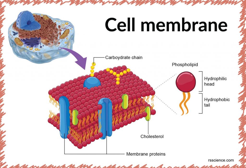
Cell membrane – definition, structure, function, and biology
The cell membrane is a lipid bilayer that separates the interior of cells from the…
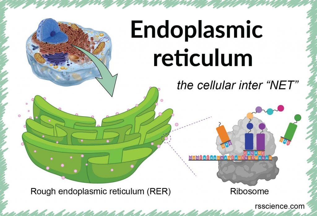
Endoplasmic reticulum – the cellular inter “NET” – definition, structure, function, and biology
Endoplasmic reticulum (ER) is an internal membrane that forms branching networks of many interconnected sacs…
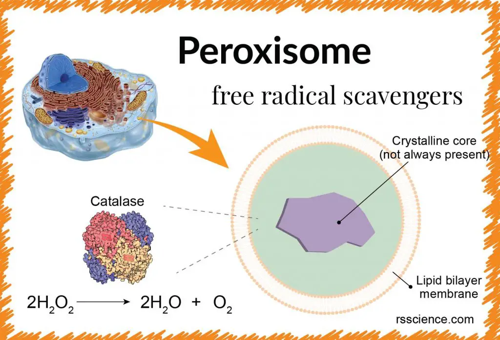
Peroxisome – free-radical scavengers – definition, structure, function, and biology
The peroxisome is a spherical membrane-bound organelle responsible for the fatty acid breakdown and the…
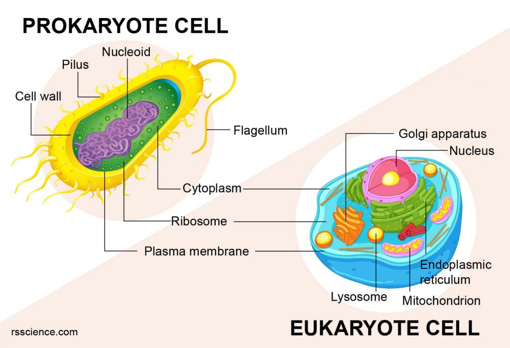
Eukaryotes and Prokaryotes – What are the Similarities, Differences, and Examples
All living organisms fall into one of two categories: Eukaryotes (plants, animals, and fungi) or…
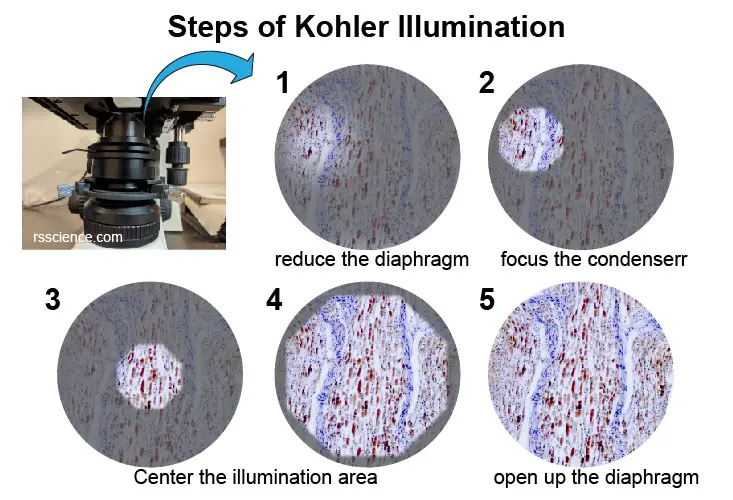
What is Kohler illumination, why is it important, and how to do it?
Kohler illumination is a technique that ensures even illumination of the specimens. It requires a…
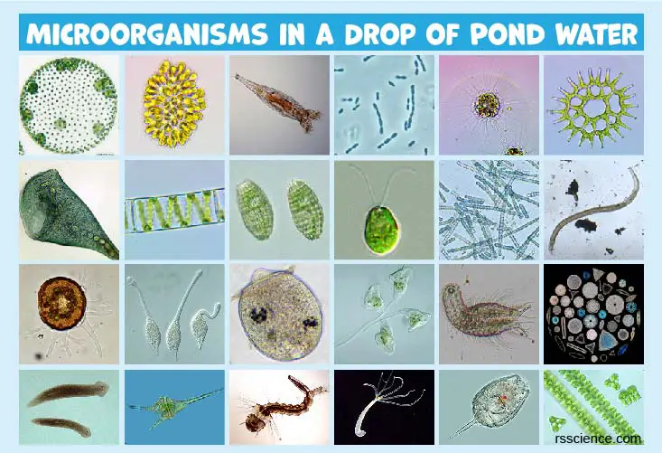
Microscopic Organisms in a Drop of Pond Water
Microorganisms are microscopic organisms that include bacteria, archaea, and protist (protozoa, protophyta, and mold). They…
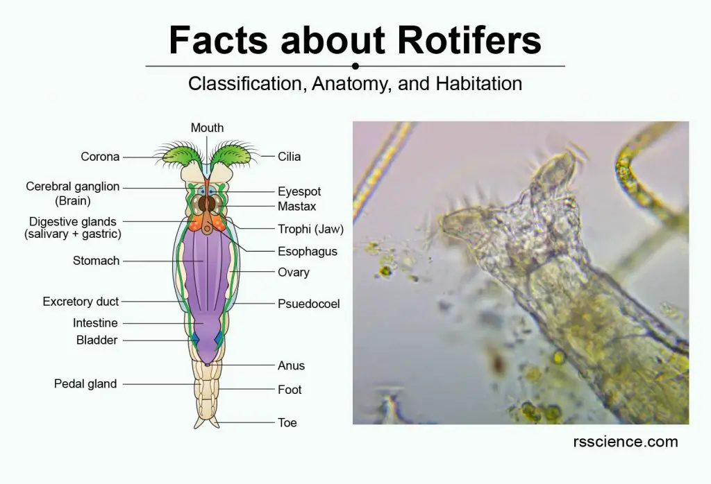
Facts about Rotifers – Amazing Microscopic Animals under the Microscope
Rotifers are multicellular (~1000 cells) animals and 100-500 μm in size. Rotifers got their name…
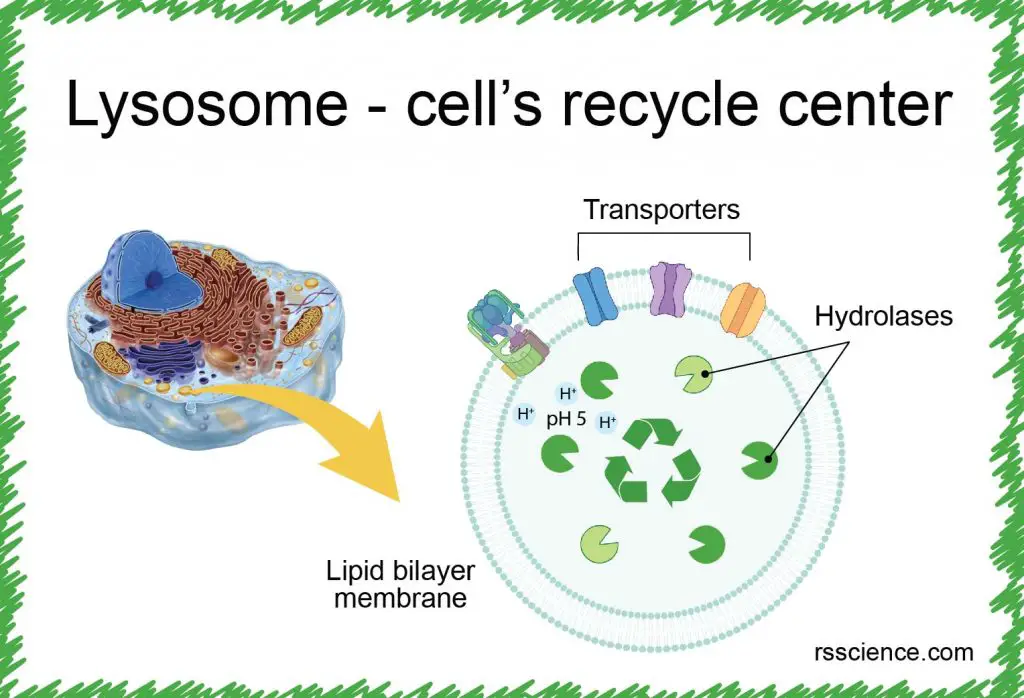
Lysosome – the cell’s recycling center – definition, structure, function, and biology
Lysosomes are small organelles that work as the recycling center in the cells. Many lysosomal…
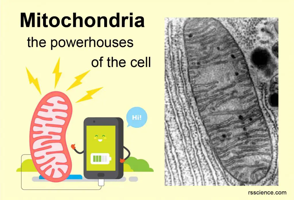
Mitochondria – the powerhouses of the cell – definition, structure, function, and biology
Mitochondrion is a rod-shaped organelle and its function is to generate energy (ATP) for the…

Different types of Microscopes – light microscope, electron microscope, scanning probe microscope.
Three types of microscopes: light microscopes (compound, stereo, inverted, fluorescence, confocal, and super-resolution microscope), electron…
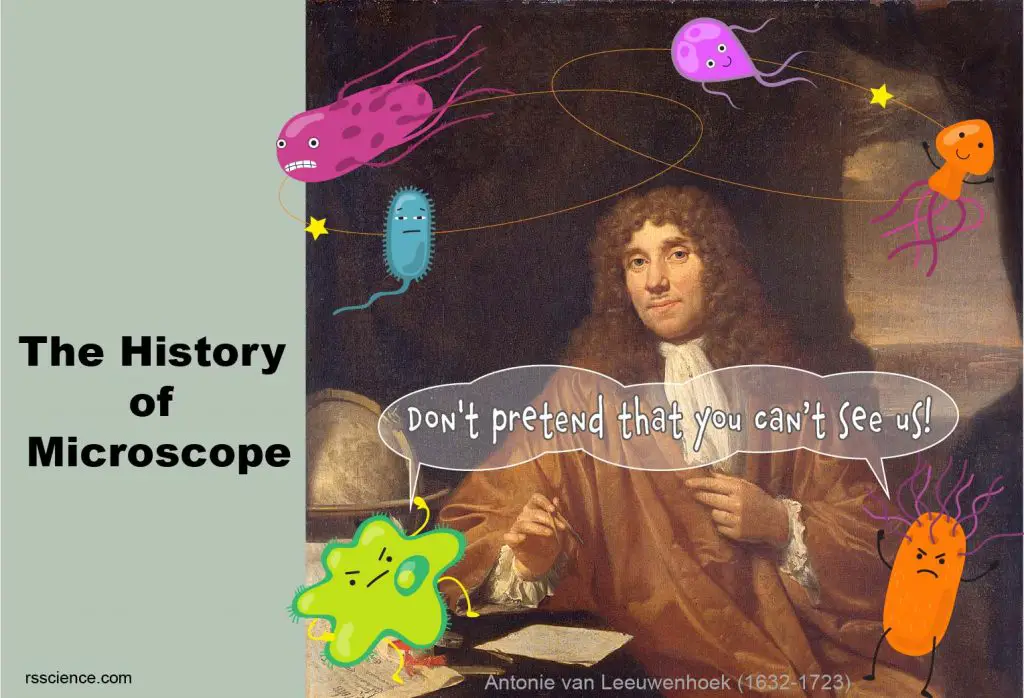
Who Invented the Microscope? History of Microscope
A simple microscope is the earliest type of microscope. It has only one lens and…
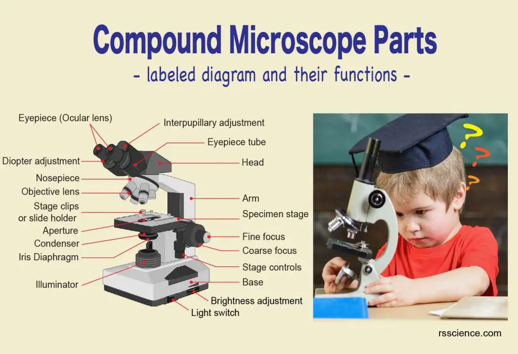
Compound Microscope Parts – Labeled Diagram and their Functions
Microscope parts include eyepiece (10x), objective lenses (4x, 10x, 40x, 100x), fine and coarse focus,…
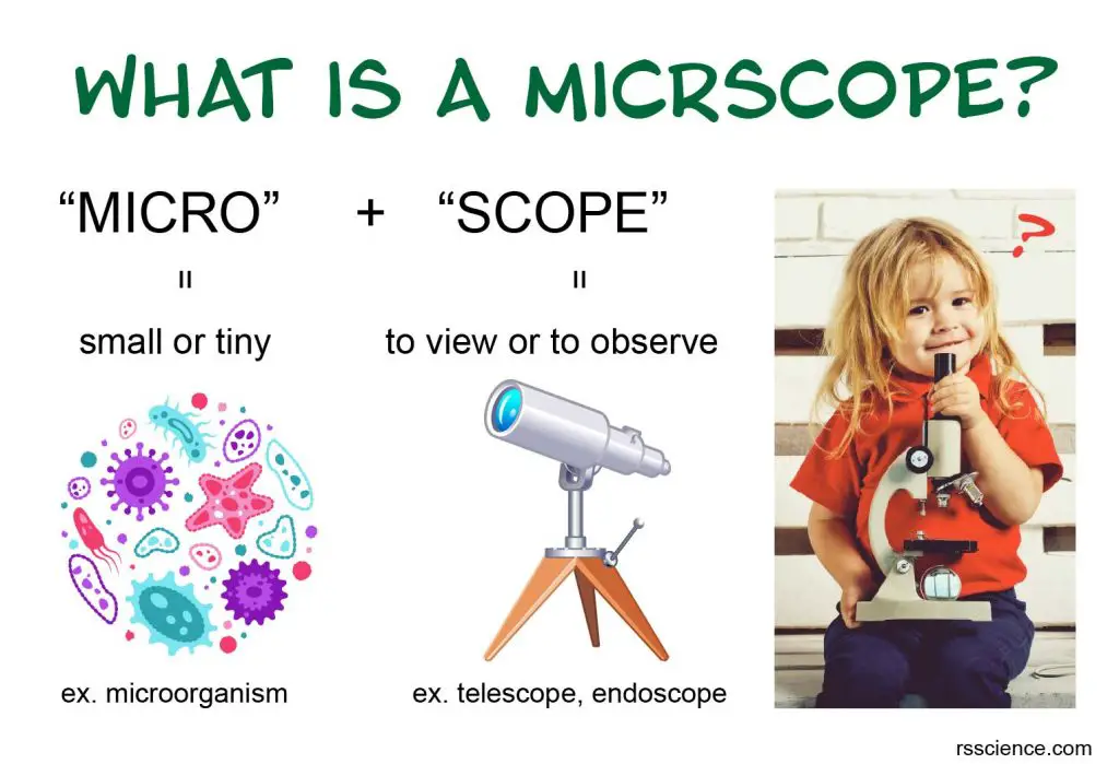
What is a Microscope? Function and Magnification
A microscope is an instrument to see objects that are too small to be seen…
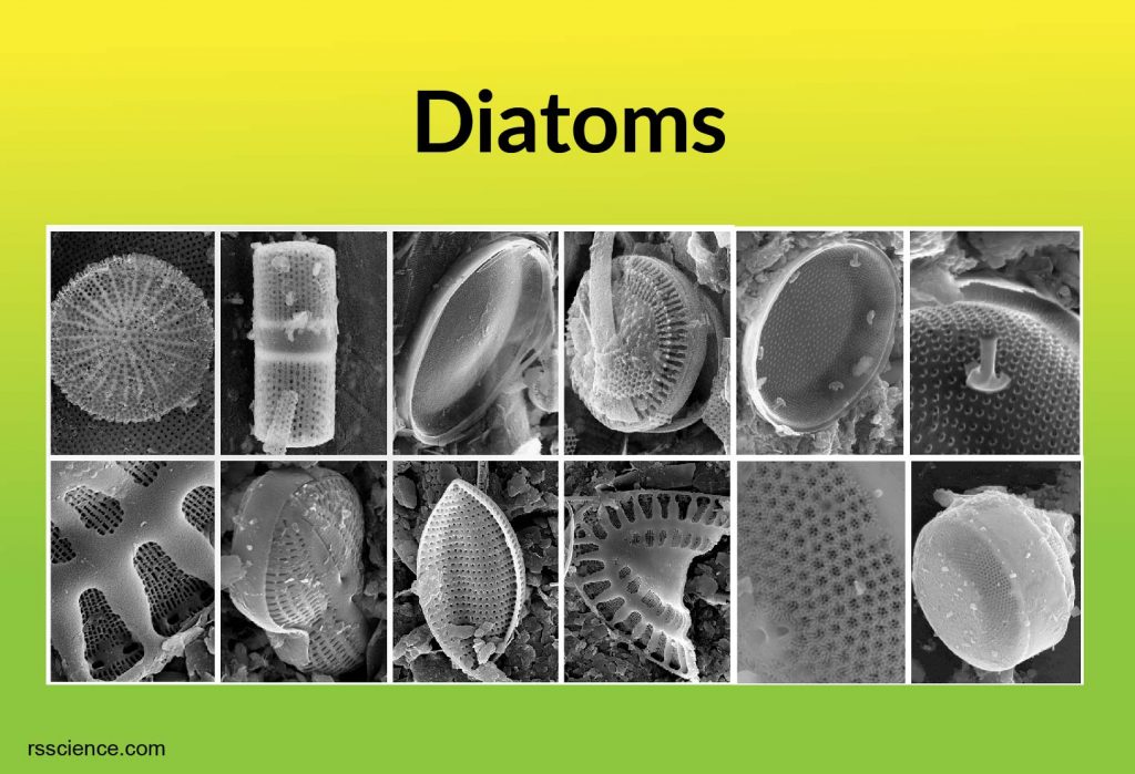
Diatoms
Diatoms are free-floating unicellular algae in oceans and freshwater. Their cell walls are made of…
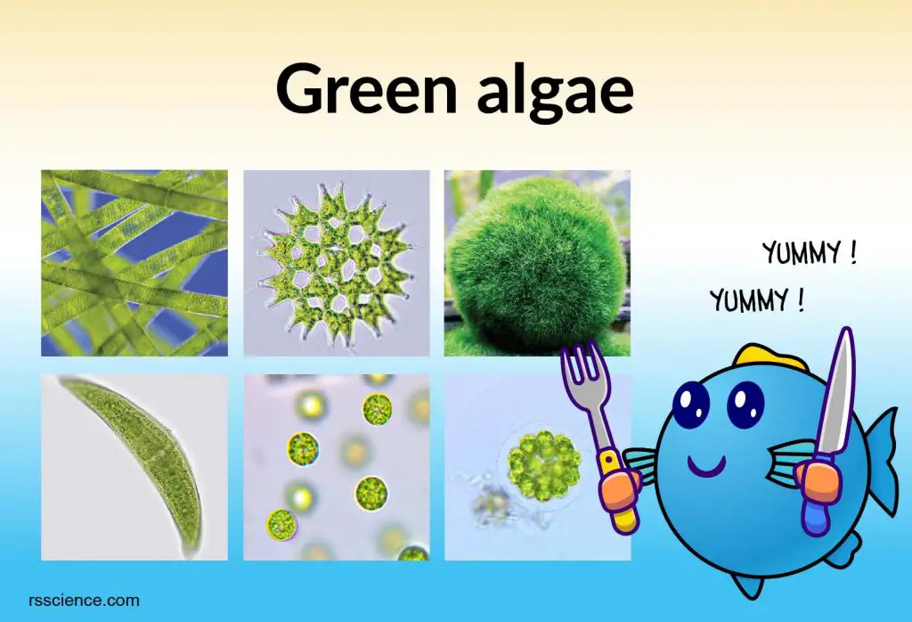
Green Algae
Green algae are a diverse group of photosynthetic eukaryotic organisms. It includes unicellular and multicellular…
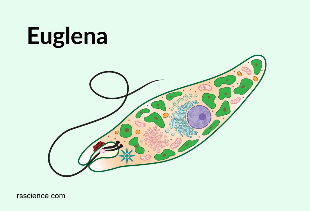
Euglena under a microscope – anatomy, reproduction & facts
Euglena is a single-celled eukaryote with flagella and it shares some characteristics of both plants…
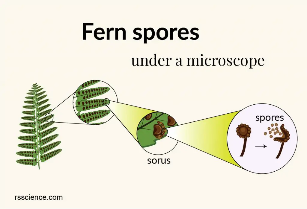
Fern Spores Under a Microscope
Ferns are vascular plants. Unlike other vascular plants that reproduce via seeds and flowers, ferns…
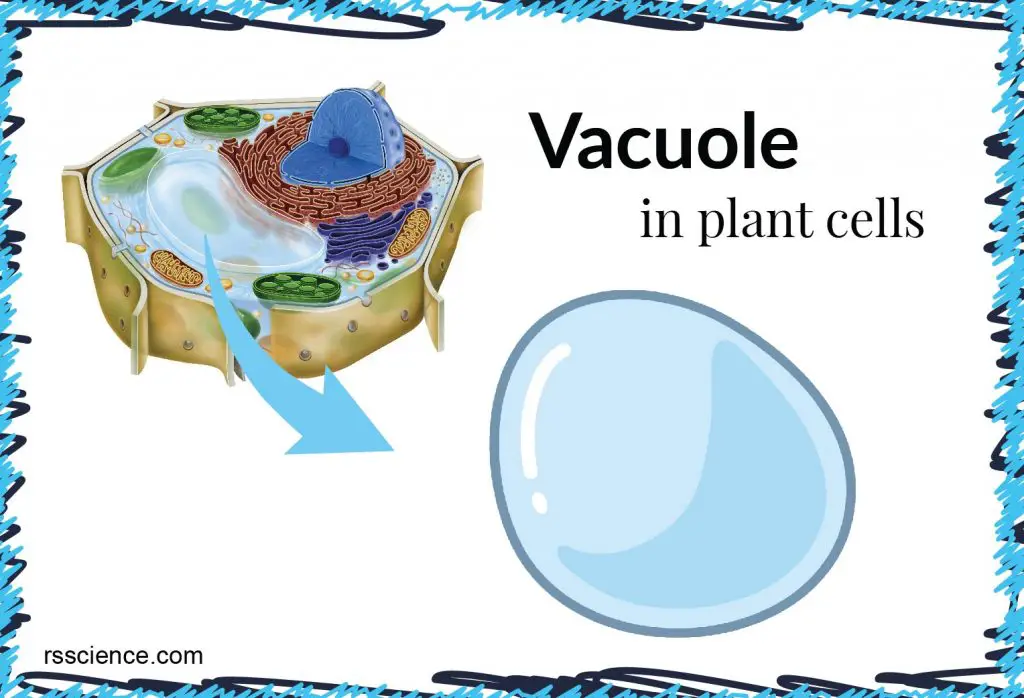
Vacuole Function and Structure – Extra Space Storage
The vacuole is a membrane-bound organelle that is present in all plant cells. The main…
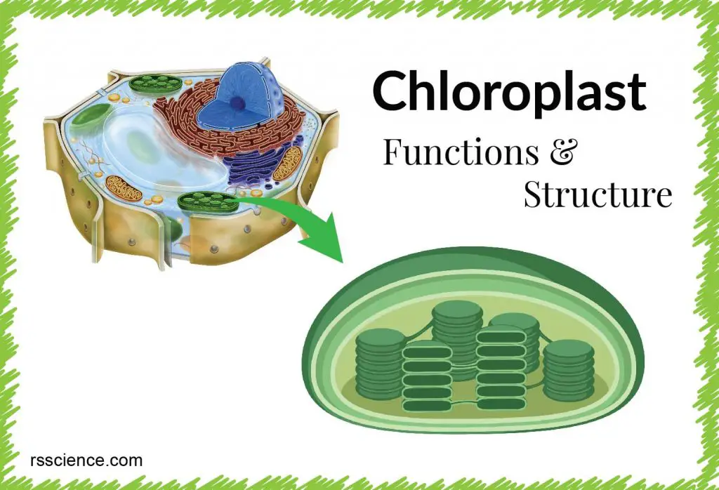
Chloroplast Function and Structure – Solar Panels
The chloroplast main function is to convert energy from the Sun into glucose for growth,…
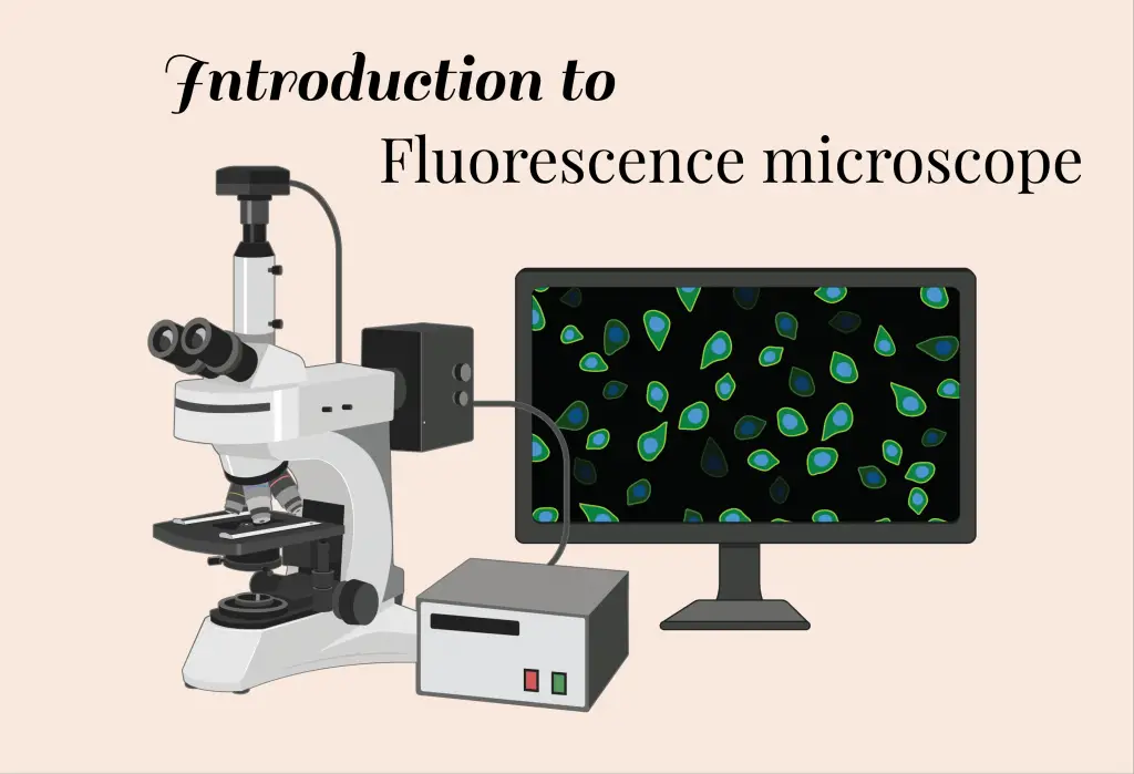
Fluorescence Microscope
A fluorescence microscope is an optical microscope that uses fluorescence instead of other light properties…
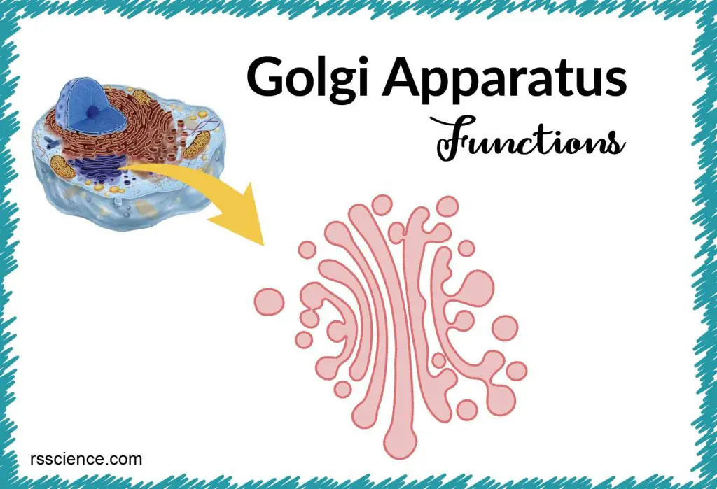
Golgi Apparatus Function – the Post Office inside the Cells
Golgi apparatus is a membrane-bound organelle found in most eukaryotic cells. The function of Golgi…
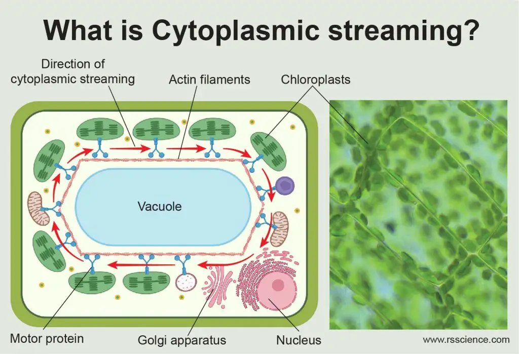
What is Cytoplasmic Streaming?
Cytoplasmic streaming is the movement of the cytoplasm within a cell. It plays an important…
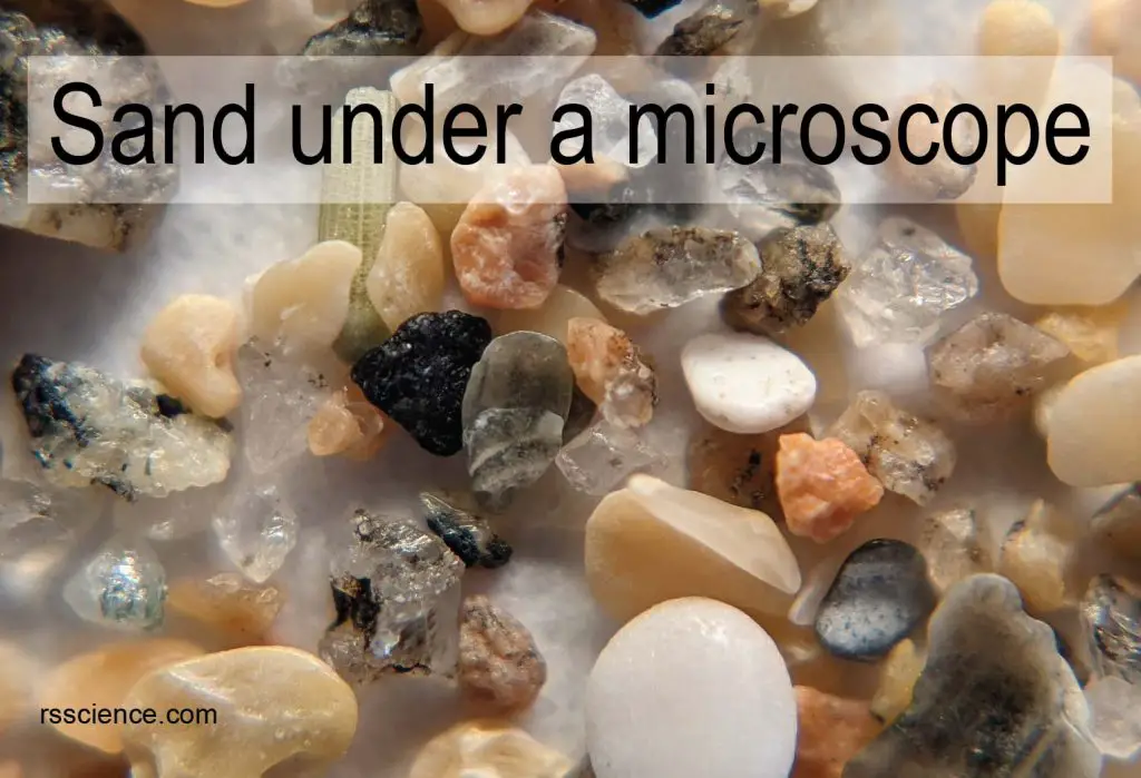
Sand Under a Microscope
Have you ever seen sand under a microscope? If not, come to join us. This…
Cell Organelles and their Functions
A cell organelle is a tiny cellular structure that performs specific functions within a cell,…
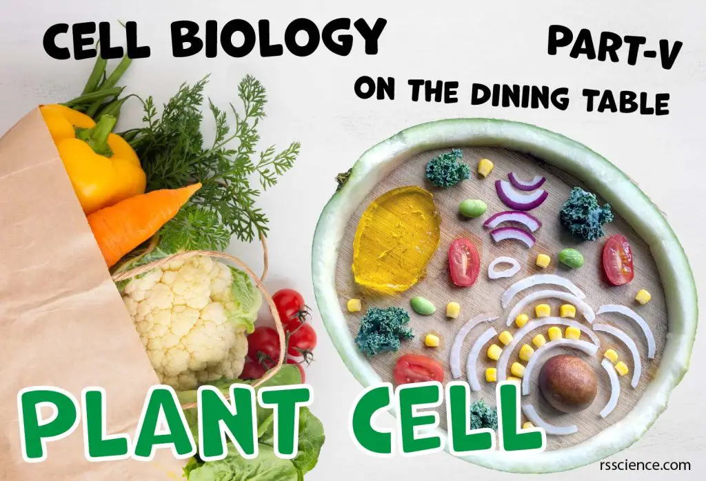
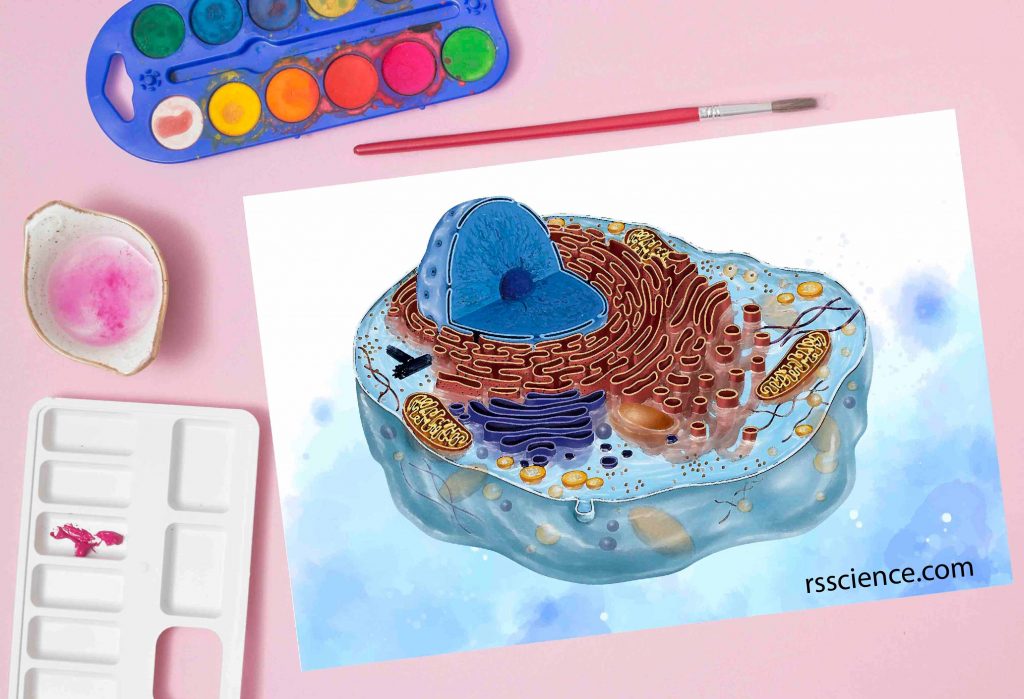
Cell Biology on the Dining Table – Plant Cell Model
Plant cells share many organelles in common with animal cells. There are three unique features…
Microscopy Stains
Microscopy stains enhance the visualization of cells and cell parts under a light microscope. Methylene…
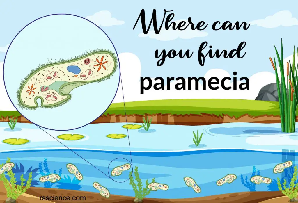
The Natural Habitation and Cultivation of Paramecium
We discuss the place where to find paramecium, how to make paramecium culture at home,…
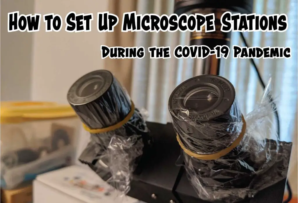
How to Set Up Microscope Stations During the COVID-19 Pandemic
Our idea is to cover the microscope parts that each user may touch with plastic…
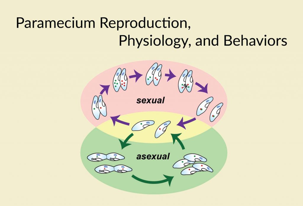
Paramecium Reproduction, Physiology, and Behaviors
Paramecium reproduction, aging, learning and memory ability, movement, sensing, feeding behaviors, and their endosymbiotic relationship…
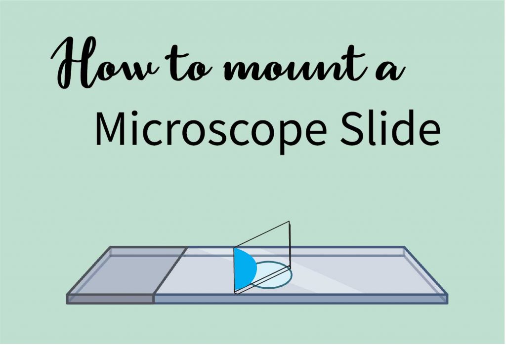
How to Mount a Microscope Slide
Carefully lower the coverslip slowly with an angle. This permits air to escape from the…
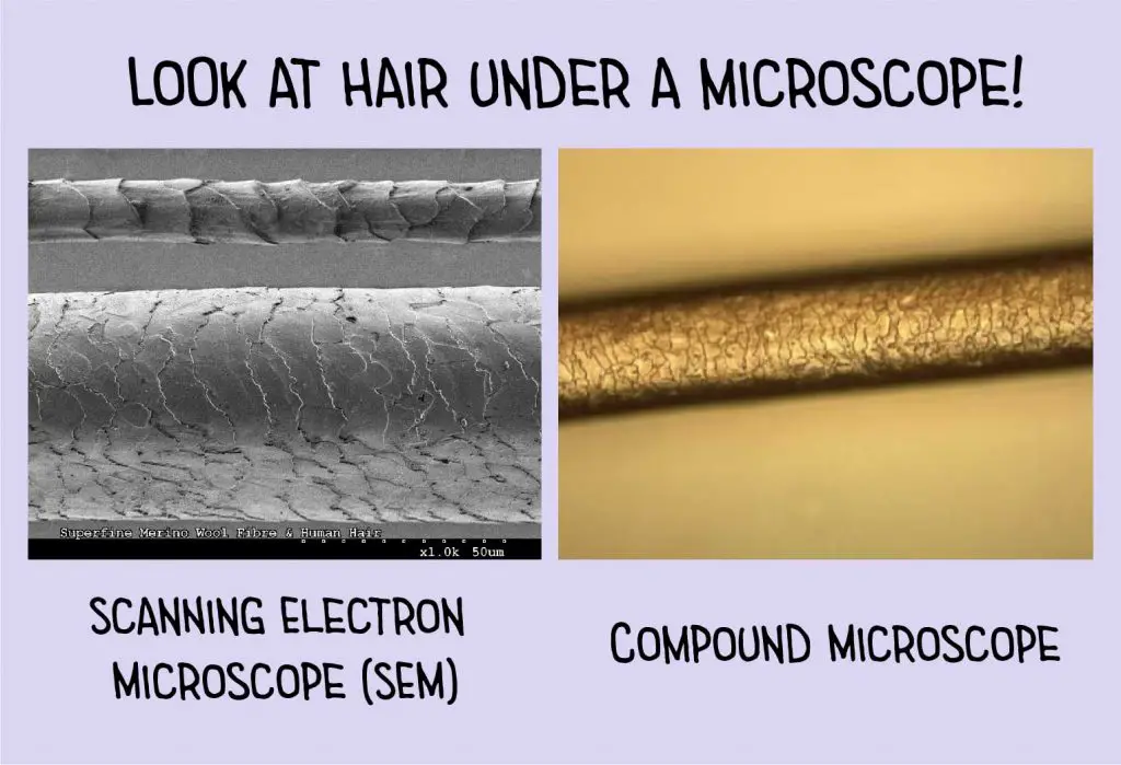
Hair Under a Microscope
This post discusses the biology, the structure, the stereo and compound microscopic view of hairs,…
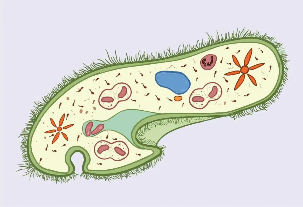
The Structure of Paramecium Cell
Paramecium uses cilia for movement. It contains two types of nuclei, Macronucleus for gene expression…
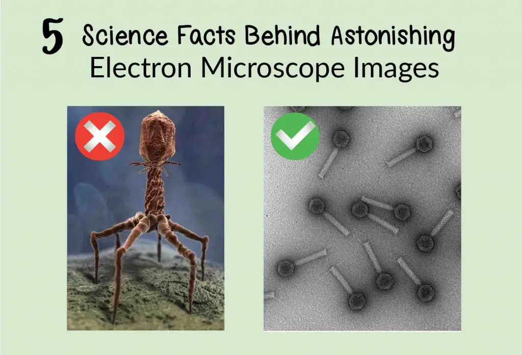
5 Science Facts Behind Astonishing Electron Microscope Images
Were images taken by electron microscopes? You will learn how to recognize images from EM…
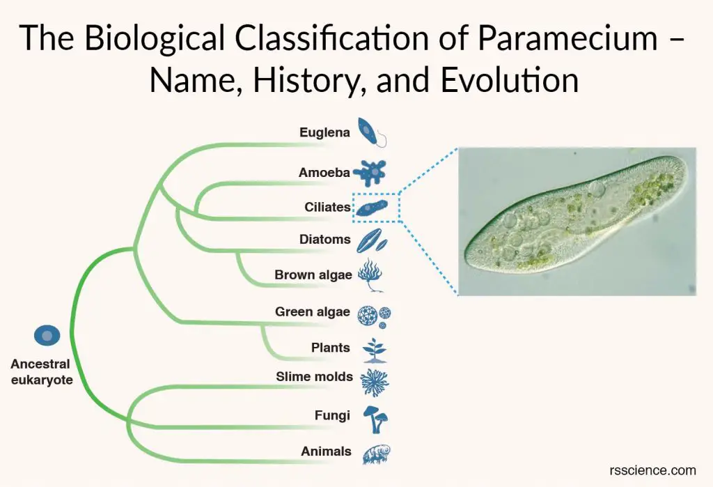
The Biological Classification of Paramecium – Name, History, and Evolution
Paramecium is a unicellular organism with a shape resembling a slipper and 50-300 µm in…
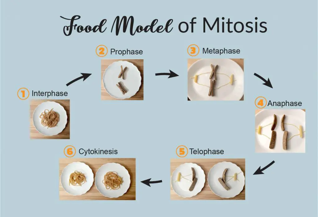
What is Mitosis (Food model of mitosis)
Mitosis is a process of cell division, one cell divides and produces two new cells…
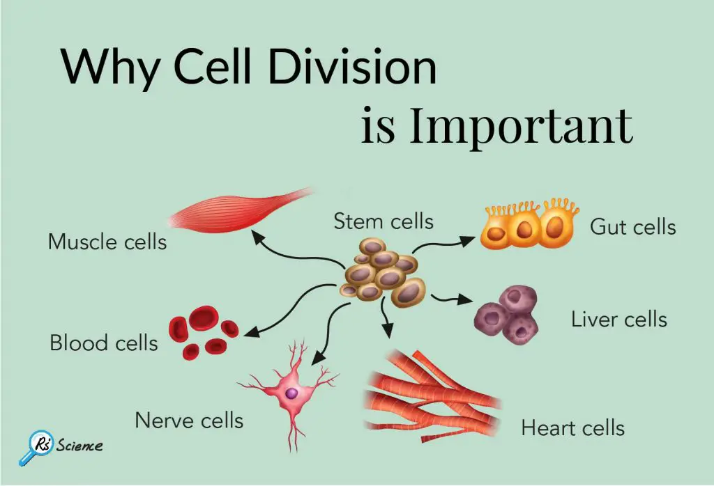
Why Cell Division is Important
Cell division is necessary for the growth of organisms, repair of damaged tissues, healing and…
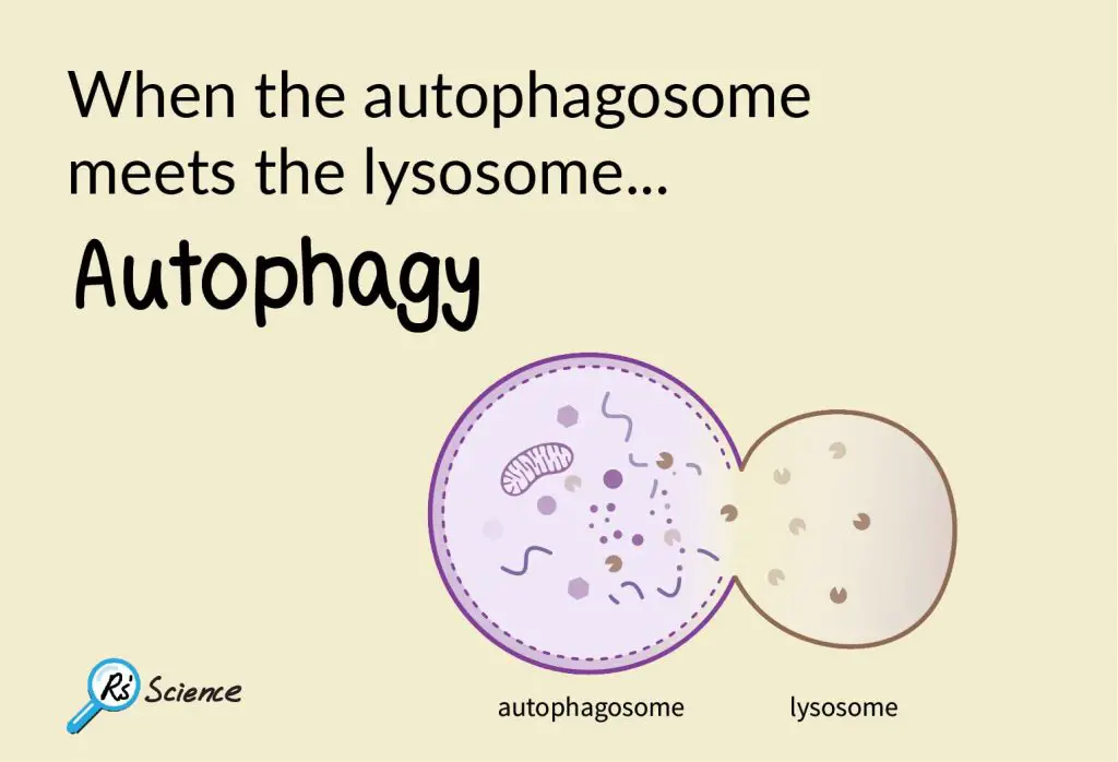
The Function of the Autophagosome and Endosome
Autophagosomes break down the misfunctioned organelles and recycle the nutrient. Endosomes bring outside material into…

Monocot vs Dicot plants
I am often amazed by the delicate structure of the cross-section of the plant stems…
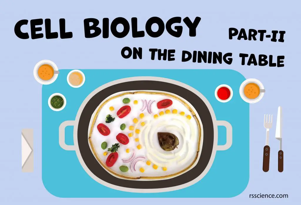
Cell Biology on the Dining Table – Animal Cell Model Part II
Use this guide to build a creative animal cell model using household items. You will…
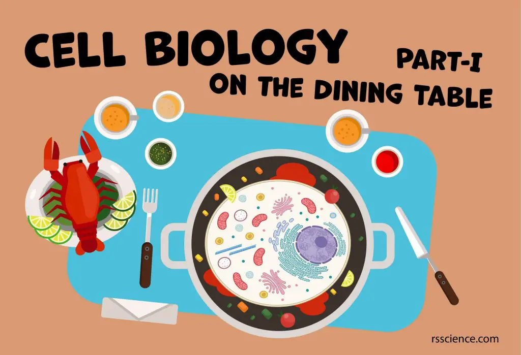
Cell Biology on the Dining Table – Animal Cell Model Part I
We choose fruit and vegetable to create our own cell model project. For example, we…
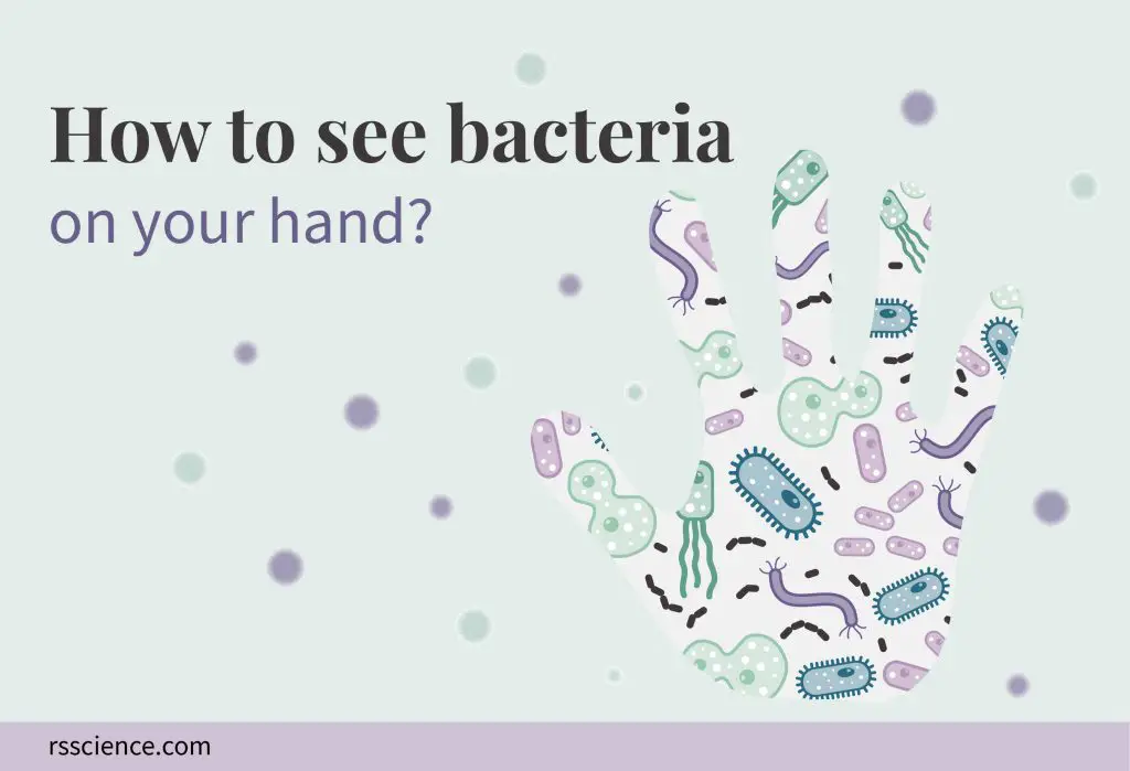
How to See Bacteria on Your Hand (Bacteria Handprint)
A step-by-step guide to make your bacteria handprint. Bacteria colony morphology differs in shape, size,…
Biological Stains for Microscope You Can Find at Home
Biological stains are used for better visualization of microscope specimens. Some pet medicine at your…
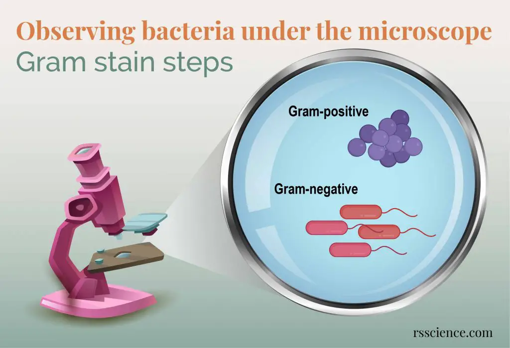
Observing Bacteria Under the Microscope – Gram Stain Steps
Bacteria are hard to see because they are tiny and colorless. One way is to…
10 Everyday Things You Should Look at Under a Microscope
They are cheek cells, onion skin, yeast cells, mold, eggshell membrane, water bears, pond water…
17 Science Facts of COVID-19
Patients with mild or no symptoms can still spread their viruses. The current diagnosis method…

How to Find Tardigrades (Water Bears)
We found water bears on the dry moss, frozen lichens on the trees and rocks….
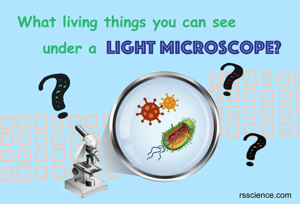
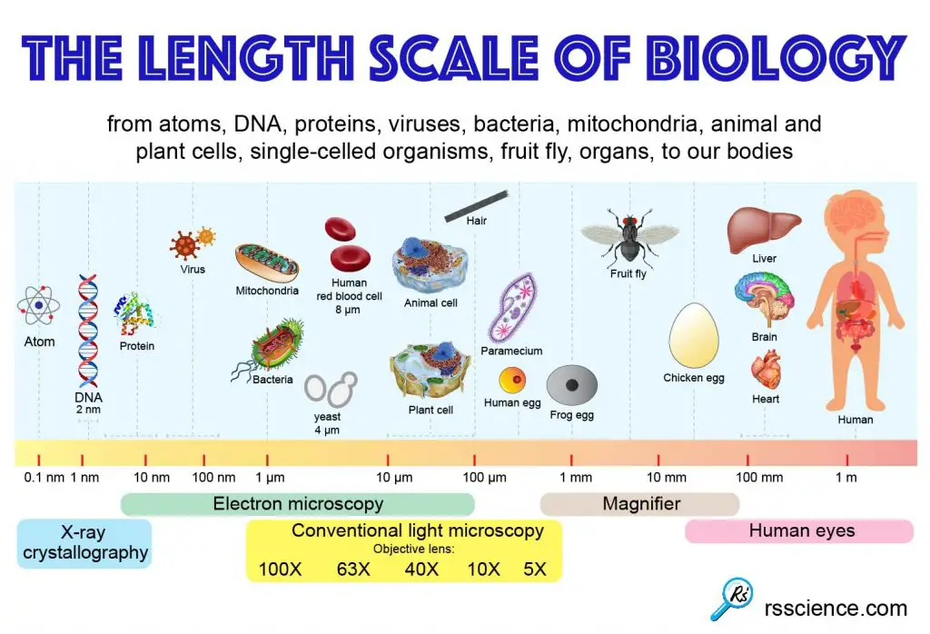
What Living Things You Can See Under a Light Microscope?
Most cells are visible under a light microscope, but mitochondria and bacteria are barely visible….
Can You See Viruses Under a Microscope?
No. Viruses are too small to be seen with an optical microscope. An electron microscope…

How to Choose the Right Microscope (Compound Microscope vs. Stereo Microscope)
How to choose the right microscope for your need. We discuss types of samples are…
13 Tips You Should Know about Taking Care of Your Microscope
Best tips you should know to take care of your microscope. This post includes how…
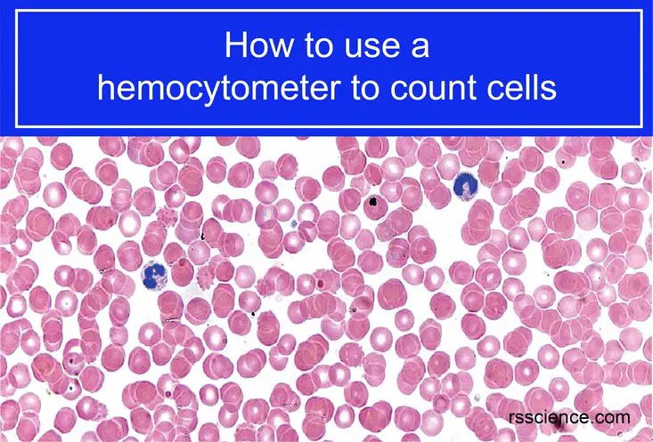
How to Use a Hemocytometer to Count Cells
Everything you need to know to use a hemocytometer to count cells, cell concentrations, cell…
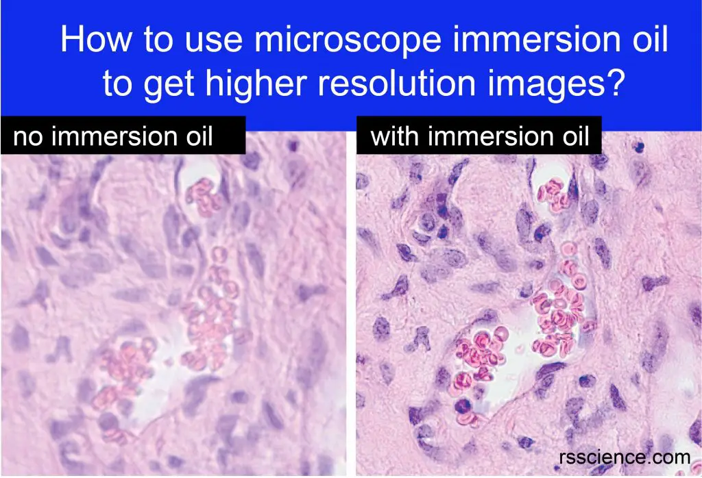
How to Use Microscope Immersion Oil to Get Higher Resolution Images
This post covers what is microscope immersion oil, why do you need it, when and…
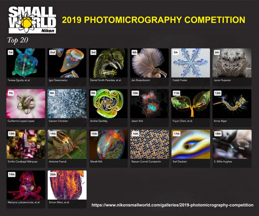
Nikon | 2019 Photomicrography Competition
Nikon is one of the world famous companies manufacturing camera and microscope. It recently announced…
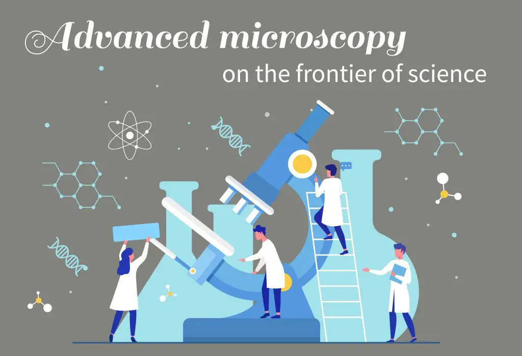
Advanced Microscopy on the Frontier of Science
Microscopes are important to study biology. We discuss light microscopes, fluorescent microscopes, and electron microscopes.
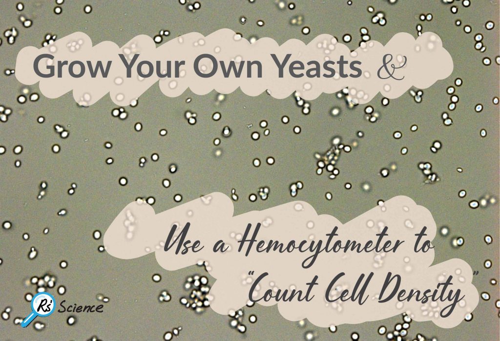
Lesson 7: Grow Your Own Yeasts & Use a Hemocytometer to “Count Cell Density”
A step by step guide for growing your own yeast and for counting the number…
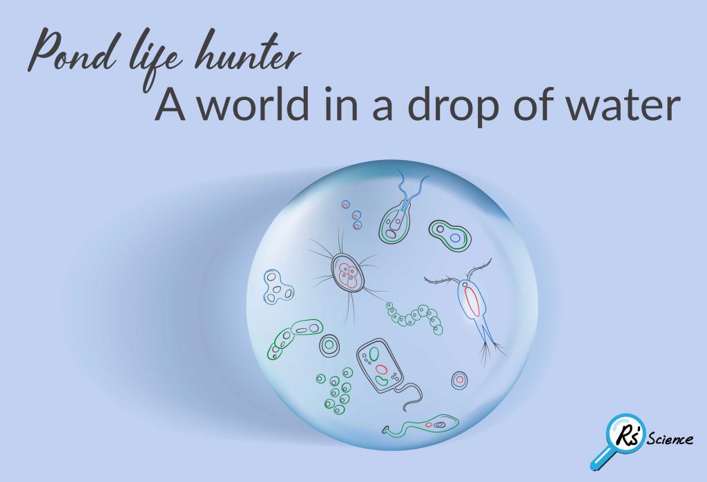
Lesson 6: Pond Life Hunter & “A World in a Drop of Water”
We saw amazing plankton tiny creatures in pond life, including ciliates, rotifers, Amoeba, Tardigrades (water…

Lesson 5: How to Do Scale Casting with Clear Nail Polish & See “Microtopography”
Guide to do scale casting with clear nail polish to see microtopography. We demonstrated plant…
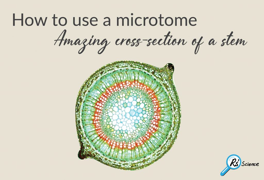
Lesson 4: How to Use a Microtome & “Amazing Cross-section of a Stem”
A step-by-step guide to use a microtome to create an amazing cross section of plant…
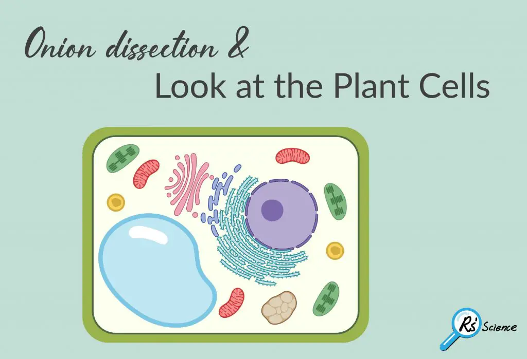
Lesson 3: Onion Dissection & “Look at the Plant Cells”
Step-by-step guide for onion dissection to get plant cells, so you can look at plant…
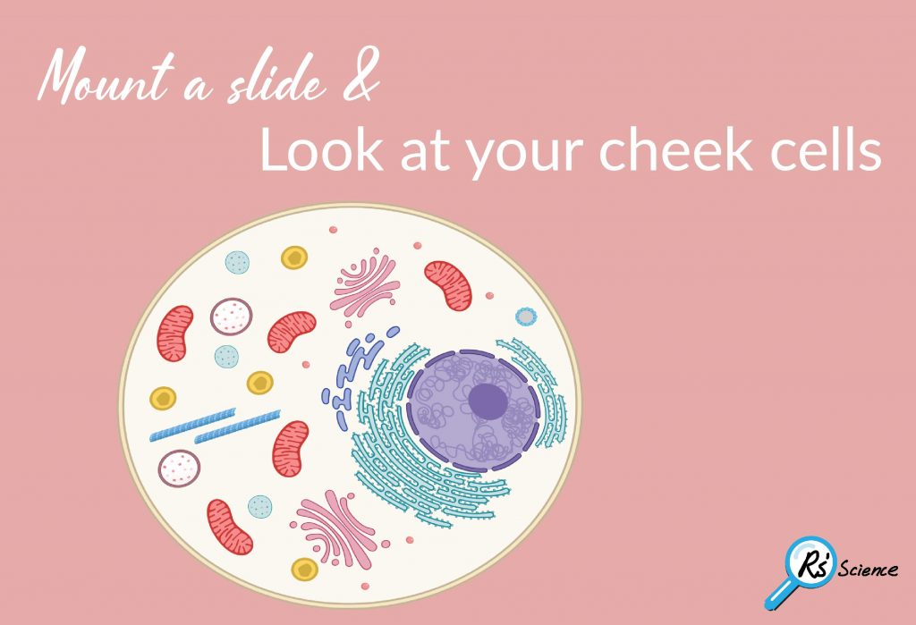
Lesson 2: Mount a Slide & “Look at Your Cheek Cells“
A step by step guide to show you how to mount a slide for microscope…

Lesson 1: Prepare a Working Space and Protect Yourself
Prepare a working space and protect yourself. This is an essential step for all science.
The Beginner’s Guide to Microscopy
A microscope guide for beginners: We discussed microscope parts; how to use a microscope; and…