
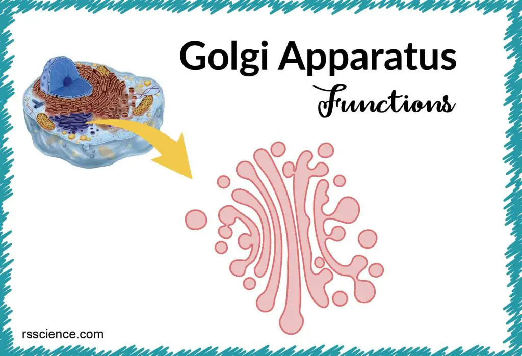
This article covers
What is Golgi apparatus?
The Golgi apparatus (also known as the Golgi complex, Golgi body, or simply the Golgi) is a membrane-bound organelle found in most eukaryotic cells. The main function of the Golgi apparatus is to process proteins and send proteins to different destinations. This is why we call Golgi apparatus the post office inside the cells.
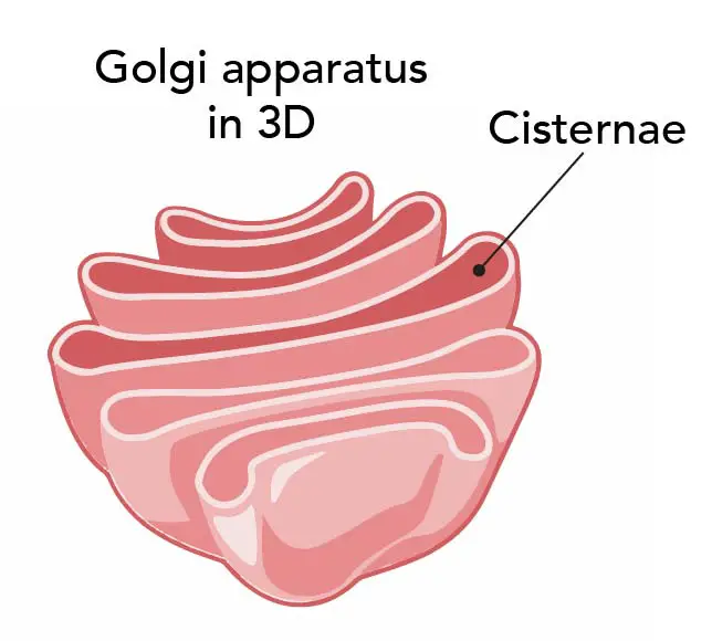
[In this figure] A 3D drawing of the Golgi apparatus.
The Golgi consists of stacks of flattened, membranous, disc-like structures called cisternae (singular: cisterna). The cisternae contain enzymes that modify proteins passing through them.
What does the Golgi apparatus look like?
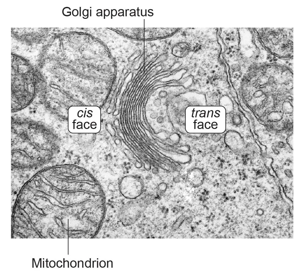
Golgi apparatus consists of several stacks of membrane-bound cisternae (sacs). Under the transmission electron microscopy (TEM), Golgi apparatus is visible as a stack of semicircular black rings surrounded by numerous circular vesicles.
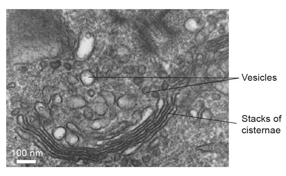
[In this figure] Transmission electron microscopic image of Golgi apparatus from a human white blood cell.
Photo credit: wiki
Where is Golgi apparatus located in a cell and how many Golgi apparatus can be found in one cell?
The subcellular localization of the Golgi apparatus may vary in different organisms. Generally speaking, Golgi apparatus stays close to the endoplasmic reticulum (ER). In mammalian cells, a single Golgi apparatus is usually located near the cell nucleus, close to the centrosome. In yeasts and plant cells, multiple Golgi apparatuses are scattered throughout the cytoplasm. One plant cell can have more than one hundred Golgi.
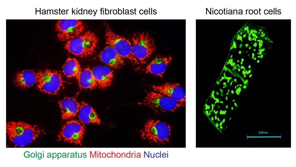
[In this figure] The number and location of Golgi apparatuses can be studied using the fluorescent microscope.
Left: Each mammalian cell (here, hamster skin cells) has one Golgi apparatus (green) near its nucleus. Right: Plant cells (Nicotiana, a species of tobacco; two cells in this image) could have many Golgi apparatuses per cell. If you are interested in how these pictures were taken, see below “How to see Golgi apparatus under a microscope”.
Photo credit: Florida State University.
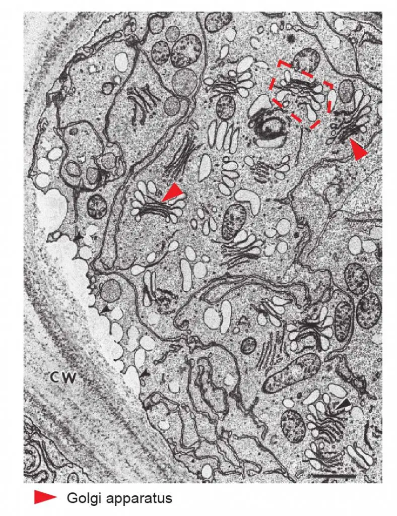
[In this figure] TEM image of many Golgi stacks in a hyper-secretory outer cap cell from maize root. Red arrowheads indicate the typical shape of Golgi in plant cells. There are at least 13 Golgi in this image. The numbers of Golgi apparatus correlate with how active the cells are in protein synthesis and secretion. (courtesy of Dr. H.H. Mollenhauer).
Image source: Biochimica et Biophysica Acta (BBA) – Molecular Cell Research
What is the biological function of Golgi apparatus?
1. Golgi is the protein delivery center inside the cells
The function of Golgi apparatus is like the post office inside the cells. It usually locates close to the endoplasmic reticulum (ER). If you think of ER with ribosomes (rough ER) as the protein factory in the cells, the Golgi then takes over the logistic work. The Golgi apparatus receives the raw protein products from the ER, modifies them (for example, adding tags made by sugar chains), and exports the proteins to a variety of destinations.
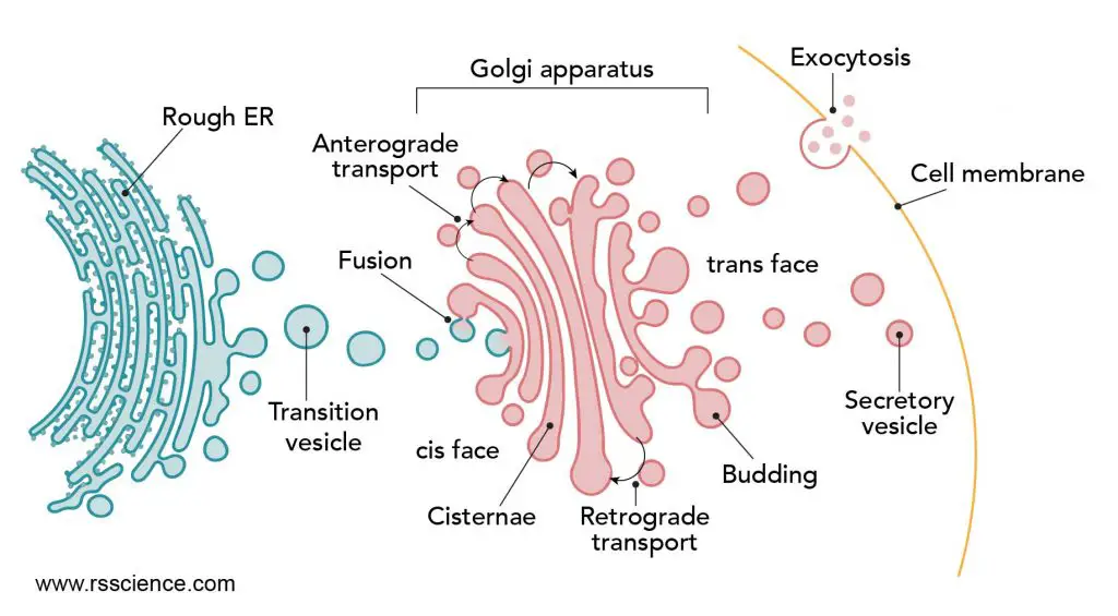
[In this figure] The journey of protein synthesis and transportation.
After proteins are synthesized in the rough ER, they traveled to the Golgi for further glycosylation. Once they are done with the modification, proteins are packed into vesicles, called secretory vesicles. Then, they travel to the membrane and release by exocytosis.
Created with BioRender.com
The Golgi apparatus is part of a manufacturing and supply chain
In order to perform its job, the Golgi apparatus consists of several stacks of membrane-bound cisternae (sacs). This unique structure is closely related to Golgi’s biological function.
Each Golgi has two “faces” – the cis face (receiving crude protein products from ER), and the trans face (exporting refined protein products to their destinations). Considering the ER is the upstream manufacturer, Golgi is working from the rough endoplasmic reticulum (RER) outwards. You can imagine dividing Golgi apparatus into three main sections of a factory:
1) Cis Golgi network (Goods inwards)
Also called the cis Golgi reticulum, it is the entry area to the Golgi apparatus.
2) Golgi stack (Main processing area)
This section is composed of a variable number, typically 3-6, of flattened cisternae. The cisternae of the Golgi stack are divided into three working areas: cis cisternae, medial cisternae, and trans cisternae.
3) Trans Golgi network (Goods outwards)
This section is where the final reactions and sorting takes place. The concentrated biochemicals are packed into sealed droplets or vesicles by budding off from the trans Golgi surface. The vesicles are then transported away for use in the cell and beyond.
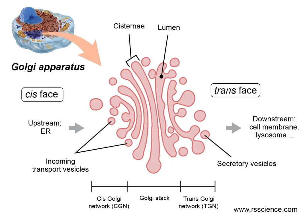
[In this figure] The structure of Golgi apparatus.
Created with BioRender.com
Cells use small lipid bubbles to shuttle proteins around ER and Golgi
The proteins are transported within small, sealed bubbles, called vesicles, which are generated by budding from the membrane of the ER and Golgi. The ER packs crude protein products into a budding vesicle, releases the vesicle, and sends it toward the Golgi for further processing.
The “cis” face of Golgi receives this package by the fusion of membrane between the vesicle and Golgi. The protein products are now inside the lumen of the Golgi apparatus. Next, the same process (budding and fusion) repeats when the proteins travel between each Golgi stack from the “cis” face to “trans” face. Within each stack, there are special enzymes modifying the proteins.
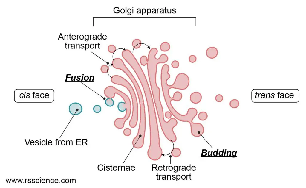
[In this figure] The transportation between ER-Golgi and within Golgi is achieved by exchanging vesicles.
Cargos are imported by “Fusion” of vesicles and membranes. Export is done by packaging cargos in new vesicles departing by “Budding”. Although the mainstream of proteins is moving from cis to trans direction, scientists also found retrograde transportation inside the Golgi.
Created with BioRender.com
Golgi products – Where do they go? How do they get there?
The final protein products will be sorted by their destinations and packaged into the secretory vesicles.
There are 3 main destinations for proteins released from the trans Golgi network. In each case, the destination is clearly linked to their functions.
Destination 1. inside the cell to other organelles (lysosomes)
Some proteins will be sent to other organelles, such as lysosomes. About 40-50 different biochemicals dispatched from the Golgi apparatus in vesicles are destined for delivery to lysosomes. Many of them are enzymes that perform the digestion of cellular wastes inside the lysosomes.
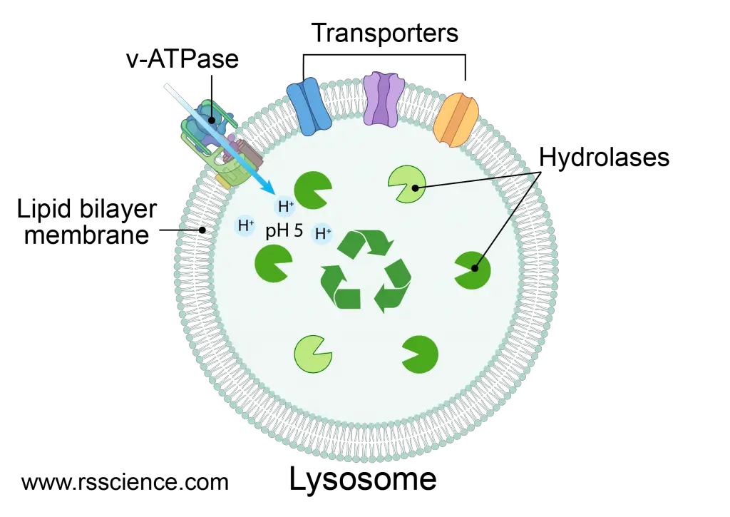
[In this figure] Lysosome contains many kinds of digestive enzymes that are responsible for the recycling of wastes inside the cells.
Created with BioRender.com
Destination 2. all the way to the cell membrane (the continuous secretion)
Some proteins will be shipped all the way to the cell membrane through the fusion between vesicles and cell membranes, to outside the cells. This type of secretion contributes to the proteins of the extracellular matrix, acting as biochemical signals to other cells, and providing proteins for the repair and replacement of the cell membrane. This constitutive (or continuous) secretory pathway is also the default pathway.
Many epithelial cells are polarized in tissues. The cell membrane of such cells is divided into two separate regions, the apical and the basolateral part, that contain specific proteins related to their functions.
For example, the apical membrane of intestinal epithelial cells faces the lumen of the intestine and is specialized for the efficient absorption of nutrients; the remaining sides of the cell is covered by the basolateral membrane. The Golgi knows how to selectively transport proteins to these distinct parts of the cell membrane. This is accomplished by the selective packaging of proteins into two types of constitutive secretory vesicles for either the apical or basolateral directions.
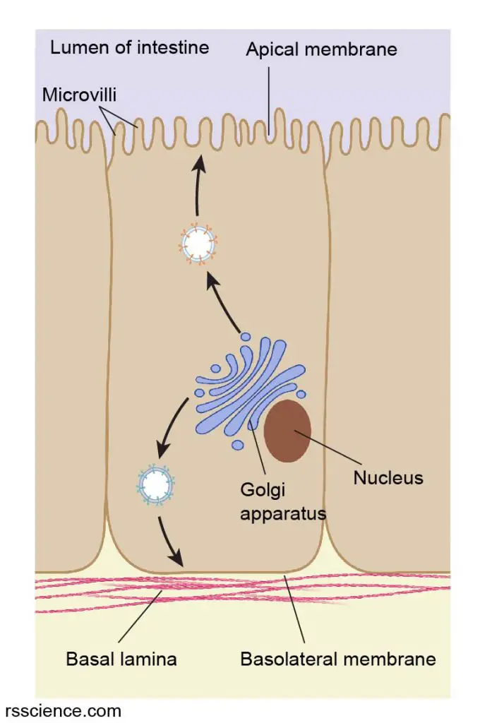
[In this figure] Intestinal epithelial cells contain polarized cell membranes: the apical and basolateral parts.
Golgi apparatus can selectively deliver proteins to either direction by using different secretory vesicles.
Created with BioRender.com
Destination 3. store close to the cell membrane until receiving a signal to release
Sometimes, the cells hold the vesicles and only secrete the proteins until the traffic light turns green. This type of vesicles is produced in specialist secretory cells (cells from glands). They move from Golgi’s trans face towards the cell membrane but accumulate in number before reaching the membrane.
Some events can trigger the fusion of vesicles with the plasma membrane and release their contents in regulated bursts from the cell surface. For example, beta cells in the pancreas are special secretory cells producing insulin. Insulin release is triggered by a rise in blood glucose levels after we finish a meal. Food intake also triggers the release of mucus and digestive enzymes into the stomach.
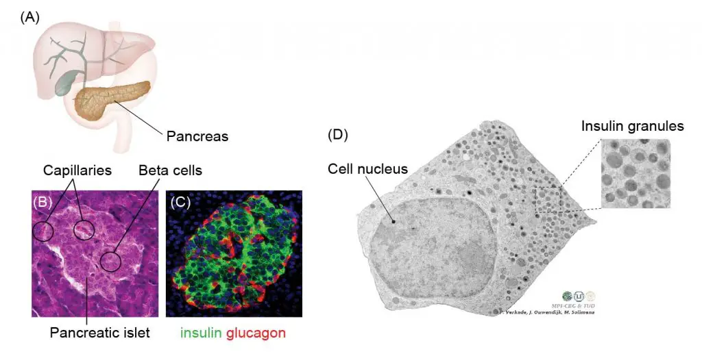
[In this figure] Biology of the pancreatic beta cells.
(A) The pancreas is an organ sitting across the back of the abdomen, behind the stomach. The beta cells form clusters of pancreatic islets inside our pancreas. Here, we use three kinds of microscopes to study the biology of beta cells. (B) Looking at a pancreatic islet with H&E (Haemotoxylin and Eosin) staining under a compound microscope. (C) A pancreatic islet under the fluorescent microscopes with antibody-labeled alpha cells red (producing glucagon) and beta cells green (insulin). (D) Electron microscopic image of a pancreatic beta cell. The beta cell senses the increased level of glucose in our blood and releases insulin to modulate the blood sugar. In this image, you can see many insulin-storing vesicles (called insulin granules) stand by near the cell membrane.
Photo credit: modified from Paul Langerhans Institute Dresden.
2. Golgi is in charge of adding sugar molecules onto proteins
Protein processing is another important function of Golgi. The raw proteins synthesized from ribosomes are purely long chains of amino acids. These proteins need to be modified by adding carbohydrate (sugar units) portions to become “glycoproteins”. The prefix “glycol-” means “sugar”.
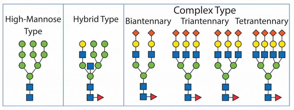
[In this figure] Type of glycoprotein.
Photo credit: BioProcess.
The process of adding sugars (called glycosylation) starts within the ER. In ER, an oligosaccharide (short chain of sugars) consisting of 14 sugar residues is added onto proteins. Following transport to the Golgi apparatus, the sugar chains of these glycoproteins are subject to extensive further modifications.
For example, the modification could be removal, addition, or substitution of sugar units, adding a new sugar branch, or adding a phosphoryl group. Different glycoproteins are modified to different extents during their passage through the Golgi, depending on both the structure of the protein and on the amount of processing enzymes that are present within the Golgi complexes in different types of cells.
Different modifications have their biological effect. For example, the addition of sialic acid can increase the stability of a protein (called half-life) after being secreted into the bloodstream. Adding oligo-mannose can tag the proteins to be delivered toward lysosomes.
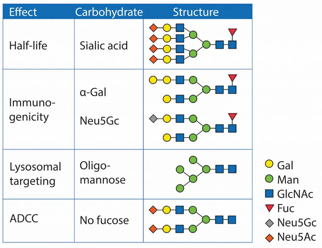
[In this figure] Glycan structures and their biological effect.
Photo credit: BioProcess.
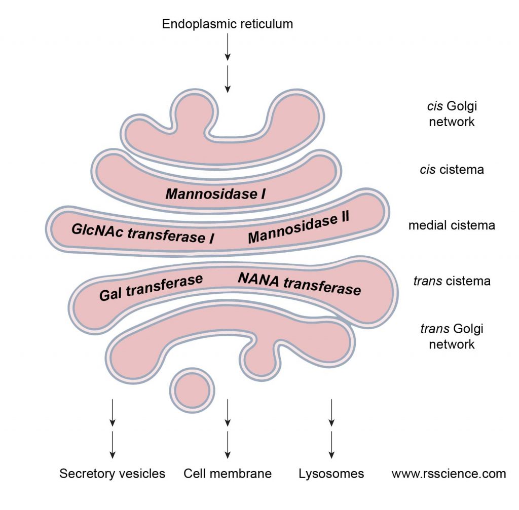
[In this figure] The step-by-step of protein processing following the protein transportation from cis to trans Golgi network.
Each cisterna has specialized enzymes to accomplish the specific steps of protein modification.
Created with BioRender.com
The correct processing in ER and Golgi (called Post-translational modification) is critical for the activity of proteins. This is a big challenge for pharmaceutical companies to produce therapeutic proteins. Many proteins can be used as drugs. For example, insulin for diabetes and erythropoietin (EPO) for anemia.
Recently, many antibodies were manufactured to fight cancers. Choosing the right cells to produce therapeutic proteins is critical if the post-translational modification of the protein is essential for its activity. Bacteria can not be used to produce glycoproteins due to the lack of a glycosylation system. Some proteins synthesized in yeasts may not work well because their glycosylation enzymes are different from humans. Many protein drugs required special mammalian cells (CHO cells) to produce.
3. Golgi also modifies lipid molecules
In addition to its role in modifying and sorting proteins, the Golgi apparatus also functions in lipid metabolism—in particular, in the synthesis of glycol-lipids (lipids with sugar modification) and phospholipids (lipids with a phosphate group). These modified lipids will become parts of cell membrane. Different cells have different composition of carbohydrates on their cell surface, which can be used as an identity in cell-cell recognition.
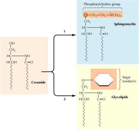
[In this figure] Ceramide (a lipid molecule synthesized in ER) will be converted either to sphingomyelin (a phospholipid) or to glycolipids in the Golgi apparatus.
Photo source: The Cell: A Molecular Approach. 2nd edition
4. Golgi synthesizes the sugar components of cell walls
In plant cells, the Golgi apparatus has an additional task of synthesizing complex polysaccharides of the cell wall. Cell wall is a unique structure that only exists in plant cells, which provides the plant cell with both structural support and protection.
The cell wall is made of three major types of polysaccharides (polymer types of sugars). Cellulose, the predominant constituent, is synthesized at the cell surface by enzymes anchored on the cell membrane (because the cellulose is a simple linear polymer of glucose residues).
However, the other two types of cell wall polysaccharides (hemicelluloses and pectins) are complex, branched-chain molecules. These two polysaccharides have to be synthesized in the Golgi apparatus and then transported in vesicles to the cell surface. The synthesis of these cell wall polysaccharides is a major cellular function, and as much as 80% of the metabolic activity of the Golgi apparatus in plant cells may be devoted to polysaccharide synthesis. This is also why plant cells have many more Golgi apparatuses compared to animal cells.
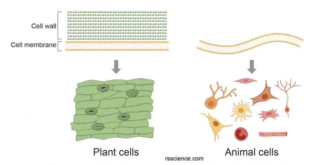
[In this figure] The difference of cell membrane between plant and animal cells.
The cell wall provides additional protective layers outside the cell membrane. Plant cells can maintain the cell shape and support the growth due to the strength of cell walls. On the other hand, animal cells only have a cell membrane. The flexibility of the cell membrane allows animal cells to move and change their cell shapes.
Created with BioRender.com
How Golgi apparatuses divide during cell division?
When a cell divides, its Golgi apparatus breaks up into small fragments. These fragments are divided more or less evenly between the daughter cells. A new Golgi apparatus grows from a fragment of Golgi apparatus from the previous cell. However, if there are no fragments, there will be no Golgi apparatus. To some extent, you can say that Golgi apparatuses propagate like “clones”.
Are there human diseases associated with Golgi apparatus dysfunction?
Yes, many diseases are associated with the disfunction of Golgi apparatus. Because Golgi apparatus plays a central role in the production and transportation of proteins, even a small error from Golgi apparatus can cause catastrophic consequences.
Several Golgi-associated diseases are inherited neurodevelopmental disorders. For example, Angelman syndrome is caused by a mutation of the UBE3A gene. Loss of UBE3A leads to an altered Golgi structure and pH, which is associated with a reduction in protein sialylation (adding sialic acids to protein, one type of glycosylation). People with Angelman syndrome have problems with speech and balance, intellectual disability, and sometimes, seizures. They often smile and laugh frequently, and have happy, excitable personalities.
Recently, scientists found that disrupted Golgi structure is linked to a variety of neurodegenerative diseases, including amyotrophic lateral sclerosis (ALS), Parkinson, Alzheimer, Huntington, and prion diseases.
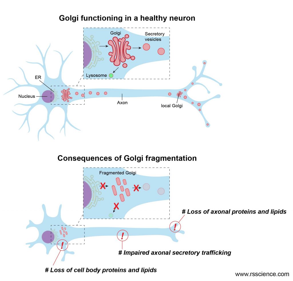
[In this figure] Golgi function in neurons.
Because neuronal cells have long projections (axons) for connecting with other cells, successful transportation of proteins and lipids to a long-distance till the ends of axons are critical for normal neuronal function. This largely relies on Golgi apparatuses. Dysfunctional Golgi (usually due to Golgi fragmentation) seriously impair the healthiness of neuronal cells, resulting in neurodegenerative diseases.
Where did the name “Golgi apparatus” come from?
The Golgi apparatus is named after the Italian histologist Camillo Golgi, who first identified the structure in 1898.
Due to its relatively large size, the Golgi apparatus was one of the first organelles ever observed. Camillo Golgi found Golgi apparatus when he investigated the nervous system by using a new staining technique he developed (which is still sometimes used today; known as Golgi staining or Golgi impregnation). Under his light microscope, Camillo Golgi noticed a thread-like cellular structure that he termed the internal reticular apparatus. Soon after he publicly announced his discovery in 1898, the structure was named after him, becoming universally known as the Golgi apparatus.
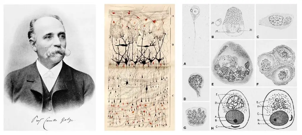
[In this figure] Left: Camillo Golgi. Middle: Original drawing by Camillo Golgi of the nerve cells of the olfactory bulb as visualized by his Golgi method. Right: Historical drawings showing the “internal reticular apparatus” published in the late 1980s.
Photo source: Histochemistry and Cell Biology.
Yet, many scientists did not believe what Golgi observed was a real organelle present in the cell and instead argued that his discovery as nothing but a staining artifact. Their microscopes were not powerful enough to identify the organelles. By the 1930s, Golgi’s description was largely rejected. The invention of the electron microscope in the twentieth century finally confirmed that the Golgi apparatus is a cellular organelle, 50 years after the great discovery.
Camillo Golgi received the Nobel Prize in 1906 with Santiago Ramón y Cajal for their work on the structure of the nervous system.
How to see Golgi apparatus under a microscope?
Without proper staining, you can not see Golgi apparatuses under regular microscopes (even phase-contrast or DIC).
Golgi’s method is a silver staining technique that is used to visualize nervous tissue under light microscopy. The method was discovered by Camillo Golgi and modified by himself as he discovered the Golgi apparatus in 1898. However, the procedure was complicated and difficult for others to repeat. This is also one of the reasons that his discovery of Golgi apparatus was not widely accepted at the beginning. Currently, Golgi’s method (also called Golgi preparation or Golgi impregnation) is used exclusively for the staining of neurons in brain tissues.
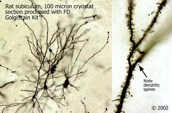
[In this figure] Stained rat neurons using Golgi’s method.
Golgi’s staining is achieved by impregnating aldehyde fixed nervous tissue with potassium dichromate and silver nitrate. Cells thus are filled by microcrystallization of silver chromate.
Photo source: FD Neuron Technologies, Inc.
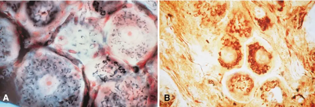
[In this figure] Histological pictures of Golgi apparatuses stained.
These are probably done by Golgi’s photographic method. However, the author and date were unknown.
Photo source: Histochemistry and Cell Biology.
There were modified histological staining methods that can stain Golgi apparatuses for light microscopy. However, it required expertise to handle toxic chemicals of osmium and silver compounds.
Immunofluorescence staining is widely used to see and study Golgi apparatuses. Immunofluorescence (IF) is a technique that permits visualization of virtually many components in any given tissue or cell type. This technique uses the specificity of antibodies to their antigen (kind of stealing the tools from our immune system). These antibodies were tagged with fluorescent dyes. Once the antibodies bind to specific biomolecule targets within a cell, we can visualize the distribution of the target molecule through the sample under a fluorescence microscope.
Unfortunately, fluorescence microscopes are pretty expensive and only found in the laboratories in the universities. It is very unlikely we can do immunofluorescence staining at home. However, you can find many beautiful fluorescent images of Golgi apparatuses (or other organelles) online. Below are some examples.
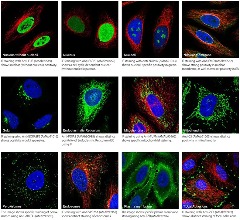
[In this figure] A gallery of immunofluorescence images showing 11 different organelles in human cells.
Antibodies were tagged with green fluorescent dyes. As a counterstain, microtubules and nuclei are visualized in red and blue respectively.
Photo source: Merck
[In this video] Golgi (green) and mitochondria (red) dynamics in a cell under the fluorescence microscope. Images were taken at time intervals of every 4 seconds.
If you want to see the structure of Golgi in an ultra-detail, transmission electron microscopy (TEM) is the only option. Of course, TEM is an equipment exclusively for research institutes and universities. However, we can enjoy these research findings through many online resources.
[In this figure] A TEM image of Golgi apparatus.
Photo source: Histology @ Yale
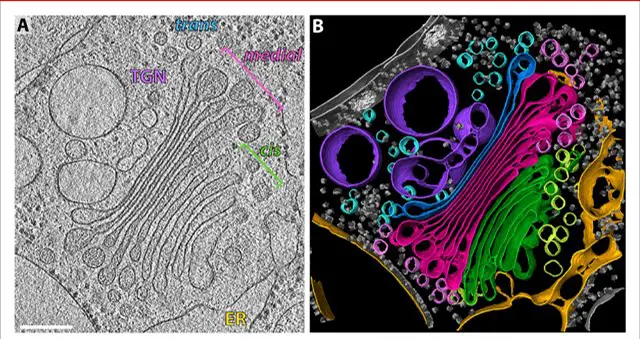
[In this figure] Molecular architecture of the Golgi apparatus and transport vesicles revealed by cryo-EM.
(A) The actual EM image. (B) The corresponding 3D segmentation showing the ER (yellow), four cis cisternae (green), four medial cisternae (magenta), the trans cisterna (blue), trans vesicles (light blue), and the TGN (purple).
Photo source: eLife
Summary
- Golgi apparatus (or Golgi) consists of several stacks of membrane-bound cisternae (sacs).
- Golgi apparatus usually locates close to the ER. It receives the raw protein products from the ER, modifies them (for example, adding sugar chains), and exports the proteins to a variety of destinations.
- The transportation of proteins is done within small bubbles, called vesicles.
- The vesicles are generated by budding from the membrane of the ER and Golgi. Once the vesicles reach their destinations, the fusion of membrane releases their protein cargos.
- There are three major destinations of proteins: (1) sent to other organelles, like lysosomes, (2) directly secreted outside the cells, and (3) stored until receiving a signal to release.
- Golgi apparatus also involves in lipid metabolism and cell wall formation in plant cells.
- Golgi apparatus was discovered by Camillo Golgi in 1898.
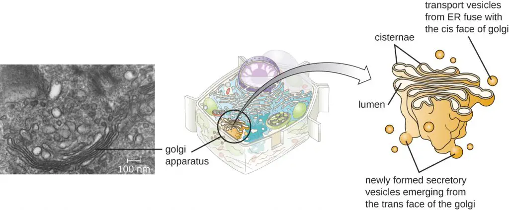
Photo source: wiki
References
“The Cell: A Molecular Approach. 2nd edition.” – Cooper GM.
“Golgi Apparatus” – British Society for Cell Biology
“Camillo Golgi and the discovery of the Golgi apparatus” – Ariane Dröscher
“Angelman syndrome” – Mayo Clinic
“Editorial: Golgi Pathology in Neurodegenerative Diseases” – Catherine Rabouille and Georg Haase
“Golgi Staining Technique” – Erica O’Neil, Sarah Taddeo
“Immunofluorescence” – Wikimedia
Related posts
Cell Biology on the Dining Table – Animal Cell Model Part II
Pingback: What happens to proteins as they pass through the Golgi apparatus? – Dr. Biology Questions and Answers
Pingback: What does the Golgi apparatus do? – Dr. Biology Questions and Answers