
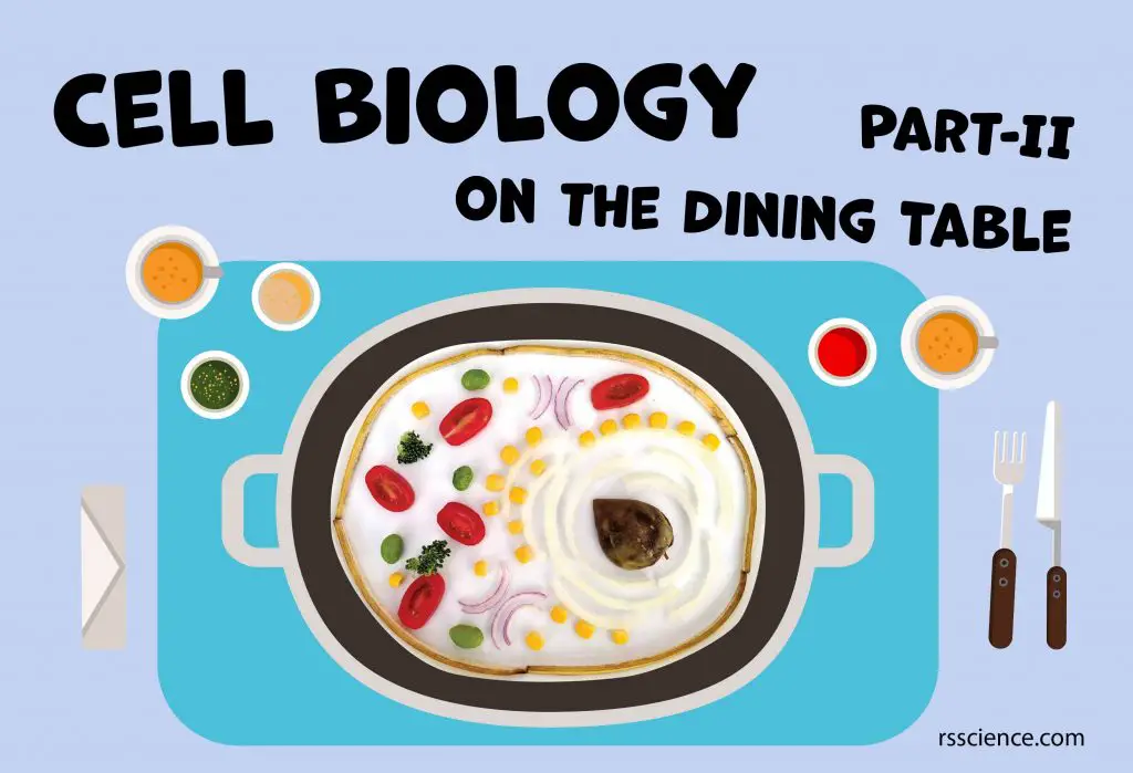
Cells are building blocks of life. Building a cell model should deepen your understanding of the cell, each organelle’s role, and how they perform their job. Some organelles are constantly present in the cell. Some organelles are temporary and only present when cells perform a particular process such as mitosis. Therefore, we have 4 blog posts series to cover the model of animal cells undergoing different processes.
- Animal Cell Model Part I – cell membrane, cytosol, nucleus, and mitochondria.
- Animal Cell Model Part II – endoplasmic reticulum, ribosome, Golgi apparatus, peroxisome, and lysosomes.
- Animal Cell Model Part III – two types of temporary organelles involving in eating behaviors, autophagosomes, and endosomes.
- Animal Cell Model Part IV – two types of temporary organelles only appearing during mitosis, centrosomes, and chromosomes.
- Plant Cell Model Part V – cell wall, vacuole, and chloroplast.
- Cell Organelles and their Functions – overview of each organelle.
This week, we are going to cover the Endoplasmic reticulum, ribosome, Golgi apparatus, peroxisome, and lysosomes. Let’s dive in!
This article covers
Endoplasmic reticulum – the cellular inter “NET”
The endoplasmic reticulum (ER) is thought to be an internal membrane that divides into a branching web throughout the cytoplasm. The ER occurs in two morphologically distinct forms: rough ER and smooth ER.
The outer side (facing the cytosol) of the rough ER is studded with ribosomes that are the sites of protein synthesis. Under the electron microscope, the dense granular ribosomes gave the name of “rough” ER. On the other hand, the smooth ER lacks ribosomes.
ER plays many important roles in our cells. In brief, rough ER coordinates protein synthesis. Smooth ER specializes in lipid synthesis, steroid hormone production, and detoxification.
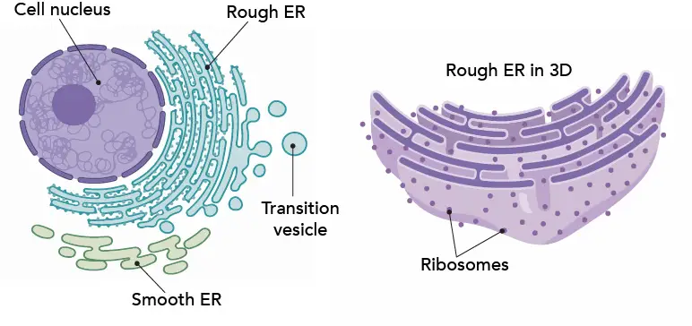
[In this figure] The anatomy of ER
Left: The relationship between the nucleus, rough, and smooth ER. The newly synthesized proteins will be transported to the Golgi apparatus (we will talk about later) in transition vesicles.
Right: A 3D view of rough ER. You can see the ER membrane actually form branching networks of many interconnected sacs and tubes.
The reason why using yellow onions
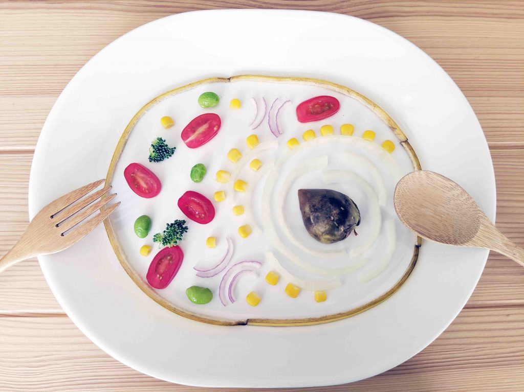
In my cell model, I use shredded yellow onion to represent the translucent looking of ER networks. I put corn kernels (as ribosomes) along some shredded onion to become the rough ER.
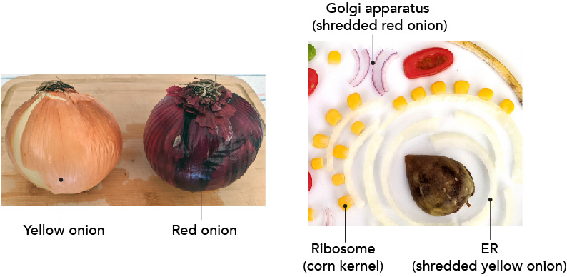
[In this figure] I shred yellow and red onion to represent two typical elongated membrane structures, ER and Golgi apparatus, in the cell model. Corn kernels are the ribosomes.
Here I made a mistake. In real cells, the rough ER sits around the nucleus (so the nucleus can have better control of protein synthesis), and the smooth ER locate away from the nucleus. However, I forgot to save the space for the ribosomes. That results in an inverted spatial relationship between the rough and smooth ER in my cell model.
Ribosome – the decrypt machine
Ribosomes are the places where proteins were synthesized in our cells. Ribosomes consist of two major components: the small and large ribosomal subunits. Scientists like to call ribosomes, the macromolecular machines, to admire how exquisite the design of ribosomes is!
In the section of the nucleus, we mention that the codes of a given gene will be transcribed into a messenger RNA (mRNA). Ribosomes bind to mRNA and use its code sequence for determining the correct order of amino acids to generate the corresponding protein.
Amino acids are carried to the ribosomes by transfer RNA (tRNA) molecules. Only the right tRNA can enter the ribosome and pair with the code on mRNA. Once the tRNA and mRNA match, the ribosome will add this amino acid onto a growing protein chain. After all the codes on the mRNA molecule successfully translate into the corresponding amino acids, the protein is synthesized. This process is called translation.
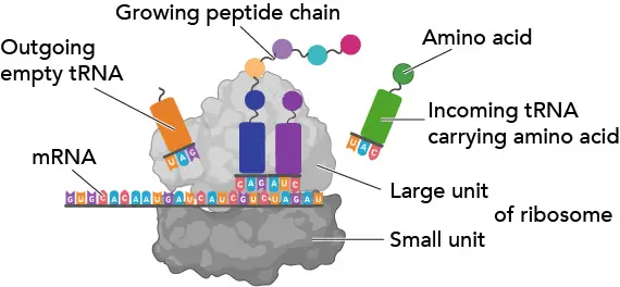
[In this figure] The ribosome works like a machine to translate the code sequence of mRNA into a protein.
The reason why using corn kernels
Considering the relative sizes, I choose corn kernels as my ribosomes. Although the majority of nutrients in corn kernels come from carbohydrates, corn kernels also provide a high-quality source of proteins. Especially plain corn is a naturally gluten-free food. It lacks the protein gluten of wheat; therefore, it can be used to make gluten-free baked goods.
Do you know mitochondria also have their own ribosomes?
Ribosomes can be found to be free-floating in the cytosol or associated with rough ER. There are also ribosomes in mitochondria for translating mitochondrial mRNAs that are encoded in mtDNA. These mitochondrial ribosomes (also called Mitoribosomes) sit inside the matrix space.
Golgi apparatus – the post office inside the cells
The Golgi apparatus (or Golgi) usually locates close to the ER. If you think of ER with ribosomes as the protein factory in the cells, the Golgi then takes over the logistic work. The Golgi apparatus receives the raw protein products from the ER, modifies them (for example, adding tags made by sugar chains), and exports the proteins to a variety of destinations.
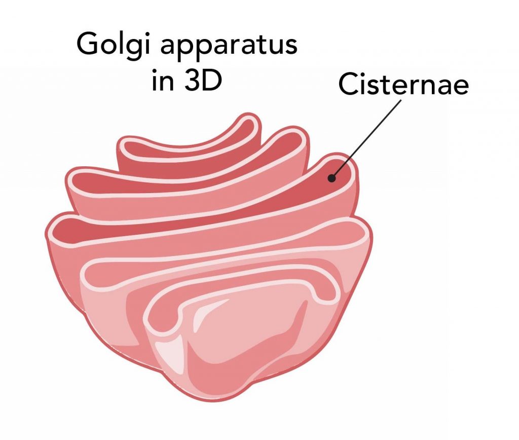
[In this figure] A 3D view of Golgi apparatus.
In order to perform its job, the Golgi apparatus consists of several stacks of membrane-bound cisternae (sacs). Each Golgi has two “faces” – the cis face (receiving crude protein products from ER), and the trans face (exporting refined protein products to their destinations).
The transportation of proteins is done within small bubbles, called vesicles. The vesicles are generated by budding from the membrane of the ER and Golgi. The ER packs crude protein products into a budding vesicle, releases the vesicle, and sends it toward the Golgi.
The “cis” face of Golgi receives this package by the fusion of membrane between the vesicle and Golgi. The protein products are now inside the lumen of the Golgi apparatus. Then, the same process (budding and fusion) repeats when the protein products travel between each Golgi stack from the “cis” face to “trans” face. Within each stack, there are special enzymes to modify the protein products.
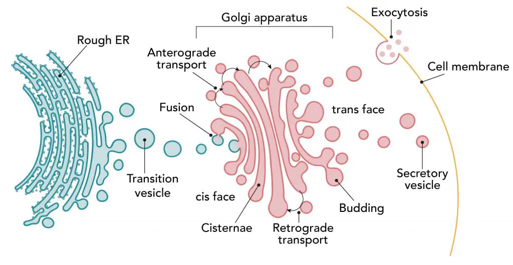
[In this figure] The journey of protein synthesis and transportation.
After proteins are synthesized in the rough ER, they traveled to the Golgi for further glycosylation. Once they are done with the modification, proteins are packed into vesicles, called secretory vesicles. Then, they travel to the membrane and release by exocytosis.
At the last steps, the final protein products will be sorted by their destinations and packaged into the secretory vesicles. Some proteins will be sent to other organelles. Some proteins will be released into the cytosol. Some proteins will be shipped all the way to the cell membrane, and through the fusion between vesicles and cell membranes, these proteins will be secreted outside the cells. Sometimes, the secretion happens right away. Sometimes, the cells hold the vesicles and only secrete the proteins until the traffic light turns green (for example, insulin is only released after we finish a meal).
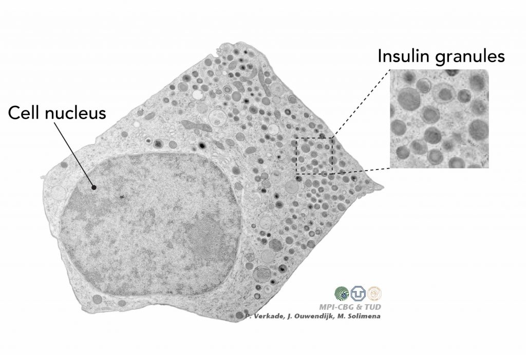
[In this figure] Electron microscopy image of the pancreatic beta cell.
The beta cells form clusters of pancreatic islets inside our pancreas. The beta cells can sense the increased level of glucose in our blood and release insulin to modulate the blood sugar. In this electron microscopy image, you can see many insulin-storing vesicles (called insulin granules) stand by near the cell membrane.
Photo credit: modified from Paul Langerhans Institute Dresden.
The reason why using red onions
Due to the consistent exchange of lipid membrane, the ER and Golgi are considered, to some degree, continuous membrane-bound organelles. For this reason, I use two kinds of onions but in different colors: yellow onion for ER and red onion for Golgi. I arrange the shredded red onion to represent the Golgi stacks.
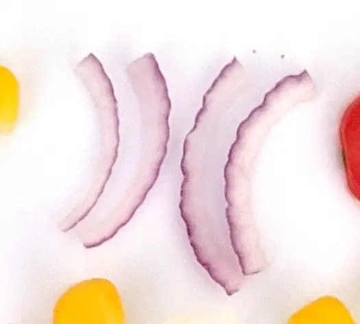
[In this figure] I arrange shredded red onion to mimic the cisternae stack of Golgi.
Peroxisome – free-radical scavengers
The peroxisome is a spherical organelle responsible for the fatty acid breakdown. Fatty acids are the basic units of fat and oil. The foods we eat include a variety of fatty acids. For example, fatty acids could be either saturated or unsaturated fats. Tey can be further classified according to their different lengths of carbon chains (from 4 to 28 carbon atoms).

[In this figure] A lipid molecule with two fatty acid chains (18 carbon atoms). One chain is saturated, and the other one is unsaturated.
To use these fatty acids, the peroxisomes have to breakdown some of the very long and branched-chain fatty acids into simpler nutrients. This process involves several special enzymes and complicated biochemical reactions.
In addition, the peroxisomes in the liver cells also handle the detoxifications of many chemicals, including alcohol and drugs. It is fair to say that peroxisome is the chemistry laboratory inside the cell.
The chemical reactions are potentially dangerous to the cells. This is why we need the peroxisomes to control chemical reactions within a membrane-bound space separated from the rest of the cells. Let me here pay tribute to those peroxisomes which handle the high-risk tasks for us.
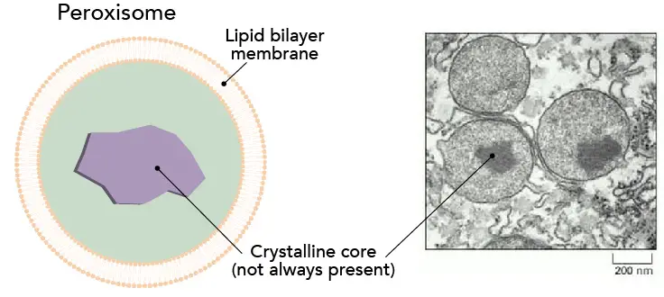
[In this figure] Peroxisomes.
Left: the structure of peroxisome.
Right: an electron microscopy image of peroxisomes. (Image from Schrader, M. and Fahimi, H. 2008. The peroxisome: still a mysterious organelle. Histochemistry and Cell Biology 129(4), pp. 421-440.)
From the chemical point of view, many enzymes inside the peroxisomes catalyze Redox (reduction-oxidation) reactions. Oxygen is an active molecule, and many chemical reactions happen base on the reactivities of oxygen.
Generally speaking, oxidation is adding an oxygen (increase in the oxidation state), and reduction is removing an oxygen (decrease in the oxidation state) from a molecule.
Typically, oxidation is burning. When a piece of wood on the fire, the oxidation state of carbon atoms in the wood increase by the reaction of oxygen gas, turning the carbon into carbon dioxide.
When a reductive reaction happens in the peroxisome, the enzyme takes away oxygen (in the form of superoxide O2•−). However, the enzyme cannot hold the oxygen forever, so the oxygen is transferred to a water molecule. As a result, the water molecule is oxidized to become a hydrogen peroxide (H2O2).
The molecules that contained chemically reactive oxygen (like O2•−, •OH, H2O2, and NO) are called reactive oxygen species (ROS) or free radicals. Many ROS are created as an unavoidable byproduct of normal cellular metabolism. These ROS need to be removed from the cells carefully; otherwise, ROS will damage the cells by unwanted reactions with DNA, lipid, and proteins.
In fact, many diseases like cancers and aging originate from the bad effects of ROS in our bodies. Radiation, tobacco, and drugs also increase the chances of damage by ROS. Antioxidants or free-radical scavengers can cancel out the effects of ROS. This is why we are encouraged to eat more healthy food that enriches natural antioxidants, like vitamins A, C, and E.
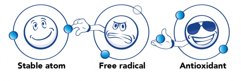
[In this figure] A stable atom has a balanced number of electrons, no more, no less. Free radicals eagerly want to steal electrons from other atoms to fulfill their unstable status. Antioxidants have free electrons that can give to free radicals to make them calm down.
As we mention, the peroxisomes generate many hydrogen peroxide molecules (H2O2) as dangerous byproducts. In fact, this is why the “peroxi”-some got its name. Fortunately, the peroxisomes are able to contain these H2O2 and break them down into water (H2O) and oxygen (O2). This critical reaction is done by another peroxisomal enzyme, called “Catalase”.
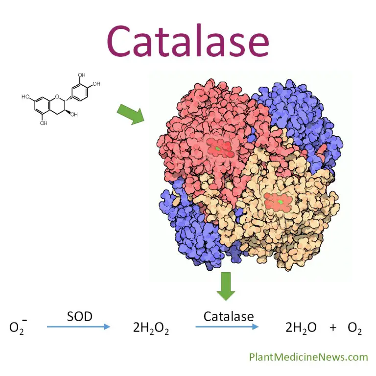
[In this figure] Catalase is an enzyme that converts harmful hydrogen peroxide into water and oxygen.
The reason why using broccolis
I use broccoli as the peroxisomes in my cell model because the broccoli contains high antioxidants. Of no doubt, broccoli is a healthy superfood, and a particularly rich source of vitamin C and vitamin K. Broccoli also contains several free-radical scavenging enzymes, including one of the highest catalase contents in vegetables.
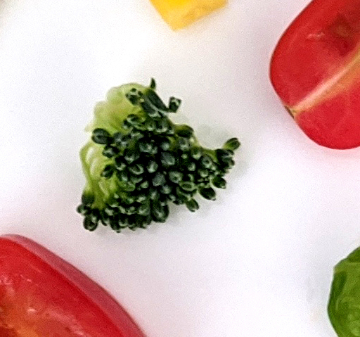
[In this figure] Broccoli, which is rich in antioxidants, is the peroxisome in my cell model.
Lysosomes – the cell’s recycling center
The lysosomes are small organelles that work as the recycling center in the cells. They are membrane-bounded spheres full of digesting enzymes. These enzymes can break down whatever substance entering the lysosomes into raw materials (like amino acids, nucleotides, lipids, and sugars), so the cell can reuse these raw materials to build new organelles.
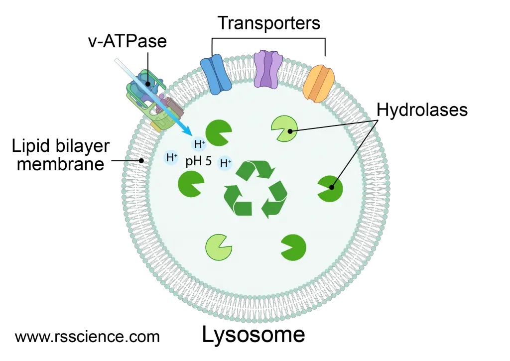
[In this figure] Lysosome is the recycling center of the cell.
The lysosome is a membrane-bound vesicle containing hydrolase enzymes that can break down old organelles and proteins into raw materials. There are v-ATPase that pump protons into the lysosome to acidify its pH value. There are also several types of transporters to bring the cell parts in for degradation.
Created with BioRender.com
As you can imagine, if the lysosomes somehow leak or burst, these enzymes could cause huge damages to the cells. Fortunately, the cells have a safety mechanism to only allow lysosomal enzymes to be active in an acidic environment, but not in the rest of the cell. These digesting enzymes will only function properly in an environment with a pH of 5 (only exists inside a mature lysosome). Comparing to the internal pH of 7 in the cytosol, the pH of 5 is two orders of magnitude more acidic. This design restricts the enzymatic activities only inside the lysosomes.
The reason why using edamame beans
In order to match the image of recycling, I choose “green” edamame beans as my lysosomes. I love edamame served as a side dish in Japanese cuisine. Edamame is also healthy food and rich in proteins, dietary fiber, antioxidants, and vitamin K. I bought precooked frozen edamame from my local Trader Joe’s. You can substitute them with other green beans and peas.
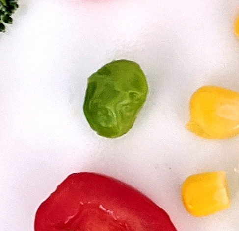
[In this figure] The edamame bean is the lysosome in my cell model.
My first edible cell model is ready to serve!
We discussed all the basic organelles and their functions in an animal cell. I hope that you get inspired to create your own cell model project. I like to encourage you to use special veggies or fruits in your local area and make a UNIQUE cell model.
It will be even more awesome if you take a “CELL-FIE” and share it with us on our Facebook page: “Totally Science”.
The story is not over yet – what about autophagosomes, centrosomes, and endosomes?
How does the cell decide what should be recycled in the lysosomes? This involves temporary organelles called “autophagosomes” and a special process called “autophagy” (aka “self-eating”).
There are several temporary but essential organelles that are rarely mentioned in other 3D cell models. Except for autophagosomes, I can also think of the “centrosomes” for cell division and the “endosomes” for cells to import outside substances.
We will continue with these temporary organelles, as well as other specialized cell types in our Cell Biology on the dining table article series. Stay tuned!
Reference
W. B. Storey. What Kind of Fruit is the Avocado? California Avocado Society Yearbook 1973-74. Pages 70-71.
https://www.britannica.com/science/endoplasmic-reticulum
https://www.histology.leeds.ac.uk/cell/cell_organelles.php
https://www.khanacademy.org/test-prep/mcat/cells/eukaryotic-cells/a/organelles-article
Related posts
Animal Cell Model Part I – cell membrane, cytosol, nucleus, and mitochondria.
Plant Cell Model Part V – cell wall, vacuole, and chloroplast.
Cell Organelles and their Functions – overview of each organelle.