
Cells are building blocks of life. Building a cell model should deepen your understanding of the cell, each organelle’s role, and how they perform their job. Some organelles are constantly present in the cell. Some organelles are temporary and only present when cells perform a particular process such as mitosis. Therefore, we have 4 blog posts series to cover the model of animal cells undergoing different processes.
- Animal Cell Model Part I – cell membrane, cytosol, nucleus, and mitochondria.
- Animal Cell Model Part II – endoplasmic reticulum, ribosome, Golgi apparatus, peroxisome, and lysosomes.
- Animal Cell Model Part III – two types of temporary organelles involving in eating behaviors, autophagosomes, and endosomes.
- Animal Cell Model Part IV – two types of temporary organelles only appearing during mitosis, centrosomes, and chromosomes.
- Plant Cell Model Part V – cell wall, vacuole, and chloroplast.
- Cell Organelles and their Functions – overview of each organelle.
This article covers
Design your own cell model project
Have you ever seen these colorful cell models while you are browsing Facebook, Instagram, or Pinterest? Of course, you can buy a plastic cell model online. However, I prefer to DIY my own unique version of the cell model that no one in the world will have the same design. It is time to be creative!
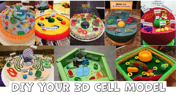
[In this figure] Various 3D cell models I saved on Pinterest.
The rational of choosing materials
I personally saved a lot of pictures of these very nice models that I found and I hope one day I can make my own version of the 3D cell model. Some of them are made of clay or styrofoam. Some of them are made of recycled cardboard and old toys. Some of them are edible and made of cakes, candies, and even pizza!
Of no doubt that all these cell models are beautiful and educational. This makes me wonder how I can add creative ideas into my original version of the cell model and make it stand out.
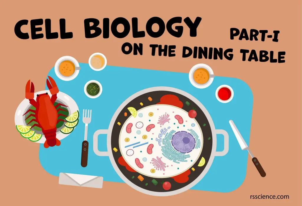
I am going to teach you “Cell Biology” on the dining table
Building a cell model should deepen your understanding of the cell. The key step of making your own cell model is picking the materials that represent different organelles (like nucleus and mitochondria). It’s also important to understand the functions of each organelle and how they work together inside the cell.
My thought is to make an edible cell model with food items I already have in my house. More importantly, I want to choose the food (most of them are fruits and vegetables) based on its “scientific meaning” that represents the biological function of that particular organelle.
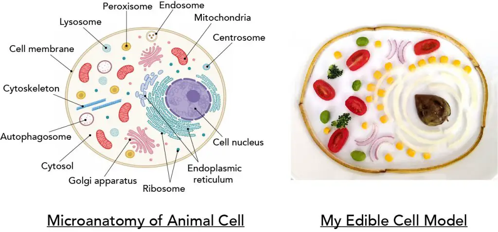
[In this figure] A side by side comparison of the cell diagram I drew and the cell model I made.
Left: the animal cell with many kinds of organelles. Each organelle performs its unique and critical function for the survival of the cell as a whole. We will cover all these organelles in these series of articles. Right: my first version of the “Edible Cell Model”. Can you guess what materials I use?
In this article, I am going to show you how I build my cell model step-by-step. At the same time, I will explain to you the biology of these organelles and the reason why I choose, for example, the cherry tomato for representing mitochondria.
Cell membrane – a balloon filled with water
Our cell is literally a balloon filled with water (70% of our body weight is water). This soft but tough balloon is made by the cell membrane (also known as the plasma membrane). The cell membrane is a biological membrane that separates the interior of cells from the outside space and protects the cell from its environment.
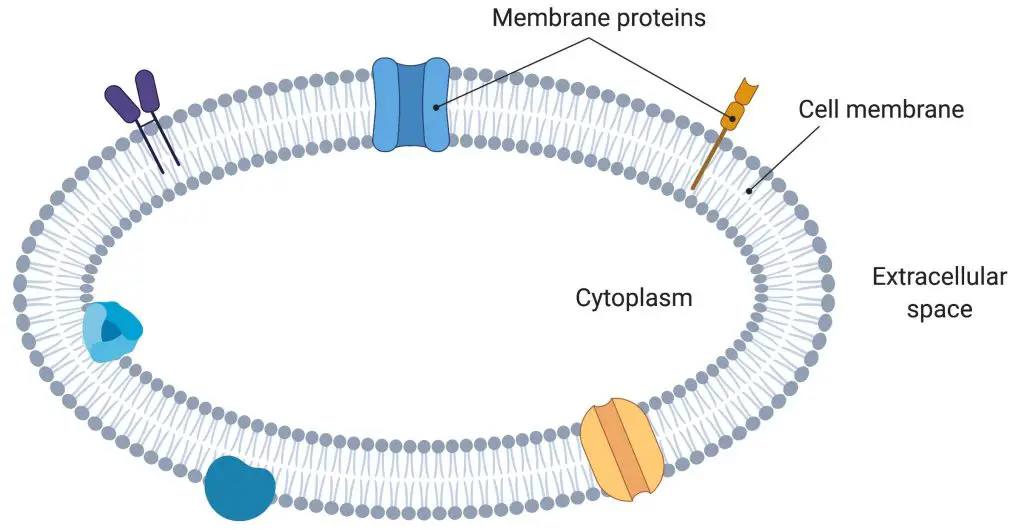
[In this figure] The cell membrane defines the inside and outside spaces of a cell.
The cell membrane is made by two layers of lipid films (oil molecules) with many kinds of proteins inserted. It controls the movement of molecules such as water, ions, nutrients, and oxygen in and out of the cell. In addition, the cell membrane also involves in cell movement and the communication between cells. Plant cells have an additional layer of cell wall around the cell membrane.
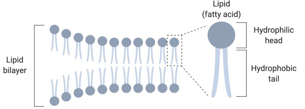
[In this figure] The cell membrane is made of two lipid films, called lipid bilayer.
The reason why using banana
I use banana peels as the cell membrane in my cell model. The banana peel, literally, is the outer skin covering of the banana fruit, whose function is similar to the cell membrane. In fact, banana peels are very nutritious and can be processed to feed animals and manufactured to produce ethanol.
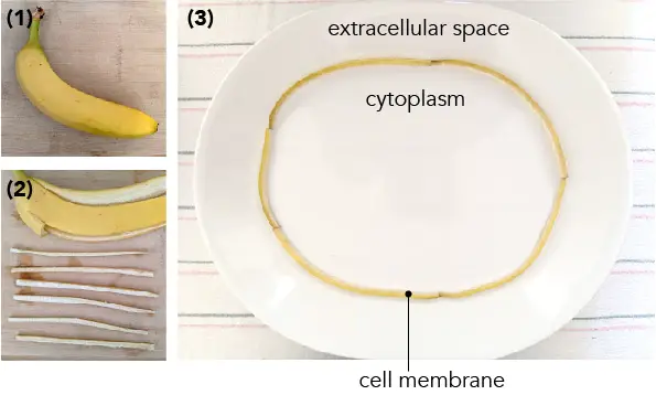
[In this figure] I cut the banana peel into strips (1-2) and arrange them into a circle to represent my cell membrane (3).
By the way, the reason why a banana peel is so slippery is because of a layer of a lubricating substance called polysaccharide follicular gel produced by the banana fruit. Some single-cell microorganisms (like ciliates) can also produce similar materials around their cell membrane to help them move or swim.
Cytosol – the mysterious cellular soup
The cytosol is like condensed soup inside the cell. It is a complex mixture of all kinds of substances dissolved in water. You can find small molecules like ions (sodium, potassium, or calcine), amino acids, nucleotides (the basic units of DNA), lipids, sugars, and large macromolecules such as proteins and RNA.
The cytosol is not a uniform pool. In fact, the concentration of a particular substance can vary a lot in different sub-regions of cytosol.
Even more surprisingly, there is a highway system inside the cytosol, called the cytoskeleton. The cytoskeleton is a dynamic network built by interlinking protein filaments. Its network reaches every inch inside the cells. Once a portion of the cytoskeleton contracts or extends, it deforms the cells and allows cells to change their shapes and movement.
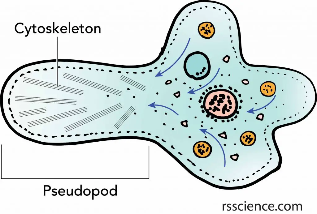
[In this figure] Amoeba is an excellent example of how cells use the cytoskeleton to move.
Moreover, the cytoskeleton serves as an intracellular transportation system. There is a group of “motor proteins” that can carry cargos while walking along the cytoskeleton. A variety of intracellular cargoes, including proteins, RNAs, vesicles, and even entire organelles, can move around in a cell by sitting on these motor proteins.
[In this video] An animation showing that the motor protein (Kinesin) carries cargo and walks on the cytoskeleton of the microtubule.
The reason why it is empty
To keep it simple, the empty space in my cell model belongs to the cytosol.
To clarify, the cytoplasm is all of the material within a cell, enclosed by the cell membrane, except for the cell nucleus. Therefore, the cytoplasm includes the cytosol and all the organelles.
Nucleus – the brain of the cell
The key feature that separates us (eukaryotic cells, include all the animals and plants) from bacteria (prokaryotic cells) is the nucleus. The nucleus (pl. nuclei) is a membrane-bound organelle that stores most of our genetic information (in the form of DNA). In contrast, the DNA is located in the cytoplasm of bacteria, which are lack of any membrane-bound organelle.
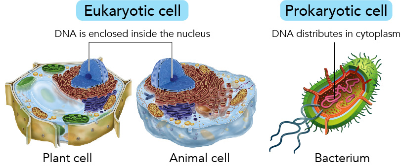
[In this figure] Eukaryotic cells v.s. Prokaryotic cell
Our genes are written as the genetic codes (A, T, G, C) in the DNA. The gene is a blueprint for making a protein. Inside the nucleus, a process that makes copies of a certain gene in the form of massager RNAs (mRNAs), called transcription. These mRNAs will be exported outside of the nucleus for the ribosomes to make proteins.
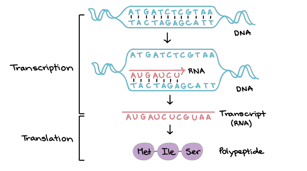
[In this figure] The decryption of genetic codes involves two steps: (1) Transcription – from DNA to mRNA, happens inside the nuclei; and (2) Translation – from mRNA to protein, occurs in ribosomes. (Source: khan academy)
If you stretch all the DNA from a single cell into a linear thread, it can be as long as 3 meters (or 10 feet) long. It is incredible how the cell can pack entire DNA into a tiny nucleus (usually the diameter of a nucleus is less than 2-3 micrometers; one micrometer = 0.000001 meters). Most of the time, we can not see the DNA molecules under a regular light microscope.
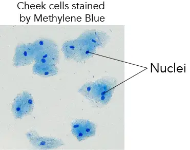
[In this figure] Although we can’t see a single DNA thread, we can stain the total DNA by Methylene Blue to visualize the nuclei. These are my cheek cells. You can stain and see your own cell nuclei by following this guide.
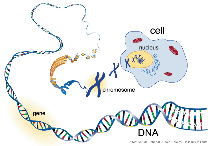
[In this figure] The relationship between DNA, gene, chromosome, nucleus, and cell.
All our genes (called the genome) are located on 46 pieces of long DNA threads. When the cell prepares for dividing, each DNA thread will be organized with special proteins, called histones, to form chromosomes (now could be visible with proper staining). We have 23 pairs of chromosomes (1-22, X, and Y), and the number will be doubled right before the cell division.

[In this figure] 23 pairs of human (male) chromosomes by a microscopic technique called Giemsa banding (G-banding) karyotype.
During division, the membrane of the nucleus (called the nuclear envelope) will temporally disappear. The duplicated chromosomes will be pulled toward the opposite ends of the cell by centrosomes to form two daughter nuclei. Then, the rest of the cell also divide into two daughter cells, each with one nucleus.
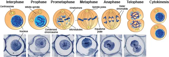
[In this figure] Mitosis in animal cells.
The eggs of parasitic nematodes, Ascaris, are a good specimen to study mitosis in animal cells. Their chromosomes are easily visible under a light microscope.
The reason why using avocado seed
What could be a good substitute for the nucleus in my cell model? I happen to have my favorite avocado toast as breakfast this morning, so why not recycle the avocado seed.
A seed, which carries all the genetic information and can grow into an entirely new plant, is, to some extent, like the nucleus.
Do you remember the cloned sheep “Dolly”? In fact, the scientists took the nucleus from a mature cell (which provide the genetic material) and planted the nucleus into an oocyte with its own nucleus removed (which provide the cytosol and other organelles). After several months, a sheep called “Dolly” was born with an identical genome of the nuclear donor.
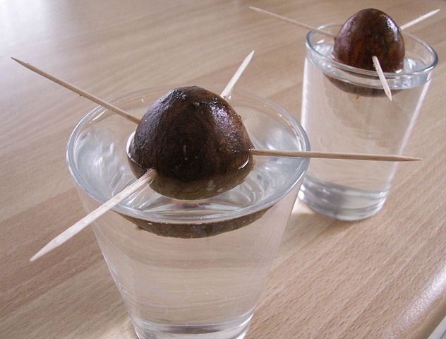
[In this figure] A common technique to germinate avocados at home is to poke the avocado with toothpicks and leave it partially submerged in indirect light. Source: https://en.wikipedia.org/wiki/Avocado
The avocado seed is a hard, massive core in the center of the fruit, just like the nucleus in a cell. If you manage to cut an avocado seed in half, you can see a white core enclosed by a thin shell. I like to imagine that the shell is a nuclear envelope, and the white core is a mass of DNA.
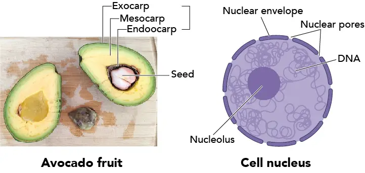
[In this figure] Avocado v.s. Cell nucleus.
The left image is an avocado fruit cut in half. You can see a massive seed in the center. The see is protected by a layer of hard skin, called the endocarp. The bulk middle part we eat is mesocarp. The outer layer, called the exocarp, is the official name of the fruit skin or rind.
The right image is a cell nucleus. The nucleus is bounded by two layers of membrane, called the nuclear envelope. There are many nuclear pores on the envelope to allow the trafficking of molecules. The nucleolus is where the ribosomes are produced.
In addition to avocado seed, I think of the mushroom as a good representative for a cell nucleus. Why? because the mushroom is rich in DNA.
Have you heard of “Gout”? A gout is a form of arthritis with symptoms like severe pain, redness, and tenderness in joints. Patients with gout are told to avoid food (like mushrooms) with high purines. The purines will be metabolized in our body to uric acids. Too many uric acids will form crystals in the joints and cause the pain. Purine is part of the DNA molecule, and the purines in foods indeed come from the nuclei.
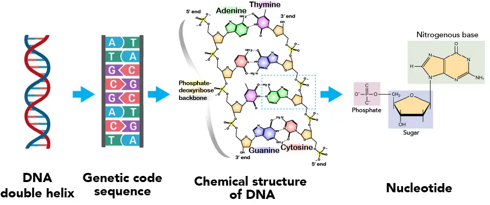
[In this figure] Step-by-step breakdown of DNA molecules.
You must hear that DNA is a double helix. The double helix can form because the genetic code sequences on each half are perfectly complementary. “A” always pairs with “T” and “G” always pairs with “C”. At the atomic scale, you can see the complementary match happens between the “Nitrogenous bases” of each nucleotide unit.
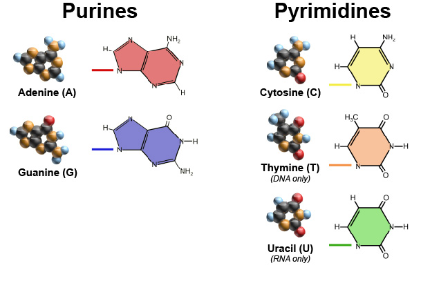
[In this figure] Purines and pyrimidines are two kinds of nitrogenous bases in our DNA and RNA molecules.
Mitochondria – the cell’s powerhouses
Mitochondria (singular: mitochondrion) are rod-shaped organelles that are considered the power generators of the cell. During the process of cellular respiration, mitochondria convert glucose and oxygen to produce adenosine triphosphate (ATP), which is the biochemical energy “currency” of the cell to do any other activities.
The numbers of mitochondria can reflect the energy demand of the cell type. For example, heart muscles host more mitochondria in order to power the heart bumping. On the other hand, our red blood cells lose their mitochondria as well as nuclei so that they can carry more oxygen.
Two unique features of mitochondria
Mitochondria are unique and quite different from other organelles in two fundamental aspects. These two features also reveal where the mitochondria came from.
Two layers of membranes
First, mitochondria have double layers of the membrane: outer mitochondrial membrane (OMM) and inner mitochondrial membrane (IMM). Between the OMM and IMM is the intermembrane space. The region inside the inner membrane is called the matrix.
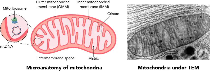
[In this figure] Microanatomy of mitochondria.
Left: Two layers of mitochondrial membrane (OMM and IMM) separate the intermembrane space and the matrix. Inside the matrix, you can find mitochondria’s own DNA (mtDNA) and ribosomes (mitoribosomes).
Right: Mitochondria under the transmission electron microscope (TEM). The folds of IMM, called cristae, are the most apparent structure to identify mitochondria.
The double-layered structure is critical for the powerhouse function of mitochondria. In fact, the mitochondria generate ATP like a hydraulic dam. At the IMM, there is a set of proteins that assemble into an energy generator called electron transport chain.
In the first three steps of electron transport, these proteins bump the protons (H+; the hydrogen ion after losing its electron) from the matrix to intermembrane space. Over time, this builds up a proton gradient across IMM. Then, at the last step, all the protons flow through a protein complex called ATP synthase, which acts as a turbine generator. ATP synthase uses the energy of proton flux to convert ADP into ATP.
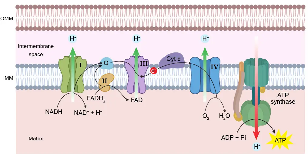
[In this figure] Electron transport chain.
The ATP generation happens on the inner mitochondrial membrane (IMM). First, protons (H+) are bumped across IMM into intermembrane space to build up a proton gradient. Then, the ATP synthase uses the energy of proton influx to drive the chemical conversion of ADP to high-energy ATP. During this process, there are two electrons (e-), which transferred between protein complexes, so-called electron transport chain.
Mitochondria have their own DNA
Second, mitochondria are the only organelles that have their own DNA other than the nucleus (in plant cells, chloroplasts have their own DNA, too). Mitochondrial DNA (mtDNA) is circular (very similar to the bacterial DNA) and stored in the matrix. Compared to nuclear DNA, mitochondrial DNA is much shorter, encoding only 13 genes. These 13 genes are coded for making the components of the electron transport chain that we mentioned above.
Endosymbiotic theory
Scientists believe mitochondria are derived from the bacteria that were engulfed by the early ancestors of today’s eukaryotic cells. This theory is called the endosymbiotic theory.
Around 1.5 billion years ago, some prokaryotes incorporated other prokaryotes into their cells. These incorporated prokaryotes then lost their ability to live independently and become integrated as part of the hosts. They later became specialized in specific functions, such as energy production in both mitochondria and chloroplasts.
Mitochondria appear to be related to Rickettsiales proteobacteria, and chloroplasts appear to be related to nitrogen-fixing filamentous cyanobacteria. Both mitochondria and chloroplasts still keep their own DNA to make some of their proteins, but the majority of their proteins still require nuclear DNA from the host cells.
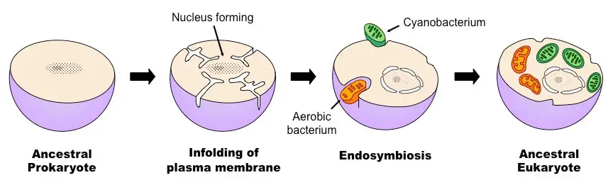
[In this figure] Overview of the process of endosymbiosis. Source: https://ib.bioninja.com.au/
The double layers of mitochondrial membranes are another evidence of endosymbiotic origin. The IMM could be the original membrane of the engulfed bacterium. The OMM was the remained vesicle when the host cell incorporated the bacterium. The engulfing process is similar to “phagocytosis” of Amoeba.
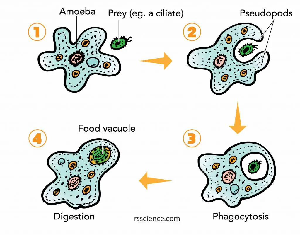
[In this figure] Phagocytosis of Amoeba cell.
The IMM forms many folds called cristae. The cristae greatly increase the surface area of IMM to allow greater capacity for ATP generation. The cristae are the key feature for scientists to identify mitochondria under an electron microscope and a must-have feature in every drawing of mitochondria.
The reason why using cherry tomato
All things considered, I choose cherry tomato to represent mitochondria in my cell model. They are both rod-shaped. The fiery red color of cherry tomato conveys an energetic image of the cell’s powerhouse. More importantly, the slices of cherry tomato also show internal folds, just like the cristae inside the mitochondria.
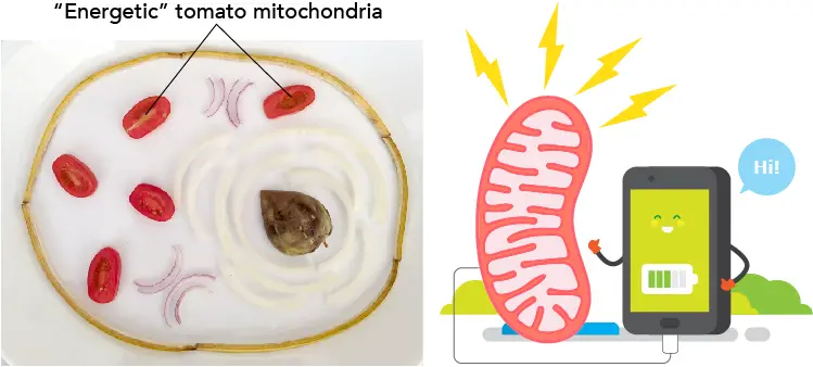
[In this figure] Cherry tomato represents the mitochondria in my cell model.
Summary
Voila! We got to learn the function of the cell membrane, cytosol, nucleus, and mitochondria this week.
Cell membrane
The cell membrane is composed of two layers of lipid films (oil molecules) with many kinds of proteins inserted. It separates the cells from the outside environment.
Cytosol
The cytosol is the substance inside the cells except for the nucleus and organelles. It is a mixture of proteins, irons, amino acids, and small molecules. Because there is so much stuff in it, you can imagine that it is like condensed soup.
Nucleus
The nucleus is the place where the genome is. i.e. the place to store an organism’s genetic information. Its function is like the “brain” of the cell because it instructs and coordinates the function of different organelles.
Mitochondria
Mitochondria are rod-shaped organelles that are considered the power generators of the cell. It generates ATP, a currency of the cellular energy.
Woo-hoo, next week, we are going to cover Endoplasmic reticulum, Ribosome, Golgi apparatus, Peroxisome, and Lysosomes! Stay tuned!
Related posts
Plant Cell Model Part V – cell wall, vacuole, and chloroplast.
Cell Organelles and their Functions – overview of each organelle.