
This article covers
How to mount a microscope slide?
This lesson will show you the basic skill to mount a slide (bubble-free) using your own cells as an example.
The material you need
- Blank microscope slides and coverslips
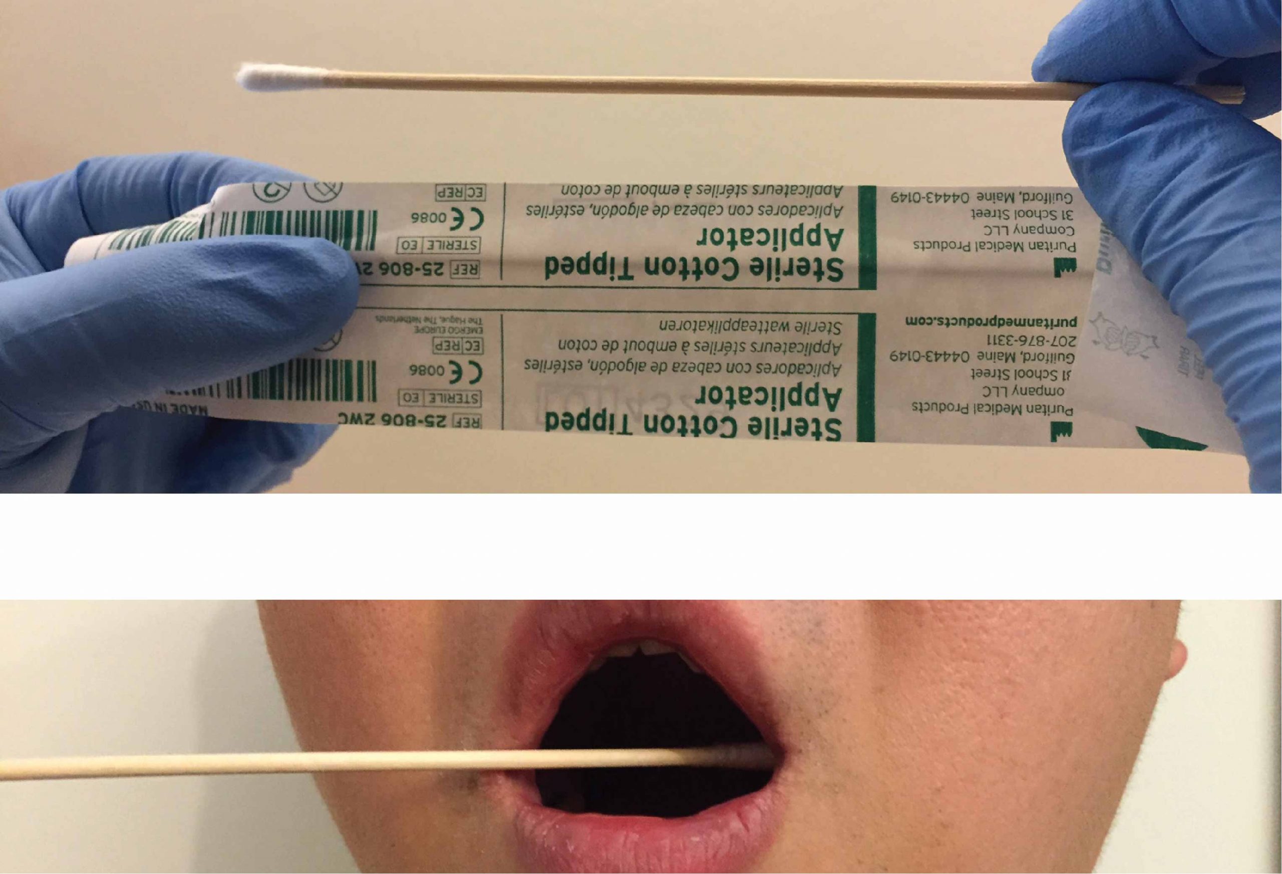
- Sterile cotton tips
- Forceps
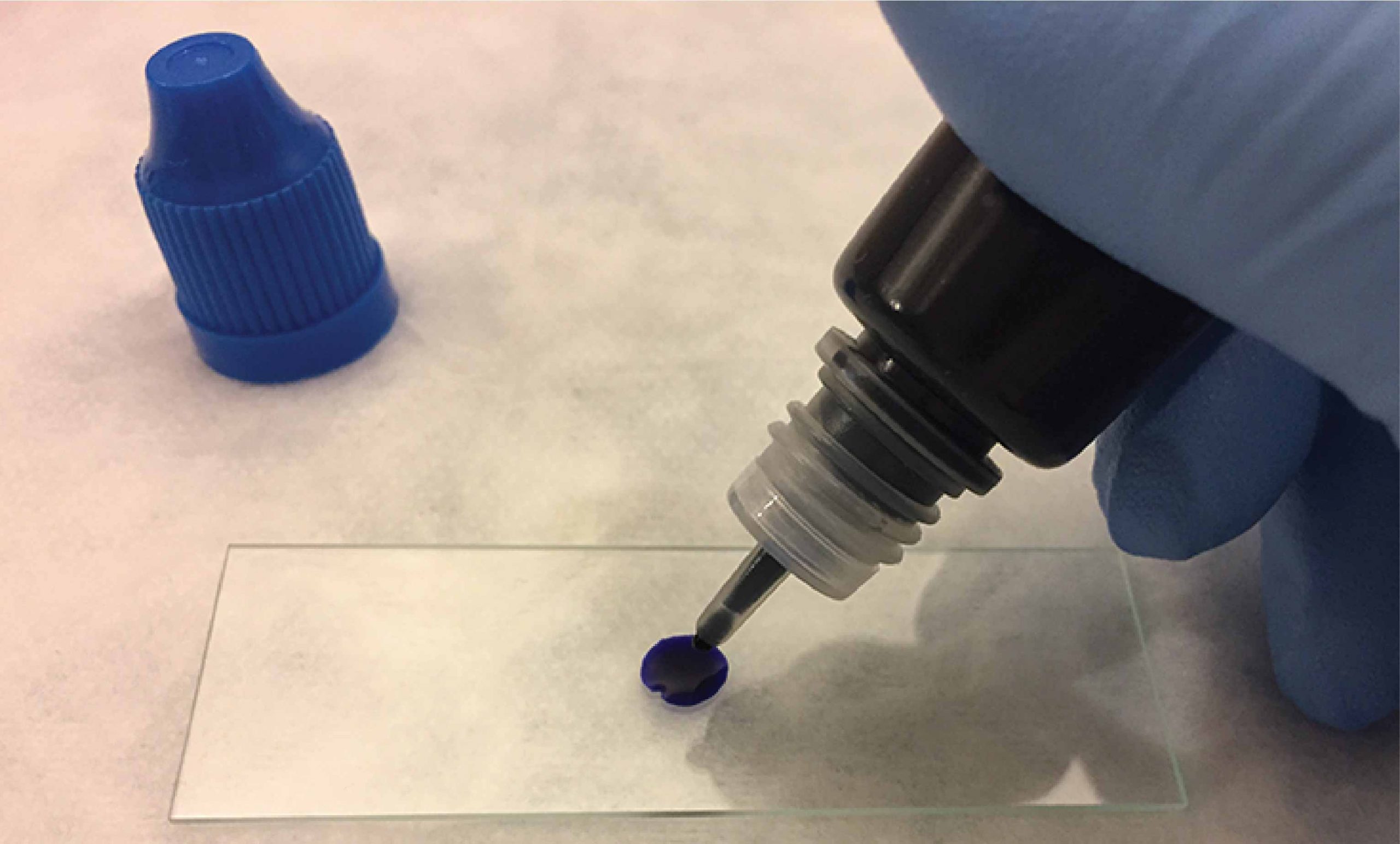
- Methylene blue staining solution
How to mount a slide for a microscope
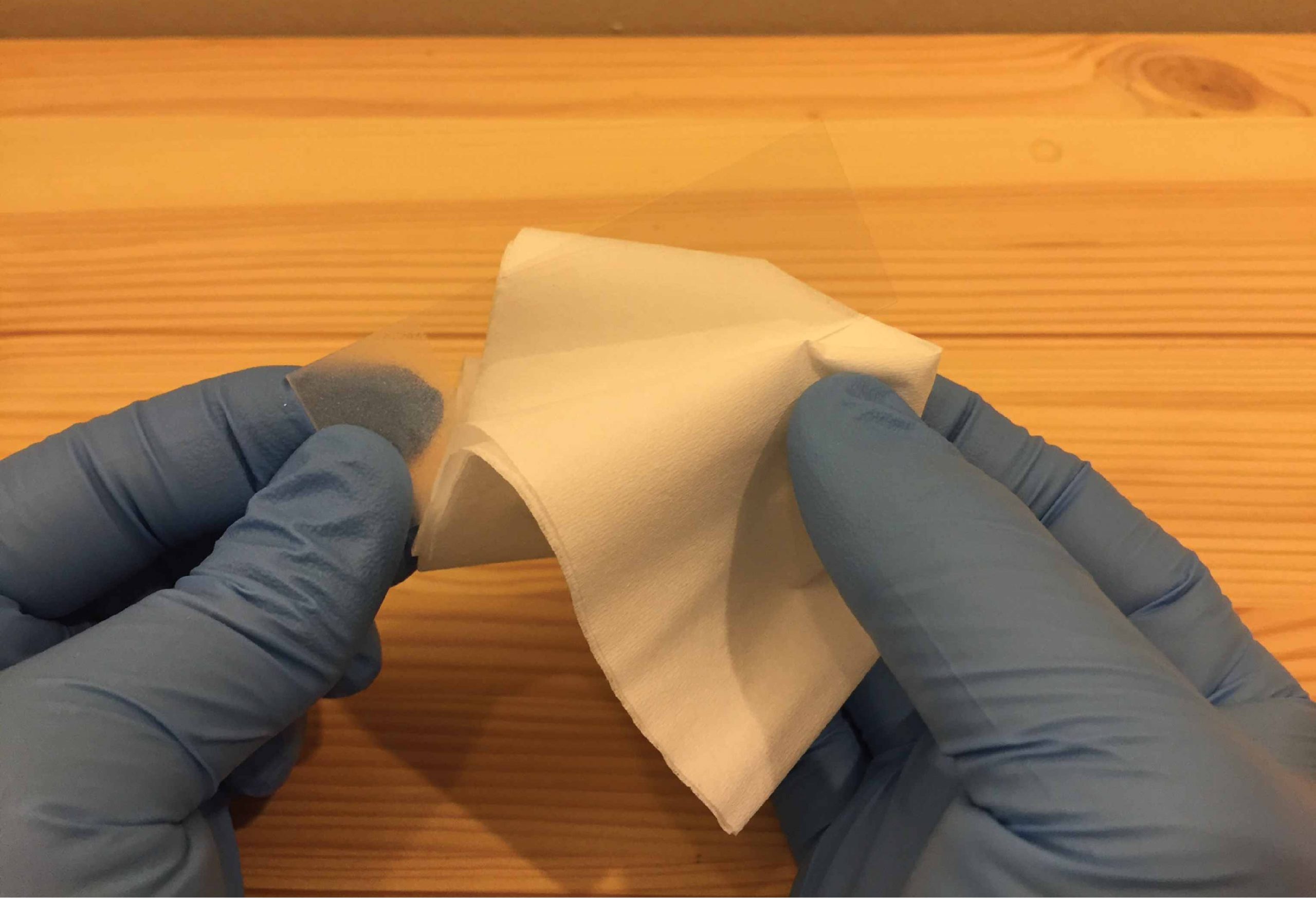
- Clean a slide with a piece of Kimwips paper.
- Take a clean cotton swab (sealed package, sterile) and gently scrape the inside of your mouth.
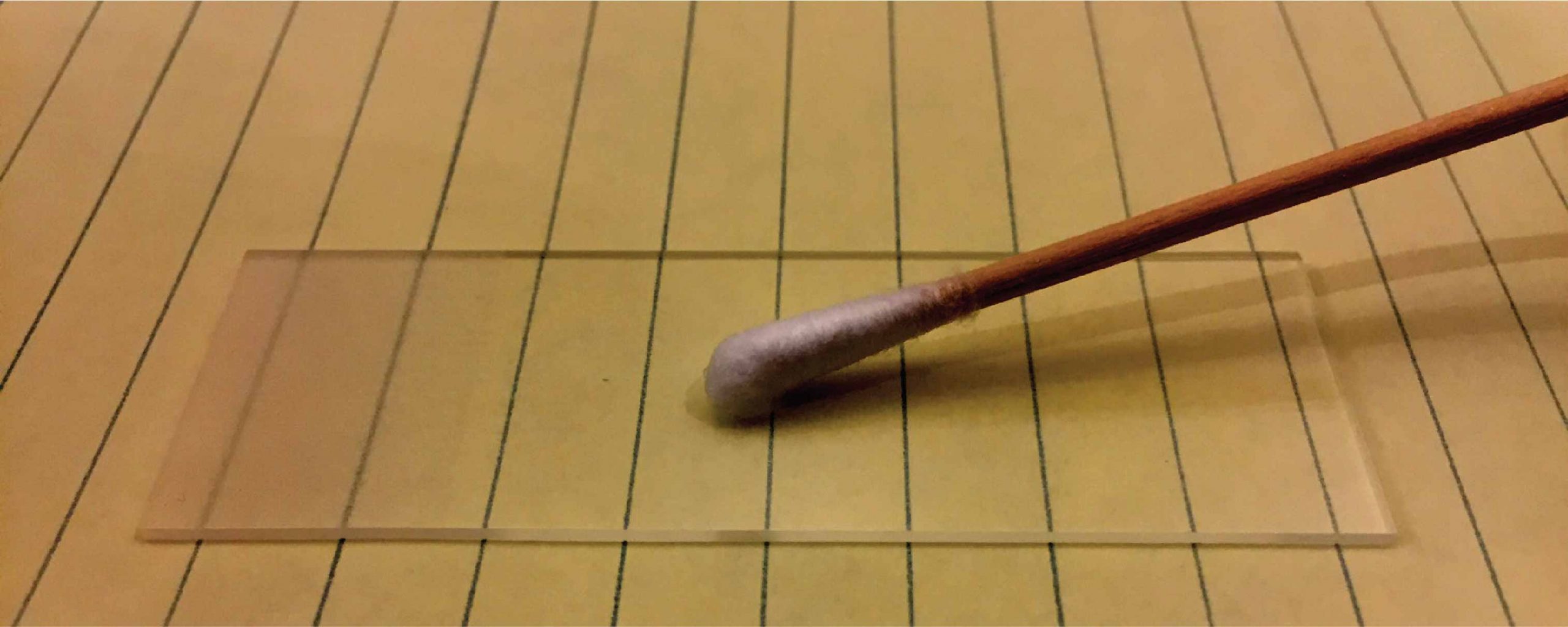
- Smear the cotton swab on the center of the slide for 2 to 3 seconds.
- Add a drop of methylene blue solution on the smear. Methylene blue solution can stain the cells with blue color so they are easier to see under the microscope.
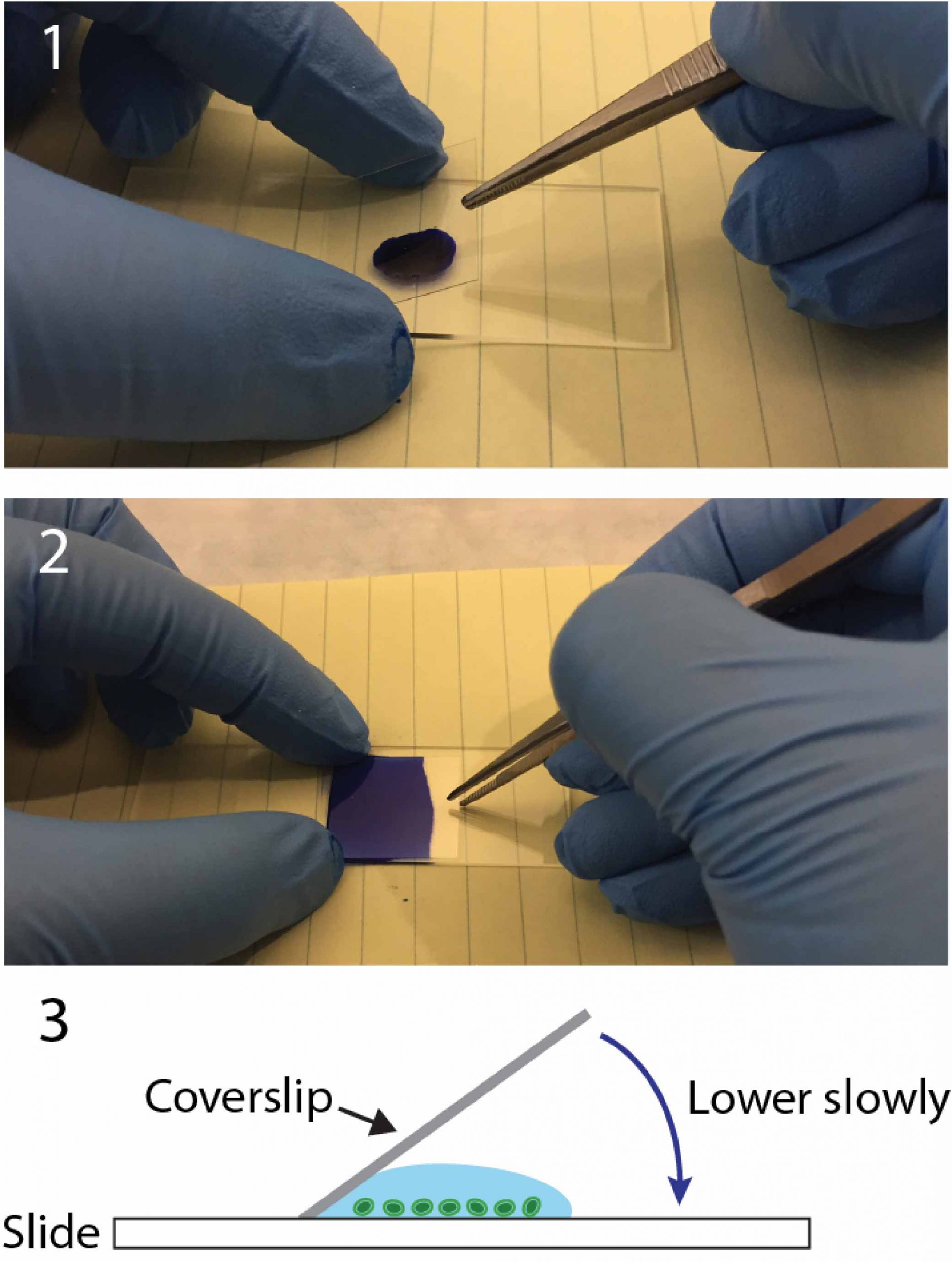
- Place a coverslip on top.
Note: Lower the coverslip slowly with an angle. Allow one side of the liquid droplet touches the coverslip first. This permits air to escape from the other side. You can use the forceps to help you control the coverslip.
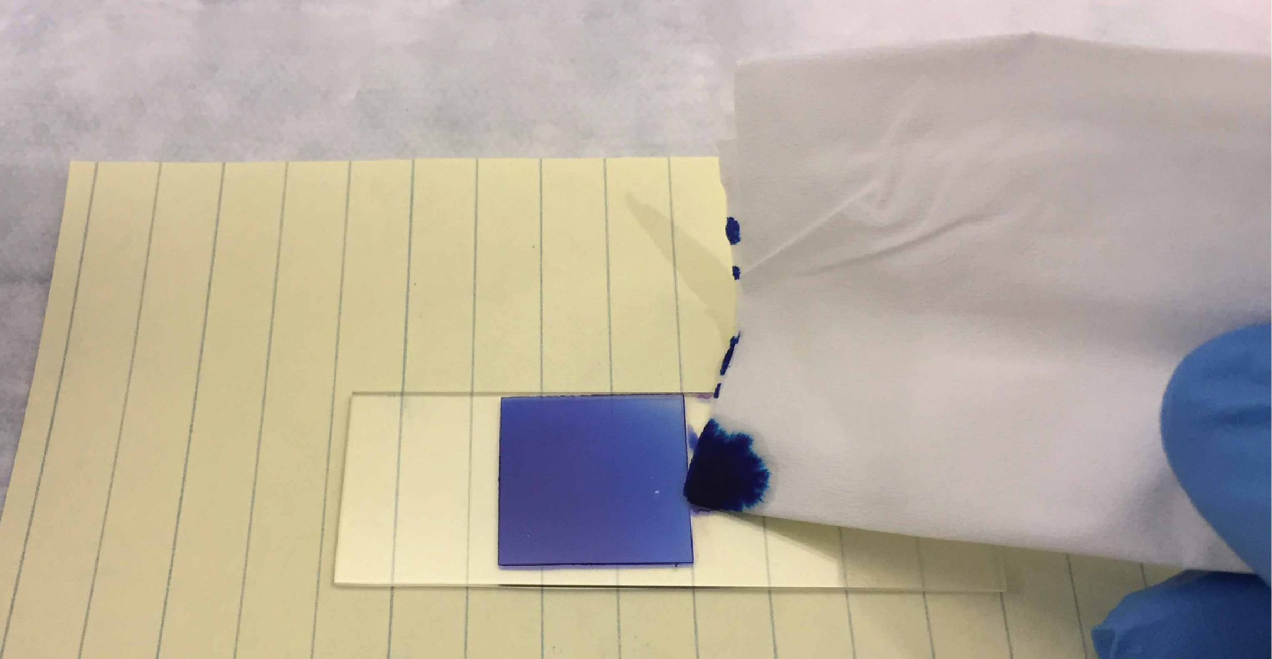
- Remove any excess solution by using a Kimwips paper to touch one side of the coverslip.
Note: You can also seal all the edges of the coverslip with super glue (or nail polish) for long-term storage.
Methylene blue
Methylene Blue is a popular alkaline stain used to stain animal cells to make nuclei more visible. Methylene blue binds DNA which is very abundant in the nucleus. Cells stained by methylene blue will show the nucleus with a deep blue color. It also helps make cells show up against their background, where their shape can help you determine what they are (their morphology).
You can substitute Methylene Blue in most staining protocols that call for carmine or Janus green B. Methylene blue is also used by aquarium hobbyists for preventing fungal disease.
Wet Mounting Medium
A glycerol-based mounting medium can provide a high refractive index than pure water. It is necessary to see certain cellular structures. If a lower refractive index is needed, then pure water could be mixed into the Glycerol solution for an appropriate refractive index.
High glycerol concentration also has a strong tendency to withdraw water from the sample. It may cause the specimen to shrink and deform, especially for algae and other water organisms, which are sensitive to dehydration. It is possible to seal the glycerin mount by applying super glue or nail polish to the sides of the cover glass. This will hold the cover glass in place for longer time periods.
What will you see?
The methylene blue solution stains the cell nuclei, so you can see the nuclei as dark blue and cytoplasm being colored as light blue.

You can also see some small rod-shaped bacteria on the right image. Don’t worry, they are normal oral microbes.
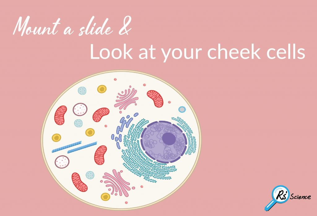
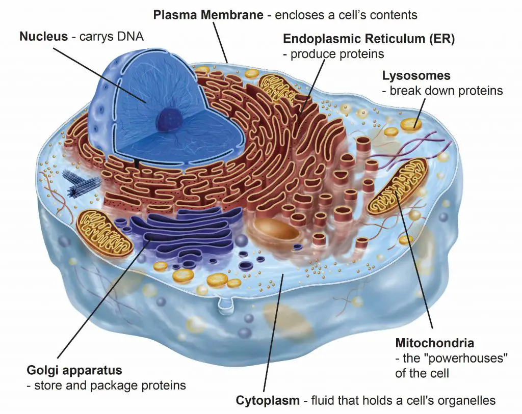
Anatomy of the animal cell
Cells are “building blocks” of life. The body of an average man contains around 30 to 40 trillion cells. Although a cell is only about 20 micrometers (one-millionth of a meter) across, there is still amazing complexity within it.
Each organelle has its own function. Interesting in what they do? Please check out our article.
What does your cell look like? Please share it with us!