
This article covers
What is cell organelle?
A cell organelle is a tiny cellular structure that performs specific functions within a cell. You can think of cell organelles as a cell’s internal organs. For example, the nucleus is the cell’s brain, and the mitochondria are the cell’s hearts. Cell organelles are often enclosed by their own membranes, which divide the cell into many small compartments for different biochemical reactions.
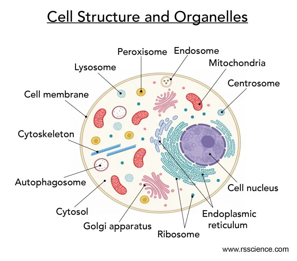
[In this figure] The anatomy of an animal cell with organelles labeled.
Cell organelles have a wide range of responsibilities, from generating energy for a cell to controlling its growth and reproduction. From this point of view, you can also think of cell organelles as different teams within the factory. Each team carries out its specific task and coordinates to make sure the entire factory works smoothly.
Below is a list of the cell organelles found in animal and plant cells, which we’ll use as our guide for this discussion.
| Cell Organelle | Biological Function | Factory Part |
| Nucleus | DNA Storage | Files and blueprints management |
| Mitochondrion | Energy production | Powerplant |
| Ribosome | Protein synthesis | Machine to product toys |
| Rough ER | Protein production and modification | Coordination of toy production line and decoration |
| Smooth ER | Lipid production and Detoxification | Accessory production |
| Golgi apparatus | Protein transportation and export | Packaging and shipping department |
| Peroxisome | Lipid breakdown; redox reactions | Hazard chemical handling |
| Lysosome | Protein destruction | Recycling |
| Cytoskeleton | Cell movement; intracellular transportation | Conveyor system |
| Cell membrane | Define the inside and outside of a cell | Factory building |
| Cell wall | Structural support and protection (plant cell) | Reinforced factory building |
| Cytosol | Cellular fluid | Internal space and floor plan |
| Chloroplast | Photosynthesis (plant cell) | Solar panels |
| Vacuole | Storage and water regulation (plant cell) | Storage spaces |
Cell organelles can be divided into three types
In this article, we are going to divide these cell organelles/structures into three types:
1. General cell organelles: they are present in both animal and plant cells all the time – cell membrane, cytosol, cytoplasm, nucleus, mitochondrion, rough and smooth endoplasmic reticulum, Golgi apparatus, peroxisome, lysosome, and the cytoskeleton.
2. Temporal cell organelles: they are only found at specific stages of the cell’s life cycle – chromosome, centrosome, autophagosome, and endosome.
3. Cell type specific cell organelles: they only exist in the plant cells – chloroplast, central vacuole, and cell wall.
Many unique cell organelles/structures only exist in specific cell types. For example, the food vacuoles in amoeba and the trichocysts in paramecia, which cannot be found in human cells. On the other hand, some human cells also have unique organelles that can’t be found anywhere else, like the Weibel–Palade bodies in blood vessel cells.
1. General cell organelles in every cell
Cell membrane
- Cell membrane is a biological membrane that separates the interior of the cell from the outside space and protects the cell from its environment.
- Cell membrane is made by two layers of lipid films (oil molecules) with many kinds of membrane proteins.
- Cell membrane controls the movement of molecules such as water, ions, nutrients, and oxygen in and out of the cell.
- Proteins on the cell membrane also involved in cell movement and the communication between cells. For example, cells received signals from the outside world through different kinds of receptor proteins inserted on the cell membrane like tiny antennas.
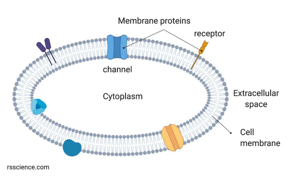
[In this figure] The cell membrane defines the inside and outside spaces of a cell. There are many proteins on or inserted in the cell membrane. They function as channels (controlling the in and out of molecules) or receptors (receiving signals from the outside world).
The image was created with BioRender.com.
Cytosol
- Cytosol is the cellular fluid inside the cell. It fills up the entire intracellular space.
- Water is the most abundant molecule inside the cells, accounting for 70% or more of total cell mass.
- Cytosol is a complex mixture of all kinds of substances dissolved in water, including small molecules like ions (sodium, potassium, or calcium), amino acids, nucleotides (the basic DNA units), lipids, sugars, and large macromolecules such as proteins and RNA.
Cytoplasm
- Cytoplasm refers to all material within a cell, enclosed by the cell membrane, except for the cell nucleus.
- Cytoplasm includes the cytosol and all the organelles.
Cytoskeleton
- Cytoskeleton is the cells’ skeleton system. Its network reaches every inch inside the cells.
- Cytoskeleton is a dynamic network built by interlinking protein filaments. It is composed of three main components, actin filaments, intermediate filaments, and microtubules.
- Once a portion of the cytoskeleton contracts or extends, it deforms the cells and allows cells to change their shapes and movement.
- Cytoskeleton also serves as a highway system inside the cytosol. Motor proteins can carry cargos while walking along the cytoskeleton. A variety of intracellular cargoes, including proteins, RNAs, vesicles, and even entire organelles, can move around inside a cell by this intracellular transportation system.
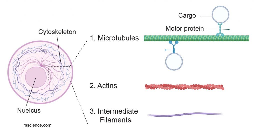
[In this figure] Cytoskeleton consists of three types of filament proteins: microtubules, actins, and intermediate filaments.
The image was created with BioRender.com.
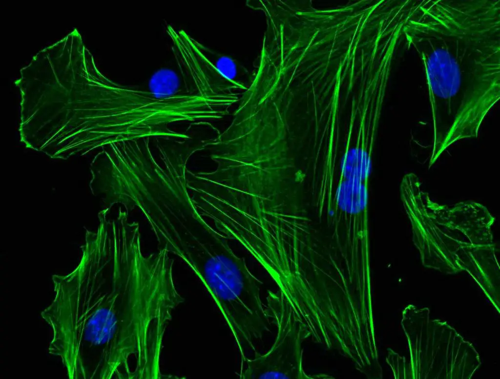
[In this figure] Fluorescent image of vimentin, an intermediate filament protein (green), in human cells. The nuclei were stained in blue color.
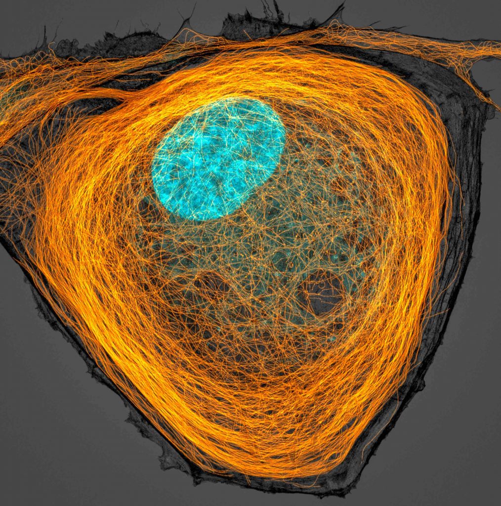
[In this figure] Fluorescence image of microtubule (orange), and the nucleus (cyan) inside a cell.
Microtubule is one type of cytoskeleton inside the cells, and it shapes cell’s morphology. Magnification, 63x.
Photo credit: Jason Kirk, 2020 photomicrograph competition.
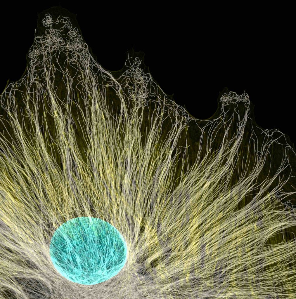
[In this figure] Fluorescence image of microtubule (yellow) and the nucleus (cyan) inside a cell.
Microtubules radiated from a tissue cell culture. Notice that the microtubules extend to the very end of the cell membrane. Magnification, 63x.
Photo credit: Jason Kirk, 2020 photomicrograph competition.
Nucleus
- The nucleus (plural: nuclei) is a membrane-bound organelle that stores most of our genetic information (genome).
- The key feature that separates eukaryotic cells (animals, plants, and fungi) from prokaryotic cells (bacteria and archaea) is the presence of a nucleus.
- The membrane of the nucleus is called the nuclear envelope. There are nuclear pores to control transportation across the envelope.
- During cell division, the nuclear envelope will temporally disappear to allow the separation of chromosomes.
- Both DNA replication and RNA transcription happen inside the nucleus. Messager RNA (mRNA) that carries the genetic information will be exported through nuclear pores into the cytosol for protein synthesis (translation).
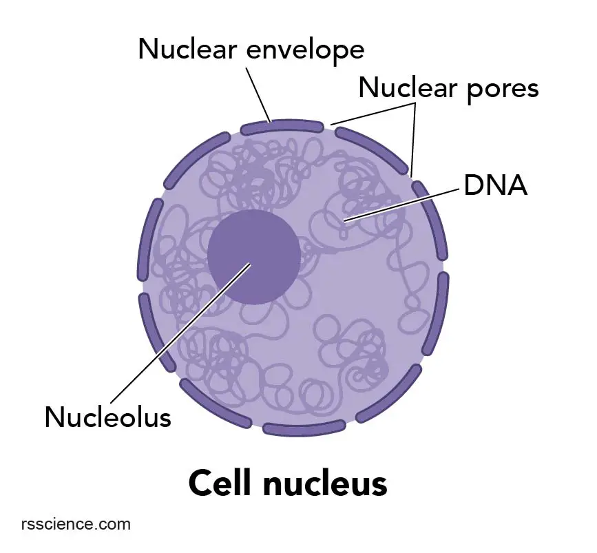
[In this figure] Cell nucleus is a membrane-bound organelle that stores DNA.
The image was created with BioRender.com.
Nucleolus
- Nucleolus (plural: nucleoli) is a structure inside the nucleus.
- Nucleolus is known as the site of ribosome biogenesis.
Mitochondrion
- Mitochondrion (plural: mitochondria) is a rod-shaped organelle that is considered the power generators of the cell.
- Mitochondrion performs cellular respiration, which converts glucose and oxygen to adenosine triphosphate (ATP). ATP is the biochemical energy “currency” of the cell for all activities.
- Mitochondrion has double layers of the membrane: outer mitochondrial membrane (OMM) and inner mitochondrial membrane (IMM). Between the OMM and IMM is the intermembrane space. The region inside the inner membrane is called the matrix.
- Mitochondrion generates ATP like a hydraulic dam. It happens via the electron transport chain across the IMM.
- Mitochondria (in plant cells, chloroplasts, too) are the only organelles that have their own DNA other than the nucleus. Mitochondrial DNA (mtDNA) is circular and encoded only 13 genes.
- Scientists believe mitochondria and chloroplasts are derived from the bacteria that were engulfed by the early ancestors of today’s eukaryotic cells. This theory is called the endosymbiotic theory.
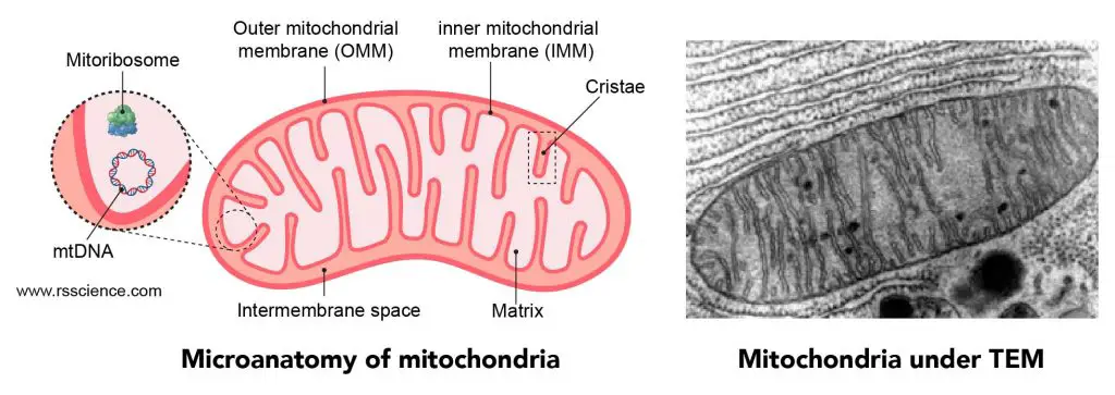
[In this figure] Left: the structure of mitochondrion showing many folds of membranes and mtDNA. Right: a mitochondrion surrounded by rough ER under a transmission electron microscope.
Endoplasmic reticulum
- Endoplasmic reticulum (ER) is an internal membrane that forms branching networks of many interconnected sacs and tubes.
- There are two types of ER: rough ER and smooth ER.
- The outer side (facing the cytosol) of the rough ER is studded with ribosomes. Under the electron microscope, the dense granular ribosomes gave the name of “rough” ER.
- Rough ER stays closer to the nucleus and coordinates protein synthesis.
- Smooth ER lacks ribosomes. It specializes in lipid synthesis, steroid hormone production, and detoxification.
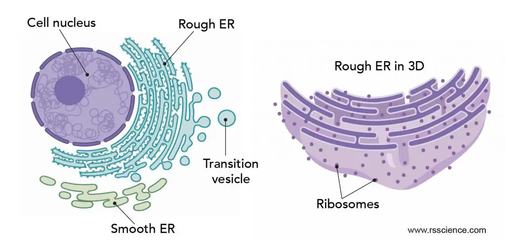
[In this figure] The anatomy of ER.
Left: The relationship between the nucleus, rough, and smooth ER. Right: A 3D view of rough ER.
The image was created with BioRender.com.
Ribosome
- Ribosomes are the places where proteins are synthesized in our cells.
- Ribosomes consist of two major components: the small and large ribosomal subunits. They are assembled by proteins and ribosomal RNA (rRNA).
- Ribosomes translate mRNA into polypeptide chains, which fold and assemble into proteins.
- Transfer RNA (tRNA) carries the corresponding amino acid. Only the right tRNA can enter the ribosome and pair with the code on mRNA. Once the tRNA and mRNA match, the ribosome will add this amino acid onto a growing polypeptide chain.
- Ribosomes can be found on the rough ER or free-floating in the cytosol.
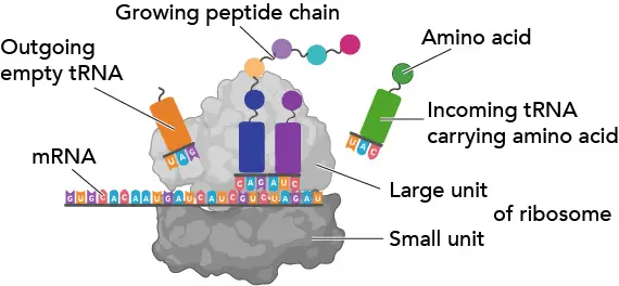
[In this figure] The ribosome works like a machine to translate the code sequence of mRNA into a protein.
Golgi apparatus
- Golgi apparatus (or Golgi) consists of several stacks of membrane-bound cisternae (sacs).
- Golgi apparatus usually locates close to the ER. It receives the raw protein products from the ER, modifies them (for example, adding tags made by sugar chains), and exports the proteins to a variety of destinations.
- The transportation of proteins is done within small bubbles, called vesicles.
- The vesicles are generated by budding from the membrane of the ER and Golgi. Once the vesicles reach their destinations, the fusion of membrane releases their protein cargos.
- There are three major destinations of proteins: (1) sent to other organelles, (2) released into the cytosol, and (3) secreted outside the cells. Secreting vesicles can also store the proteins until they receive a signal to release at a specific event.
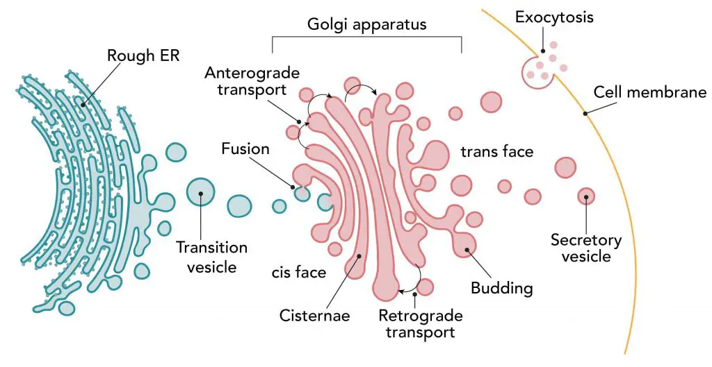
[In this figure] The journey of protein synthesis and transportation.
After proteins are synthesized in the rough ER, they travel to the Golgi for further modification. Then, proteins will be packed into vesicles and travel to their final destination.
Peroxisome
- Peroxisome is a spherical organelle responsible for the fatty acid (oil molecule) breakdown in order to generate energy.
- Peroxisomes in the liver cells also handle the detoxification of many chemicals, including alcohol and drugs.
- Many enzymes inside the peroxisomes catalyze Redox (reduction-oxidation) reactions, which will generate hydrogen peroxide (H2O2) as a dangerous byproduct.
- Peroxisomal enzyme, called “Catalase”, can convert H2O2 into water (H2O) and oxygen (O2) to keep the cell safe.
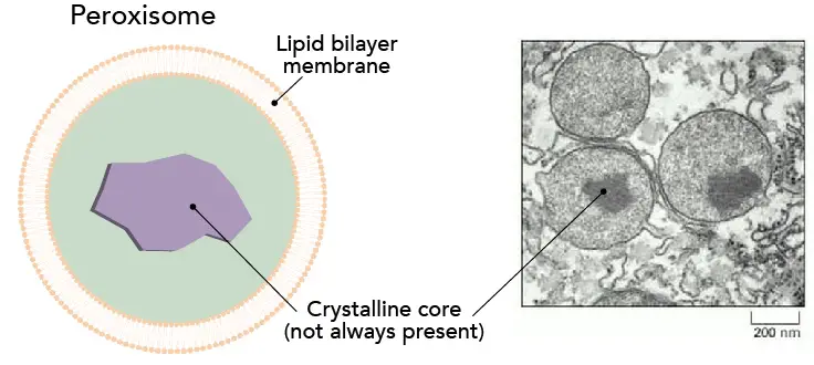
[In this figure] Peroxisomes.
Left: the structure of peroxisome. Right: an electron microscopy image of peroxisomes. (Image from Schrader, M. and Fahimi, H. 2008. The peroxisome: still a mysterious organelle. Histochemistry and Cell Biology 129(4), pp. 421-440.)
Lysosomes
- Lysosome is a membrane-bounded sphere full of digestive enzymes and works like a recycling center in the cell.
- These enzymes can break down whatever substance entering the lysosomes into raw materials (like amino acids, nucleotides, lipids, and sugars), so the cell can reuse these raw materials to build new organelles.
- Inside the lysosome is an acidic environment (pH 5), which activates the digestive enzymes. These enzymes won’t be active in the cytosol (pH 7). This is a safety mechanism in the cell in case the lysosomes somehow leak or burst.
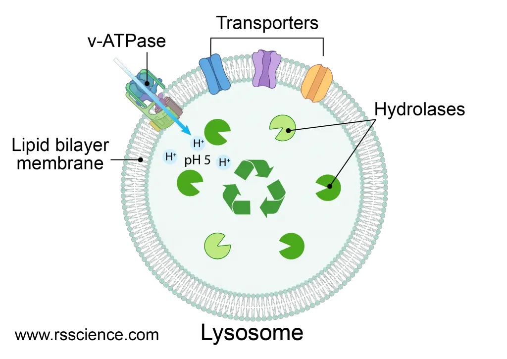
[In this figure] Lysosome is the recycling center of the cell.
2. Temporary cell organelles for specific tasks
Autophagosome
- Autophagosome is a temporary organelle for autophagy.
- Autophagy (aka “self-eating”) is a process that cells recycle some of their existed proteins and organelles due to the shortage of nutrient supply.
- Damaged proteins or organelles will be put on a “garbage tags”. The cell recognizes the tags and packs these recycle materials into autophagosomes.
- Autophagosomes carry the cellular garbage to lysosomes for degradation.
- Special autophagy to degrade bad mitochondria is named “mitophagy.”
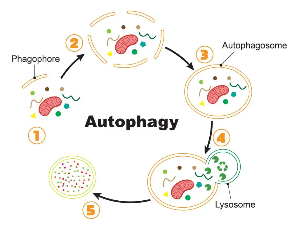
[In this figure] The process of autophagy.
Endosome
- Endosome is a membrane-bound temporary organelle for engulfing the stuff outside of the cell.
- Endosomes are formed by the invagination of the cell membrane, a process called “endocytosis.”
- After endocytosis, the endosome can carry its cargo to different places in the cell.
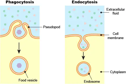
[In this figure] Phagocytosis vs. Endocytosis.
Chromosome
- When the cells prepare for the cell division, each DNA thread is organized into a much compact structure, called “chromosome”.
- Every human cell has 23 pairs of chromosomes (1-22, and X or Y).
- A chromosome is formed by wrapping DNA around histone proteins into a core complex, called a nucleosome.
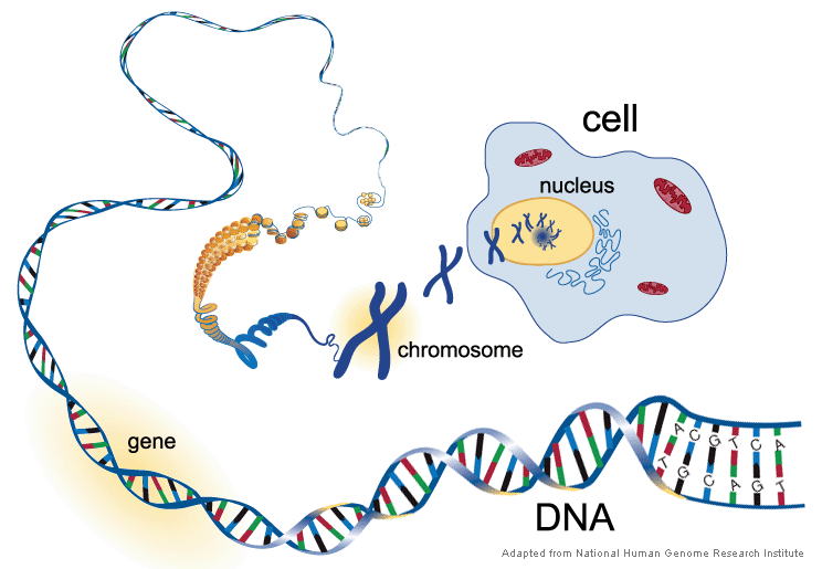
[In this figure] In order to handle the long DNA molecules, our cells pack DNA threads into many compact structures, called “chromosome”.
Sister chromatids
- Sister chromatids are X-shaped chromosomes that remain attached at a centromeric region (centromere) after DNA duplication.
- Sister chromatids will be split into two identical chromosomes during mitosis.
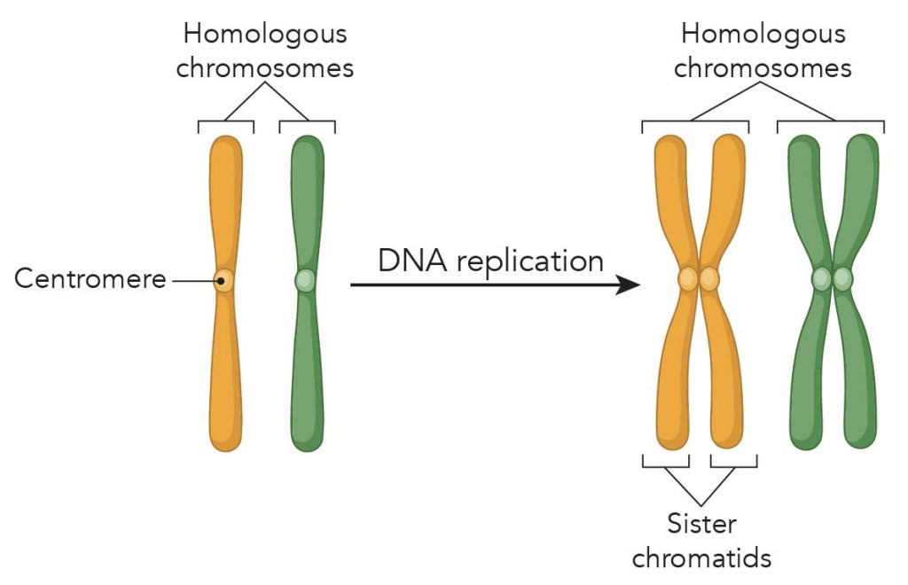
[In this figure] Chromosome replication forms sister chromatids.
Centrosomes
- Centrosomes are organelles that only appear during mitosis and serve as the main microtubule organizing center (MTOC).
- Each cell has two centrosomes. They move toward the opposite positions of the cells when the mitosis starts.
- The microtubules extend from the centrosome and attach to the centromeres of sister chromatids. Both centromeres retrieve their microtubule at the same time to split the sister chromatids apart and move into new cells.
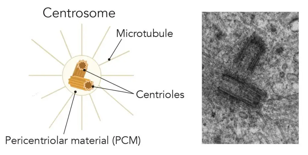
[In this figure] Illustration and electron micrography of the centrosome.
3. Unique cell organelles in the plant cells
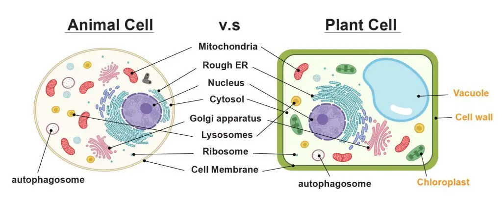
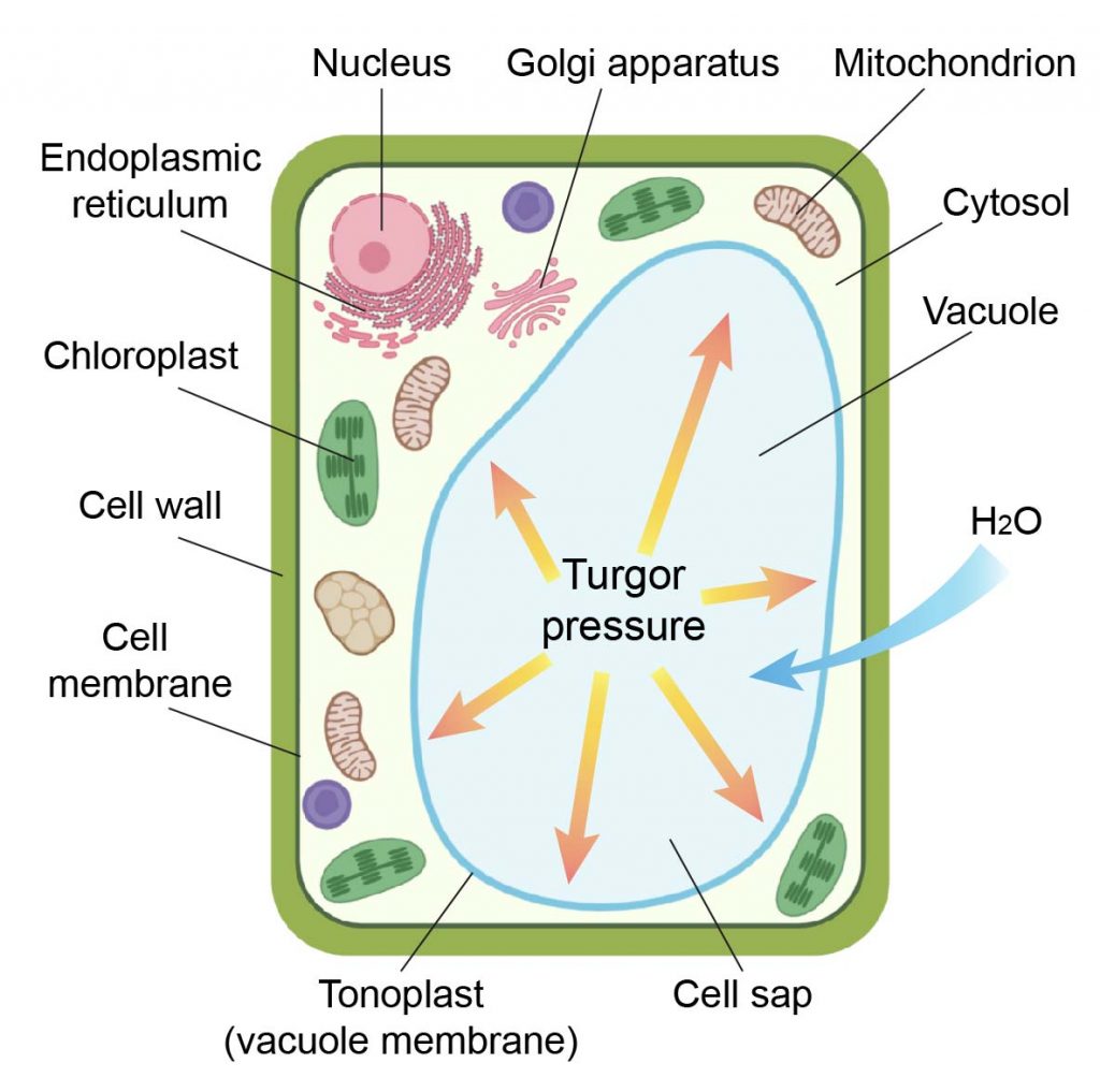
[In this figure] The cell anatomy of animal and plant cells.
The animal cell and plant cell share many organelles in common, such as a nucleus, ER, cytosol, lysosomes, Golgi apparatus, cell membrane, and ribosomes. The organelles that are unique for plant cells are Vacuole, Cell wall, and Chloroplast (shown in orange text).
Cell wall
- Cell wall is an extra layer of structural support and protection outside the cell membrane of plant cells.
- Cell wall is made of cellulose, a polymer type of sugars.
- The structural support of cell walls allows plants to grow to great heights (like pine trees). Wood is made of the reminded cellulose fibers of cell walls after the death of matured xylem tissues of woody plants.
- When Robert C. Hooke came up with the term “Cell” in the 1660s, he was actually looking at the dead plant cells’ cell walls in a thin cutting of cork.
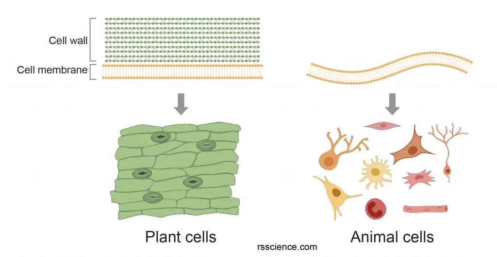
[In this figure] Cell wall provides additional protective layers outside the cell membrane.
Vacuole
- Vacuole is a membrane-bound organelle that contains a mass of fluid.
- Large, central vacuole is only present in the plant cells.
- Vacuole serves as a storage space for plant cells. It can store a variety of nutrients (including sugars, minerals, amino acids, nucleic acids, ions, and special chemicals) that a cell might need to survive.
- Vacuole also functions as a reservoir for the cell to store excess water. The amount of water in the vacuole will determine the cell’s turgor pressure (the hydrostatic pressure against the cell wall). A drooping plant has lost much of its water, and the vacuoles are shrinking.
[In this figure] Drawing of a plant cell showing a large vacuole.
Chloroplast
- Chloroplasts are organelles that conduct photosynthesis and produce energy for the plant cells.
- Chloroplasts convert the light energy of the Sun into sugars (a process called “photosynthesis”) that can be used by cells. At the same time, the reaction produces oxygen (O2) and consumes carbon dioxide (CO2).
- Chloroplasts consist of many stacks of sac structures, called thylakoid system. The molecules (Chlorophyll) that absorb the energy of the Sun locate inside the thylakoid sacs.
- Chloroplast plays an important role in plant innate immunity.
- Chloroplasts and mitochondria share many in common. They both have two layers of membranes, their own DNA and ribosomes. They are believed to be derived from endosymbiotic bacteria engulfed by the early ancestors of today’s eukaryotic cells.
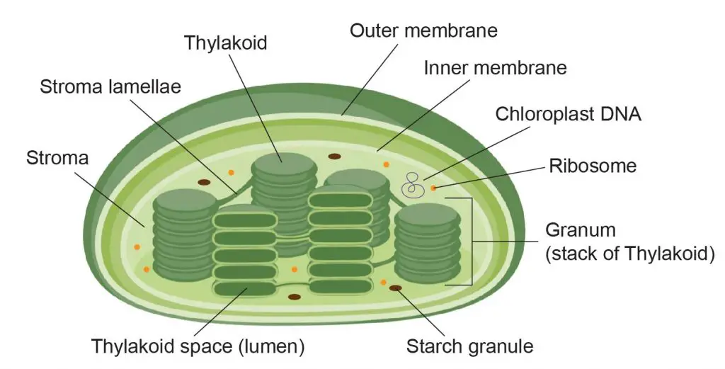
[In this figure] The structure of chloroplast.
Related posts
Animal Cell Model Part I – cell membrane, cytosol, nucleus, and mitochondria.
Animal Cell Model Part II – endoplasmic reticulum, ribosome, Golgi apparatus, peroxisome, and lysosomes.
Animal Cell Model Part III – two types of temporary organelles involving eating behaviors, autophagosomes, and endosomes.
Animal Cell Model Part IV – two types of temporary organelles only appearing during mitosis, centrosomes, and chromosomes.
Plant Cell Model Part V – cell wall, vacuole, and chloroplast.
Excellent overview of cellular anatomy.
Pingback: What is a ribosome? – Dr. Biology Questions and Answers
Pingback: Which organelle often takes up much of the volume of a plant cell? – Dr. Biology Questions and Answers
Pingback: Which organelle makes the proteins that are needed by the cell? – Dr. Biology Questions and Answers
Pingback: How many organelles are in a cell? – Dr. Biology Questions and Answers