
This article covers
What is cell nucleus?
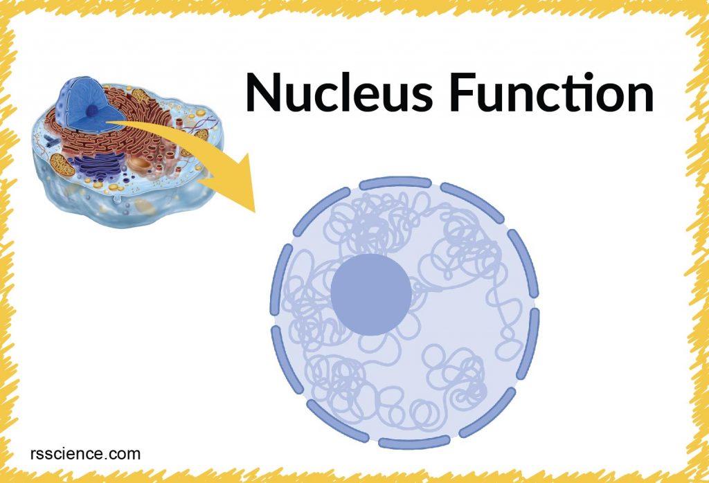
The nucleus is a specialized organelle that contains double-layer membranes with pores. The main function of the nucleus is to govern cell activities and to carry genetic information to pass to the next generation. This is why we call the nucleus the brain of the cells.
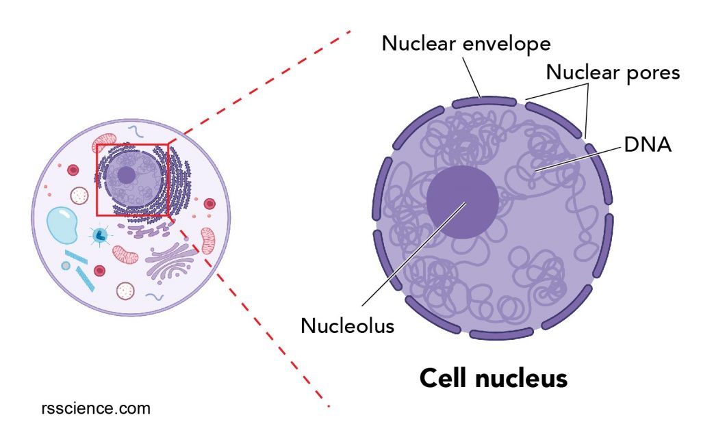
[In this figure] The cell nucleus diagram and its structure.
The nucleus consists of the nuclear envelope like double-layer membranes with pores ( nuclear pores), DNA, nucleolus (a site for ribosome synthesis, plural: nucleoli), nucleoplasm (like cytoplasm of a cell), and the nuclear matrix, a supportive structure like the cytoskeleton supports cells.
Created with BioRender.com
The nucleus is a key feature that distinguishes eukaryotic cells, including all animals and plants, from prokaryotic cells (bacteria and archaea). The nucleus (plural: nuclei) stores most of the cell’s genetic information in the form of DNA, although mitochondria also contain their own DNA in a very small percentage relative to the nucleus. In contrast, the prokaryotic cells’ DNA is located in the cytoplasm of bacteria, lacking any membrane-bound organelle.
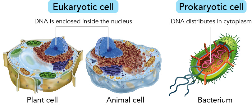
[In this figure] Eukaryotic cells v.s. Prokaryotic cell.
The DNAs of eukaryotic cells reside in the nucleus, while the DNAs of prokaryotic cells are freely distributed in the cytoplasm.
The structure of cell nucleus
The double membrane system of the nuclear envelope (outer and inner membranes) is discovered by the transmission electron microscopic image below. You can see some gaps between the nuclear envelope; these are nuclear pores, like channels that allow transportation of molecules such as RNAs between the nucleus and the cytoplasm.
The darker region next to the nuclear envelope is heterochromatin, which is a tightly packed form of DNA. The genes located in the tightly packed form of DNA are less active in their transcription (ie. less expressed). In contrast, the genes located in the euchromatin, which are lighter regions in the electron microscopic image, are more active in transcription.
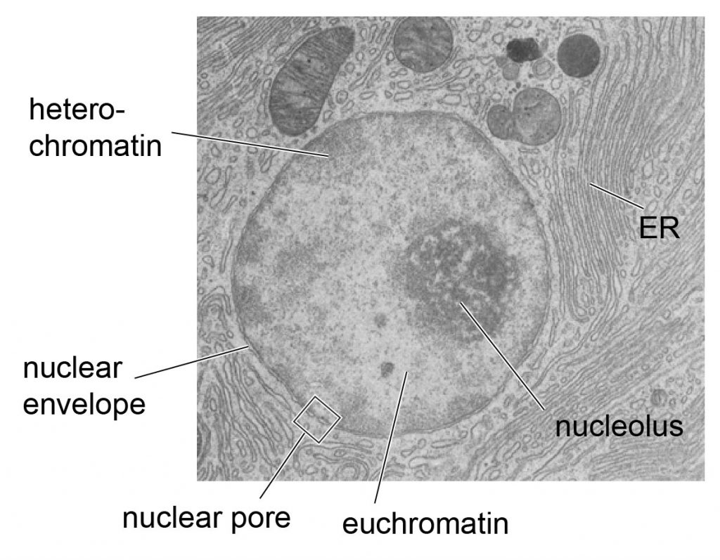
[In this figure] The transmission electron microscopic image of nuclei.
With the discovery of an electron microscope, we now know that the nuclear envelope is a double-layer membrane. The ball-like structure of the darker region inside the nuclei is the nucleolus, a place where ribosomes are made.
Photo credit: modified from https://www.stolaf.edu/people/giannini/cell/nuc.htm
Where is the nucleus located in a cell, and how many cell nuclei can be found in one cell?
In an animal cell, the nucleus is typically located in the central region of the cell. In contrast, the nucleus of a plant cell is located on one side of the cells. Because the large vacuole in a plant cell occupied so much volume, the nucleus is squeezed to the periphery.
Most of the cells have one nucleus; however, there are some exceptions. For example, our red blood cells have no nuclei at all. Our skeletal muscle has many nuclei because many myoblasts (baby muscle cells) fuse together to form a long muscle fiber.
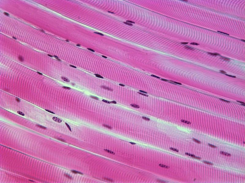
[In this figure] Skeletal muscle with multiple nuclei.
There are also many exceptions in biology!!
Paramecium, a single-cell organism has two nuclei, one micronucleus, and one macronucleus. The micronucleus is diploid and contains all paramecium’s DNA. This DNA is passed from one generation to another generation during reproduction.
In contrast, the macronucleus contains a subset of DNA from the micronucleus. These DNA fragments are copied from the micronucleus because they carry genes that are frequently needed by the paramecium. Genes in the macronucleus are actively transcripted to mRNA and then translated to proteins. The macronucleus is polyploid and contains multiple copies of each chromosome, sometimes up to 800 copies.
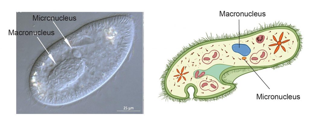
[In this figure] Dual nuclei in paramecium.
The macronucleus and micronucleus can be seen under a light microscope (left image). The right image is a cartoon illustration.
Photo source: MDPI
What is the biological function of cell nucleus?
The main function of the nucleus is to store genetic information (DNA), passing this information precisely to its daughter cell through cell division, and coordinating the whole activities through controlling gene expression and protein synthesis.
Can a cell survive without a nucleus? No, a cell requires protein synthesis to maintain its activities and functions. Without a nucleus, protein synthesis is messed up.
Regulate gene expression and protein synthesis
Our genes are written as the genetic codes (A, T, G, C) in the DNA. The gene is a blueprint for making a protein. There are two steps for making proteins from genes. First, Inside the nucleus, a process that makes copies of a certain gene in the form of massager RNAs (mRNAs), called transcription. Then, these mRNAs are exported outside of the nucleus to the cytoplasm for ribosomes to make proteins/polypeptides.
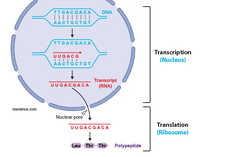
[In this figure] The process of gene expression.
From a gene to a protein, there are two steps, transcription and translation. The DNA needs to be transcripted to mRNA using complementary base pairing (i.e., A pairs with U; T pairs with A; C pairs with G; G pairs with C). Next, the mRNAs are exported to the cytoplasm through nuclear pores and are translated to proteins through ribosomes.
Store genetic information
All of our genes are stored in the nucleus, located on 46 pieces of long DNA threads. If you stretch all the DNA from a single cell into a linear thread, it can be as long as 2-3 meters long. It is incredible how the cell can pack entire DNA into a tiny nucleus (1 million times compacted, the diameter of a nucleus is less than 2-3 micrometers; one micrometer = 0.000001 meters). To do this, there are special proteins called histones that can compact DNA into the nucleus, and the resulting histone-DNA complex is called chromatins.
If you ever tried to store sewing thread, you know that it is much easier to coil the thread on a cylindrical roll. You Imagine that DNAs are like sewing threads and the histones are like cylindrical rolls. You got my idea.
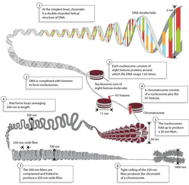
[In this figure] Chromosomes are composed of DNA tightly wound around histones.
Photo source: Scitable by natureEducation.
When cells are ready for division, the chromatins can be further compressed to chromosomes, which can be visible with proper staining (we can not see the DNA molecules under a light microscope). We have 23 pairs of chromosomes (1-22, X, and Y), and the number will be doubled right before the cell division.
How does the nucleus divide during cell division?
During division, the nuclear envelope temporally disappears. The replicated chromosomes are first aligned in the middle of the cells and then are pulled toward the opposite ends of the cell by microtubules. Once the chromosomes are equally separated into two daughter cells, the division of the cytoplasm occurs. After that, the nuclear envelop reforms and appears.
For more detail, check the phases of mitosis.
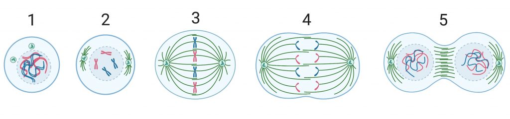
[In this figure] Chromosomes separation during cell division.
When the cells are ready for division, the chromosomes form (1), and the nuclear envelope disappears (2). The sister chromosomes are aligned in the middle of cells (3) and then are pulled toward the opposite side of the cells (4). Then the cytoplasm is separated into two daughter cells, and the nuclear envelope reform (5).
Created with BioRender.com
Are there human diseases associated with nucleus dysfunction
The nucleus is the hallmark of eukaryotic cells; therefore, the structural proteins in the nuclear envelope are important for maintaining the nuclear function. The mutations in the nuclear envelope such as lamins cause a number of human diseases, including Emery-Dreifuss muscular dystrophy (EDMD), dilated cardiomyopathy, familial partial lipodystrophy (FPLD), and the premature aging disease Hutchinson-Gilford progeria syndrome (HGPS).
How to see the cell nucleus under a microscope
There are two microscope lesson activities in this blog for you to see the nuclei in animal cells and plant cells. It is easier to see nuclei under a light microscope with staining such as methylene blue.
Lesson 2: Mount a Slide & “Look at Your Cheek Cells“
Lesson 3: Onion Dissection & “Look at the Plant Cells”

[In this figure] Cheek cells stained with Methylene Blue.
The nuclei were stained with a dark blue (because Methylene Blue stains DNA strongly). The cell membrane acts like a balloon and holds all the parts of a cell inside, such as a nucleus, cytosol, and organelles.
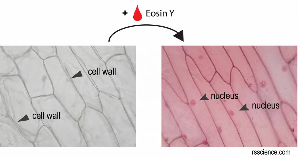
[In this figure] Onion skin stained with Eosin Y.
Without stains, you can only see the cell walls of onion cells. By staining of Eosin Y, now you can see a nucleus inside an onion cell.
Modern technology, such as immunofluorescence staining allows seeing many molecules and organelles in great detail. Immunofluorescence utilizes fluorescent-labeled antibodies to detect specific target antigens. Because of its specificity, you can detect molecules of your interest and see their subcellular localization in cells. Below is an example of the immunofluorescence image.
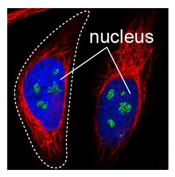
[In this figure] Immunofluorescence staining of nuclei.
DAPI stains the nuclei strongly, shown in blue color. The microtubule and nucleoli probes are visualized in red and green, respectively. The white dash line outlines the cell shape.
Photo source: Sigma
Summary
- The nucleus is a double-layer membrane organelle. It consists of the nuclear envelope, DNA (chromatin), nucleolus, nucleoplasm, and the nuclear matrix.
- The main function of the nucleus is to control cell activities and carry genetic information to pass to the next generation.
- A eukaryotic cell typically has only one nucleus. However, some cells do not have nuclei, such as our red blood cells. In contrast, some cells have multiple nuclei, such as paramecium, which has one micronucleus and one macronucleus.
References
Nuclear Mechanics in Disease
Sigma
https://www.stolaf.edu/people/giannini/cell/nuc.htm