
Cells are building blocks of life. Building a cell model should deepen your understanding of the cell, each organelle’s role, and how they perform their job. Some organelles are constantly present in the cell. Some organelles are temporary and only present when cells perform a particular process such as mitosis. Therefore, we have 4 blog posts series to cover the model of animal cells undergoing different processes.
- Animal Cell Model Part I – cell membrane, cytosol, nucleus, and mitochondria.
- Animal Cell Model Part II – endoplasmic reticulum, ribosome, Golgi apparatus, peroxisome, and lysosomes.
- Animal Cell Model Part III – two types of temporary organelles involving in eating behaviors, autophagosomes, and endosomes.
- Animal Cell Model Part IV – two types of temporary organelles only appearing during mitosis, centrosomes, and chromosomes.
- Plant Cell Model Part V – cell wall, vacuole, and chloroplast.
- Cell Organelles and their Functions – overview of each organelle.
This article covers
What is mitosis
Mitosis is a process of cell division in which one cell (the mother) divides and produces two new cells (the daughters) that are genetically identical.
In this Cell Biology on the Dining Table series Part IV, we are going to learn two types of temporary organelles only appearing during mitosis. They are centrosomes and chromosomes!
Why mitosis is important
Mitosis is a type of cell division that is necessary for the growth of organisms, repair of damaged tissues, healing and regeneration, and reproduction.
Don’t mess up with DNA
Like we mentioned in the section of “cell nucleus”, the DNA molecule is a very, very long thread. If you stretch all the DNA of a single cell into a linear thread, it can be as long as 3 meters (or 10 feet) long. This linear form of DNA is impossible to be probably separated during the cell division.
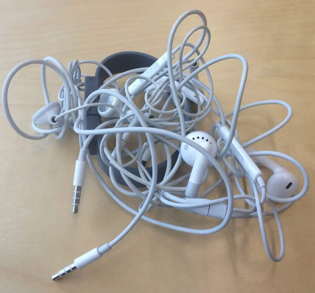
[In this figure] Tangled earphones are one of the most annoying things in our life. In our cell, the DNA threads are even longer than the earphone’s wire.
How cells pack such long DNA into an extremely tiny space (the cell nucleus) without tangling?
To solve this problem, when the cells prepare for the cell division, each DNA thread is organized into a much compact structure, called “chromosome”. All our genes (called the genome) are located on 46 pieces (or 23 pairs) of long DNA threads. As a result, we have 23 pairs of chromosomes (1-22, and X or Y).
[In this figure] In order to handle the long DNA molecules, our cells pack DNA threads into many compact structures, called “chromosome”.
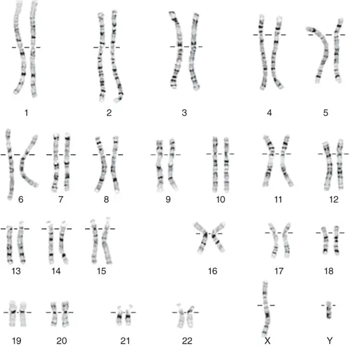
[In this figure] 23 pairs of human (male) chromosomes by a microscopic technique called Giemsa banding (G-banding) karyotype.
Pack DNA into chromosomes
Cells need a group of proteins, called “histones”, to organize the DNA threads into chromosomes. In brief, eight histone proteins form a core complex, called nucleosome. You can imagine that DNA wraps around the nucleosome like yarn wraps around a ball. Each nucleosome allows two turns of DNA wrapped. Under an electron microscope, the DNA with nucleosomes looks like many beads on a string.
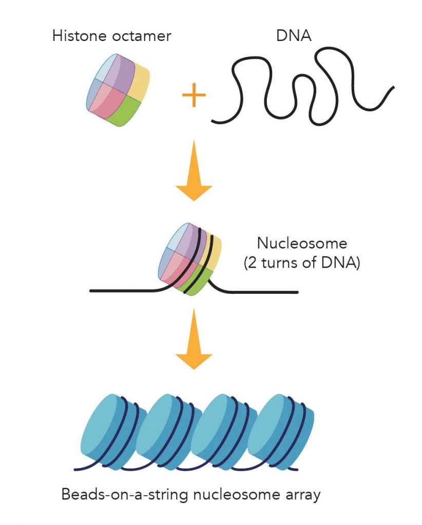
[In this figure] Illustration of the package of DNA threads by histone.
Eight core histone proteins form a histone octamer. About 147 base-pairs of DNA wraps around the histone octamer (~ two turns) to form a nucleosome. Nucleosomes arrayed along the DNA molecules look like beads on a string.
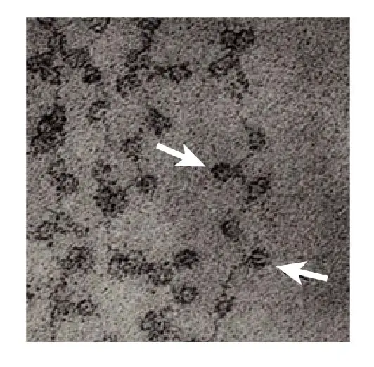
[In this figure] Electron microscopic image of DNA wrapping around the nucleosomes like the beads on a string.
The nucleosomes are indicated by arrows. Image modified from Olins, D. E. & Olins, A. L. Chromatin history: our view from the bridge. Nature Reviews Molecular Cell Biology 4, 811 (2003).
Packaging DNA into nucleosomes shortens the DNA thread length about sevenfold. These nucleosomes then are further packaged into the most compact rod-shaped structures of chromosomes. These chromosomes are visible under a regular light microscope with proper staining.
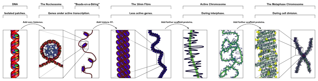
[In this figure] The process of chromosome package.
The DNA wrapped around histones and together with scaffold protein to form a chromosome. Currently, scientists only have a good idea about how DNA organizes into nucleosomes. Beyond that, we still don’t know exactly how chromosomes assemble. Image credit: wiki
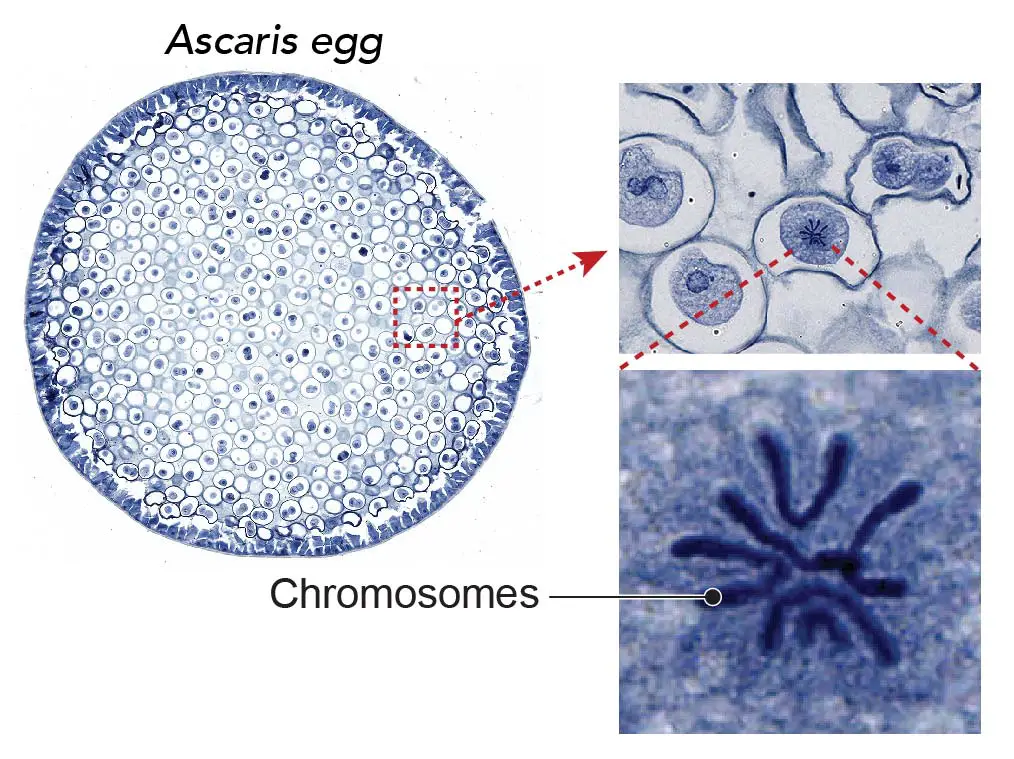
[In this figure] The chromosome is visible during the cell division.
The chromosomes become visible under the light microscope with 40x-100x objective lens. Methylene blue stains the DNA contents of chromosomes with dark blue color. This specimen is Ascaris (parasitic nematode worm) eggs that contain active mitotic cells. This specimen is included in Rs’ Science 25 Premade slide set.
X-shaped sister chromatids
Right before the mitosis, the DNA replicates to form two identical copies. At this moment, the chromosomes change from a rod shape to an “X” shape. The two identical “sister chromatids” remain attached at a centromeric region, called “centromere”.
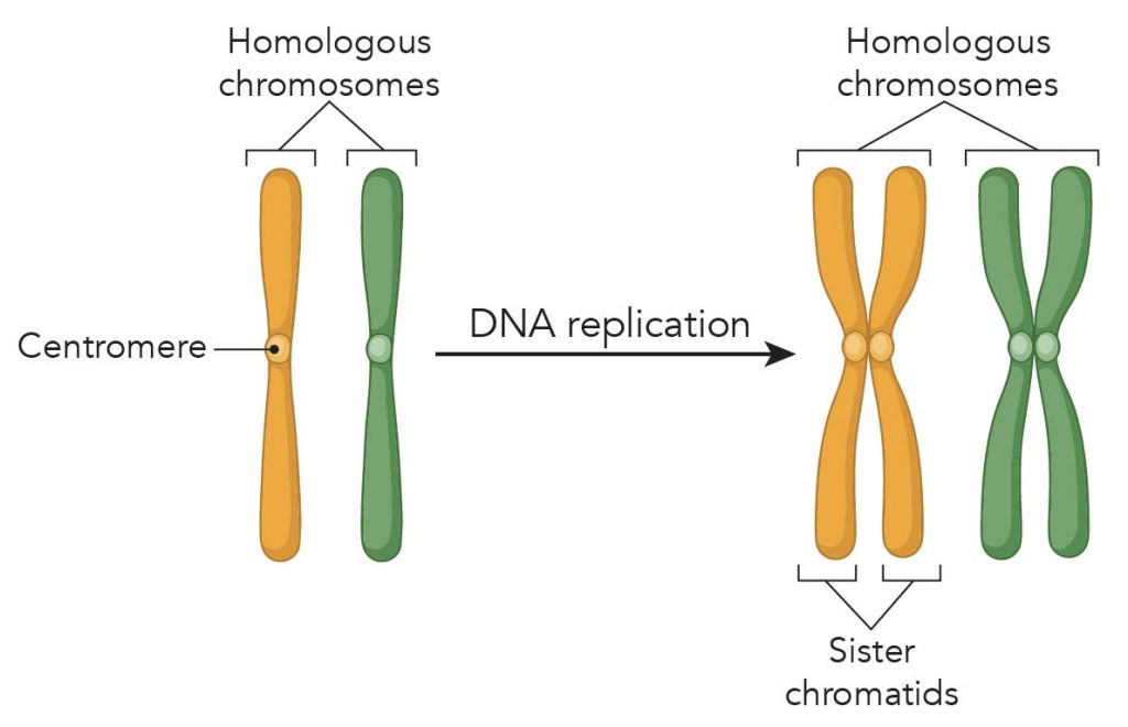
[In this figure] Chromosome replication before mitosis.
Our cells are “diploid”, meaning they have two copies of genetic materials. Before mitosis, the two copies of genetic materials form 23 pairs of homologous chromosomes. After DNA replication, each chromosome becomes an X-shaped with 4 arms attached to a central point, called centromere. The two identical parts of the chromosome called the “sister chromatids”.
The phase of mitosis
Mitosis is a process of cell division in which one cell (the mother) divides and produces two new cells (the daughters) that are genetically identical. The goal of mitosis is to make sure that each daughter cell gets a perfect and fully equal set of chromosomes. If daughter cells have either more or missing genes due to uneven separation of chromosomes, the function of the cells will totally be messed up. These cells will very likely become cancer cells!
To ensure cells split up their duplicated chromosomes equally, our cells developed Standard Operating Procedures (SOP). We call this carefully organized series of steps – the phases of mitosis. Many proteins that involve in mitosis perform their jobs at exact orders and timing. There are checkpoints between each phase during the mitosis. The cells are allowed to process to the next step only if the previous step has been checked for error-free.
Below are the sequential phase of mitosis
(1) Interphase – Cells are replicating their DNA in the nucleus.
(2) Prophase – DNA condenses to form chromosomes. Centrosomes move toward the opposite sides of the cells and form a mitotic spindle.
(3) Prometaphase – Microtubule threads project from the centrosomes and capture the chromosomes. The point that chromosomes attach to microtubules called kinetochore.
(4) Metaphase – The spindle has captured all the chromosomes and lined them up in the middle of the cell (called equatorial plate).
(5) Anaphase – The microtubules start to pull apart the sister chromatids toward the opposite spindle poles.
(6) Telophase – The cell is nearly done dividing, and it starts to re-establish its normal structures as cytokinesis (the division of the cell contents) takes place. Two new nuclei form, one for each set of chromosomes.
(7) Cytokinesis – The division of the cytoplasm and cell membrane to form two new cells.
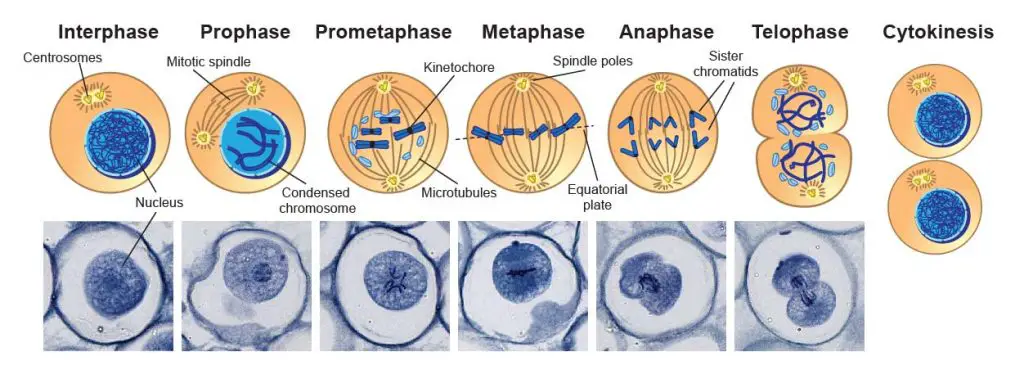
[In this figure] Mitosis phases under the microscope.
Mitosis consists of five basic phases: prophase, prometaphase, metaphase, anaphase, and telophase. These phases occur in a strictly sequential order. This specimen is Ascaris eggs (parasitic nematode worm) that contain active mitotic cells and can be purchased from Rs’ Science 25 Premade slide set.
Centrosomes and Microtubules – Amazing molecular machine
As you can see in the phases of mitosis, the sister chromatids have to be pulled apart and separated equally into two new nuclei. Cells do this job exactly like how we pull a car out of mud using a cable and a winch.
Microtubules – cable
The “cable” inside our cells is called “microtubule”. In the previous section of the cytosol, we mentioned the network of cytoskeleton inside the cell. The microtubule is one of the cytoskeleton proteins that assemble to form filaments like highways for the transportation of molecules and organelles inside the cell. During mitosis, the microtubule threads are used to pull chromosomes apart.
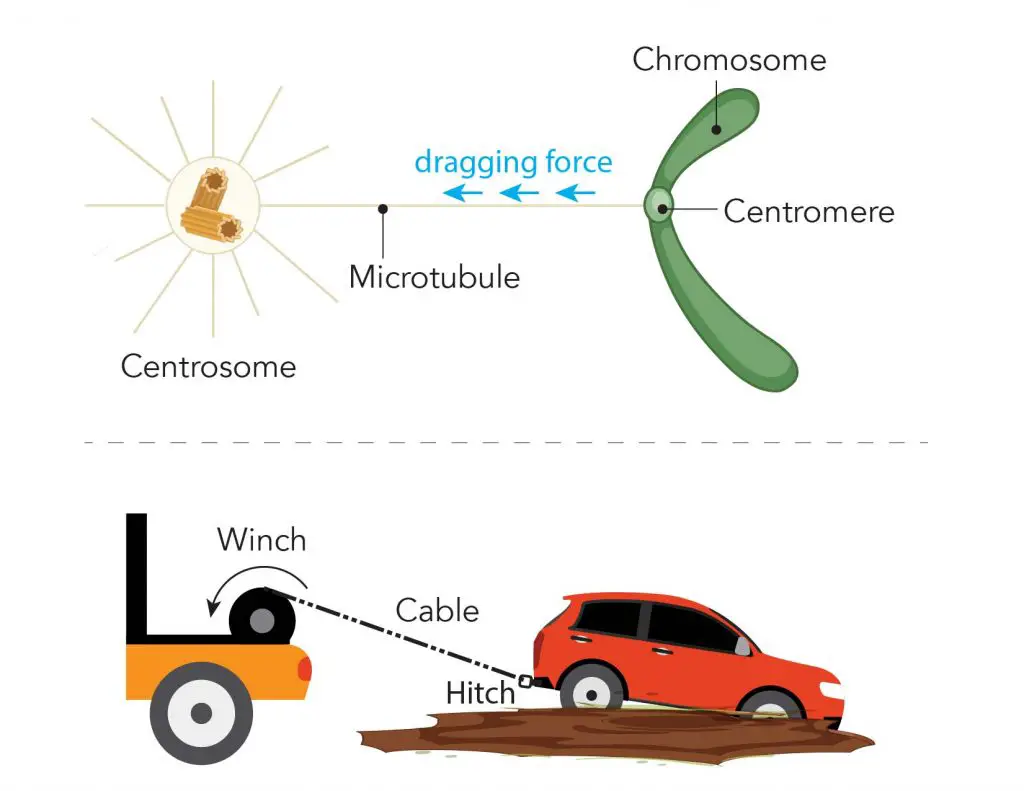
[In this figure] Illustration of microtubule, centromere, and centrosome during mitosis.
During mitosis, one end of the microtubule attached to the centromere regions of each chromosome and the other ends are controlled by a special organelle, called “centrosomes”. You can imagine that the centrosome like the winch pulls the sister chromatids (the car) apart by dragging the microtubule (the cable) anchored to the centromere (the tow hitch).
Centrosomes – winch
Centrosomes are organelles that only appear during mitosis and serve as the main microtubule organizing center (MTOC). Each cell has two centrosomes. When the mitosis starts, two centrosomes move toward the opposite positions of the cells, and the membrane of the nucleus (called the nuclear envelope) temporally disappears.
The microtubules extend from the centrosome and attach to the centromeres of sister chromatids. The coordinate action of both centrosomes allows all sister chromatids aligned in the middle of the cell, forming a structure called equatorial plate.
Both centromeres retrieve their microtubule at the same time. The contraction force separates the sister chromatids apart from the attachment points of centromere; then moves toward the opposite direction. Once all sister chromatids are equally separated into two new nuclei, the cytoplasm and cell membrane will also divide into two new cells.
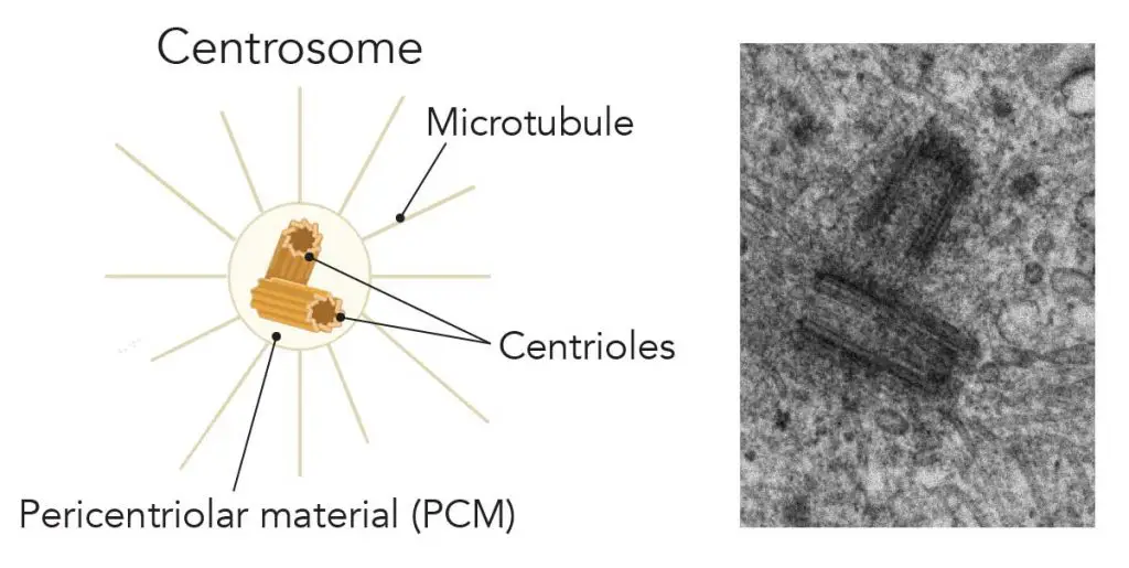
[In this figure] Illustration and electron micrography of the centrosome.
Left: Centrosomes are composed of two centrioles arranged at right-angles to each other and surrounded by proteins called the pericentriolar material (PCM). Microtubule fibers grow from the PCM. Right: Electron microscopic images of centrioles. (Image: johan-nygren)
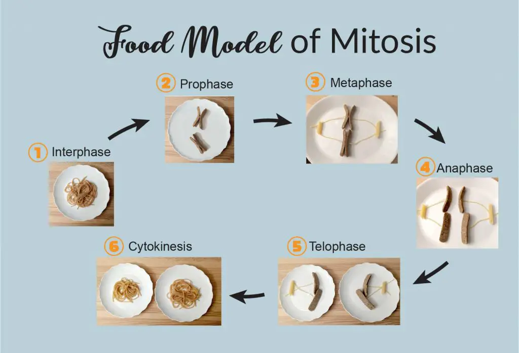
Food Model of Mitosis
The process of mitosis sounds a little bit complicated. Don’t worry. I can explain mitosis to you in a very simple and fun way – on your dining table!
Mitosis interphase
First, I cooked some whole wheat spaghetti noodles and served on a white plate. This is my cell at the Interphase. The whole wheat spaghetti noodles represent the dispersed DNA strings before packing into chromosomes. I choose the whole-wheat spaghetti for its brownish color. You will see the reason later.
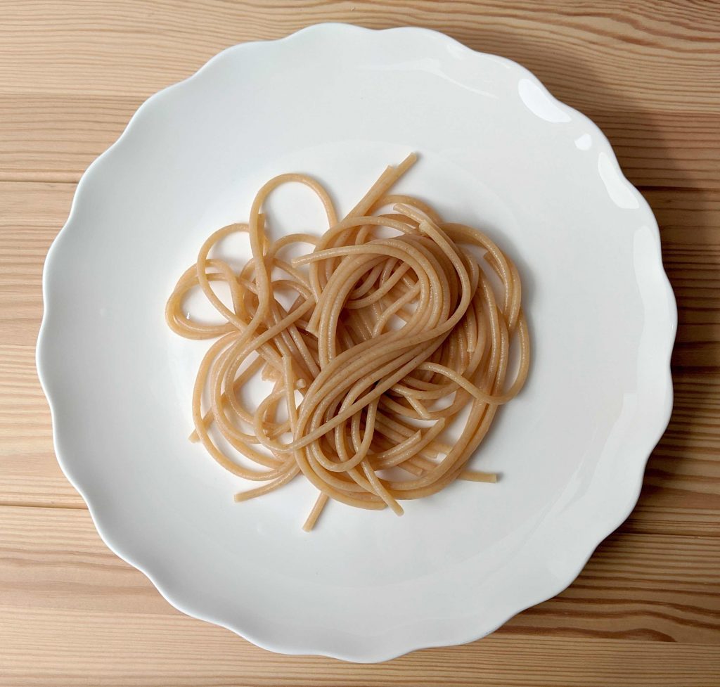
[In this figure] The food model of interphase.
The whole wheat spaghetti noodles represent the unorganized DNA strings during the interphase. The plate represents a cell.
Then, I need short sausages to represent my chromosomes. I found that Trader Joe’s Maple Chicken Breakfast Sausage is a good size (~ 2 inch long). If you have bigger plates, longer sausages may also work for you.
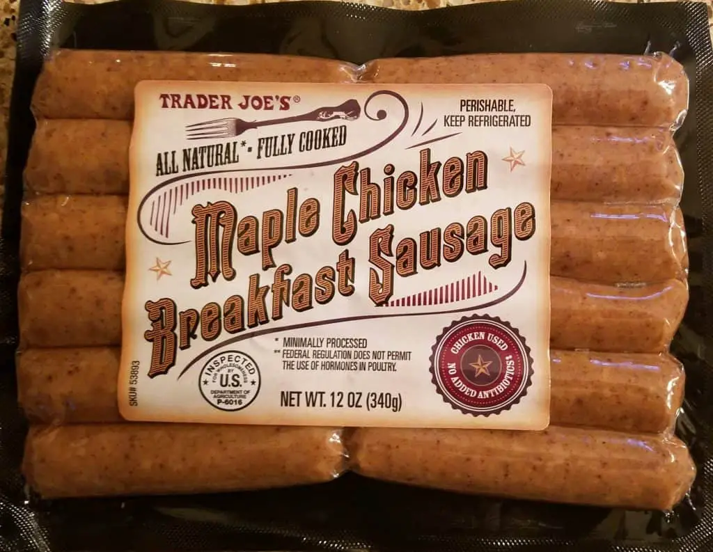
[In this figure] The food model of chromosome
Chicken sausages represent chromosomes in my food model.
I cut both ends of the sausages in half and keep the middle part (about 1/3 inch) remaining attached.
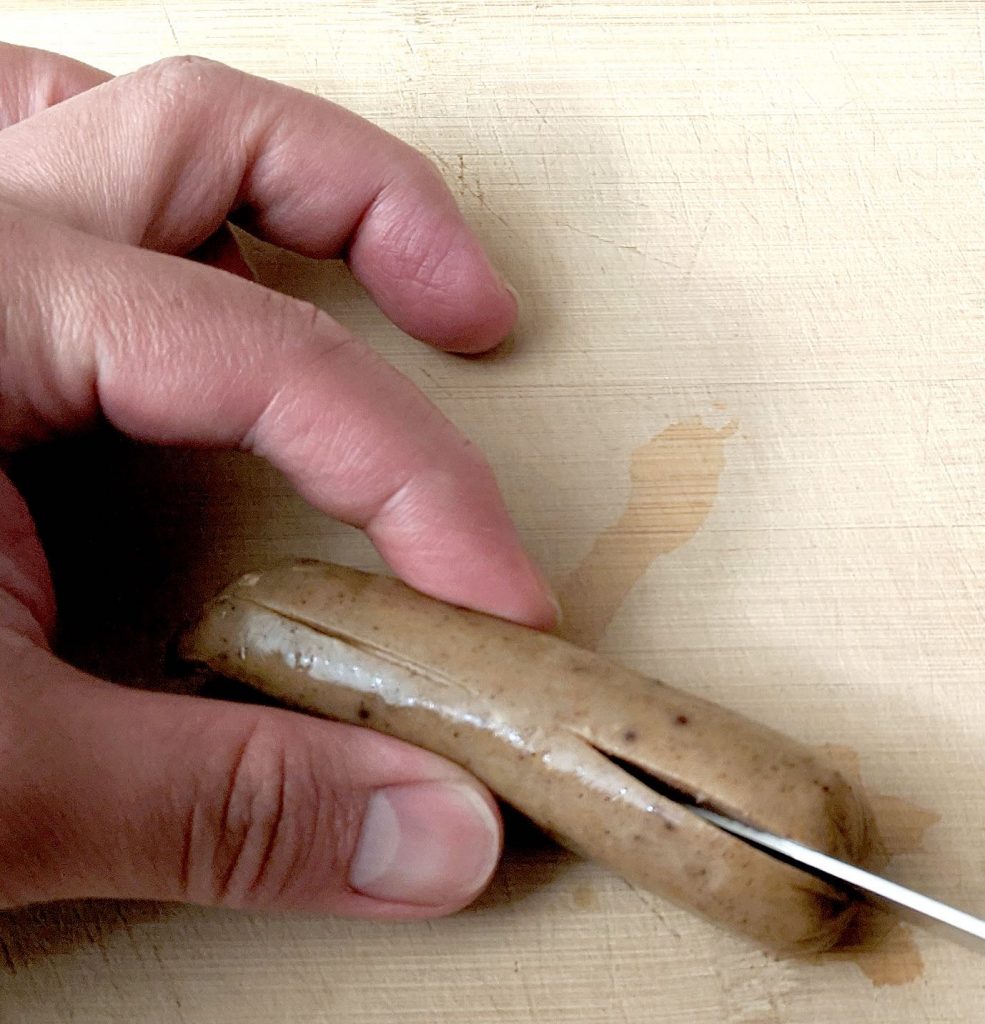
[In this figure] The food model of sister chromatids.
The sausage was cut in half and the middle parts remained connected. This represents two sister chromatids that are attached together.
Then, I carefully pierce a regular spaghetti noodle through the middle of the sausage. Of course, only “uncooked” spaghetti noodle is stiff enough to do so.
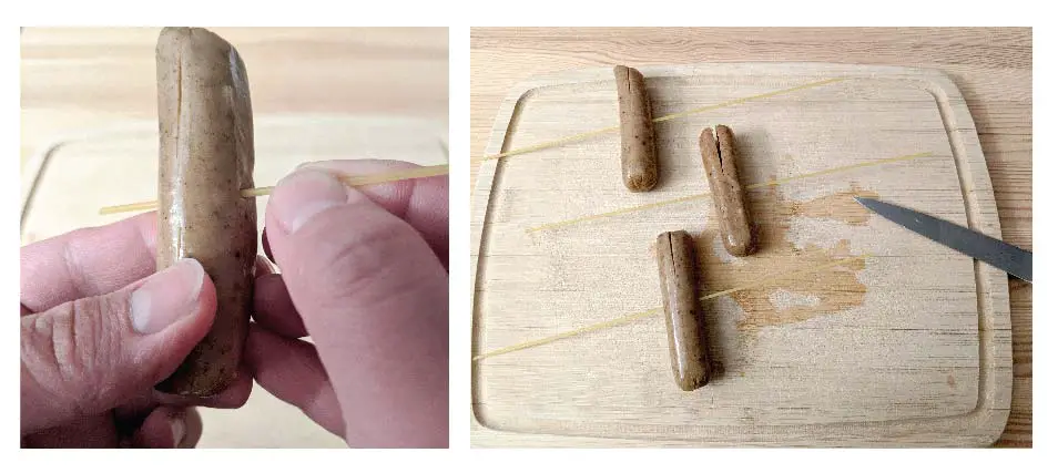
[In this figure] The food model of sister chromatids and microtubule.
The regular spaghetti noodle attached to the sausage represents the microtubules attached to the sister chromatids.
Then, I cook the sausages alone and sausages with spaghetti in a pan.
And, the magic happened!
As you can see below, the cut sausages bend after cooking and form an “X” shape. They are a perfect food model of “sister chromatids”! The brownish color of sausages matches the color of whole wheat spaghetti noodles, indicating the chromosomes are the condensed form of DNA strings.
You may also notice that the pieces of regular (white) spaghetti inserted through the sausages become soft after cooking. These regular (white) spaghetti noodles are “microtubule” anchored to the sister chromatids.
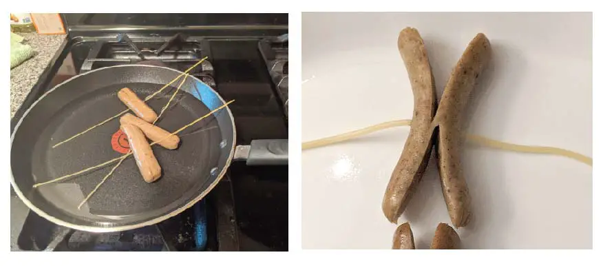
[In this figure] The food model of X- shaped sister chromatids and microtubules.
The cut sausage bends two ends after cooking, which represents X-shaped sister chromatids. The cooked spaghetti noodles become soft and represent microtubules attached to the sister chromatids.
Mitosis prophase
The key characteristic of prophase is that DNA condenses to form sister chromatids that are visible under the microscope.
I put a pair of X-shaped sausages (sister chromatids) on the plate to represent prophase during mitosis.
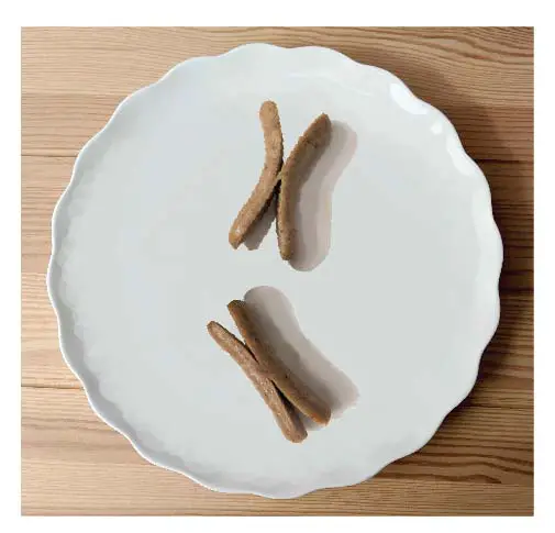
[In this figure] Food model of mitosis prophase.
Mitosis metaphase
The key characteristic of metaphase is that the spindle has captured all the chromosomes and the chromosomes lined up along the equatorial plate.
I placed two sister chromatids in the middle of a plate with their microtubules pointing to the opposite sides. The two sausages were well-aligned to represent the chromosomes arranged as “equatorial plate”. I placed two pieces of penne at the ends of microtubules spindle to represent “centrosomes”. Now, I have a perfect model of metaphase.
If you have a larger plate, try to place more sausage-spaghetti sets to make it look closer to a “mitotic spindle”.
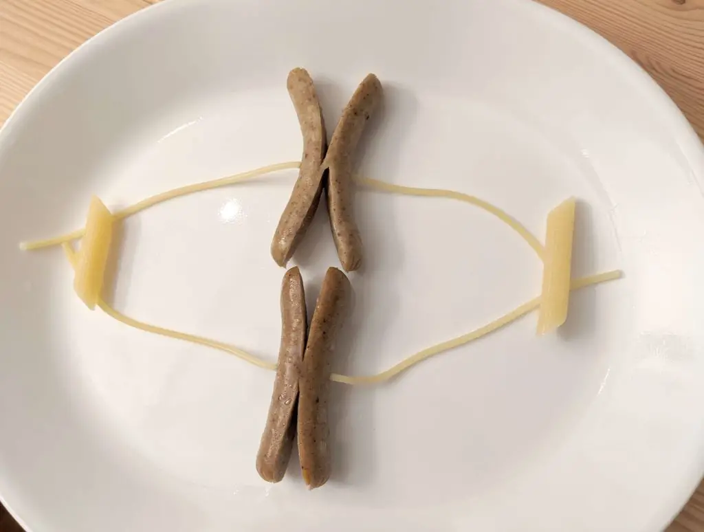
[In this figure] Food model of mitosis metaphase.
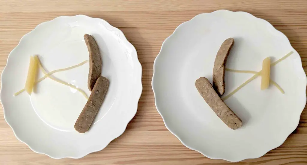
Mitosis anaphase
The key characteristic of anaphase is that microtubules start to pull apart the sister chromatids toward the opposite spindle poles.
When the mitosis processes to the anaphase phase, the force of microtubules dragging causes the separation of sister chromatids from the middle of centromeres.
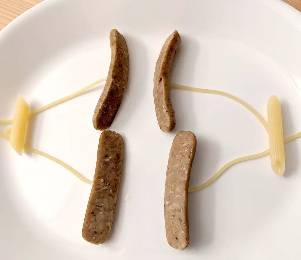
[In this figure] Food model of mitosis anaphase.
Mitosis telophase
The key characteristic of telophase is that the sister chromatids reach opposite poles and the nuclear starts to reform.
In my model of telophase, the X-shape sausages become bar-shaped, representing that the chromosomes equally distribute to two daughter cells.
[In this figure] Food model of mitosis telophase.
After mitosis, chromosomes decondense like spaghetti.
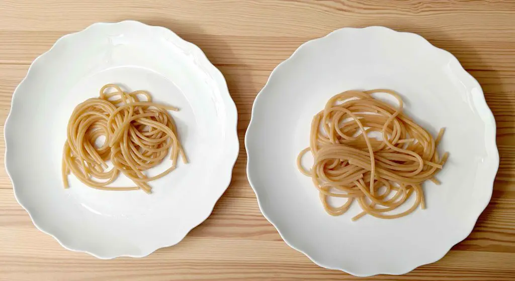
[In this figure] Food model of two newly formed cells after mitosis.
Finally, we have a full phases of mitosis cycle in a dish!
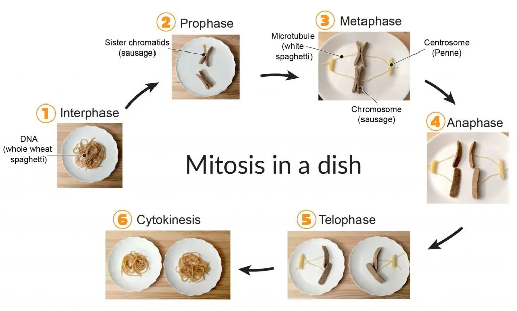
[In this figure] Food model of phases of mitosis.
Reference
“Revised Estimates for the Number of Human and Bacteria Cells in the Body” Ron Sender, Shai Fuchs and Ron Milo. PLOS Biology (2016)
“Electron micrograph of chromatin: the beads on a string” Olins, D. E. & Olins, A. L. Chromatin history: our view from the bridge. Nature Reviews Molecular Cell Biology 4, 811 (2003).
“Basic units of chromatin structure” David O Morgan. (2007) The Cell Cycle. Principles of Control.
“Throwing this idea out there: are the centrioles in the centrosome orbiting one another?” Johan-nygren.
Related posts
Animal Cell Model Part I – cell membrane, cytosol, nucleus, and mitochondria.
Plant Cell Model Part V – cell wall, vacuole, and chloroplast.
Cell Organelles and their Functions – overview of each organelle.