
Ah-choo! Oh, excuse me, now is the allergy season, and I couldn’t help but sneeze.
Pollen allergy, also known as hay fever, affects millions of people (myself included!). It happens when your body reacts too much to the pollen in the air. This causes you to sneeze, have a runny nose, itchy eyes, and feel congested. But even though pollen allergies can be annoying, we can’t afford a world without pollen. These tiny grains play a crucial role in the survival of our planet. Without them, we wouldn’t have flowers, plants, fruits, or even life as we know it.
This article covers
What is pollen?
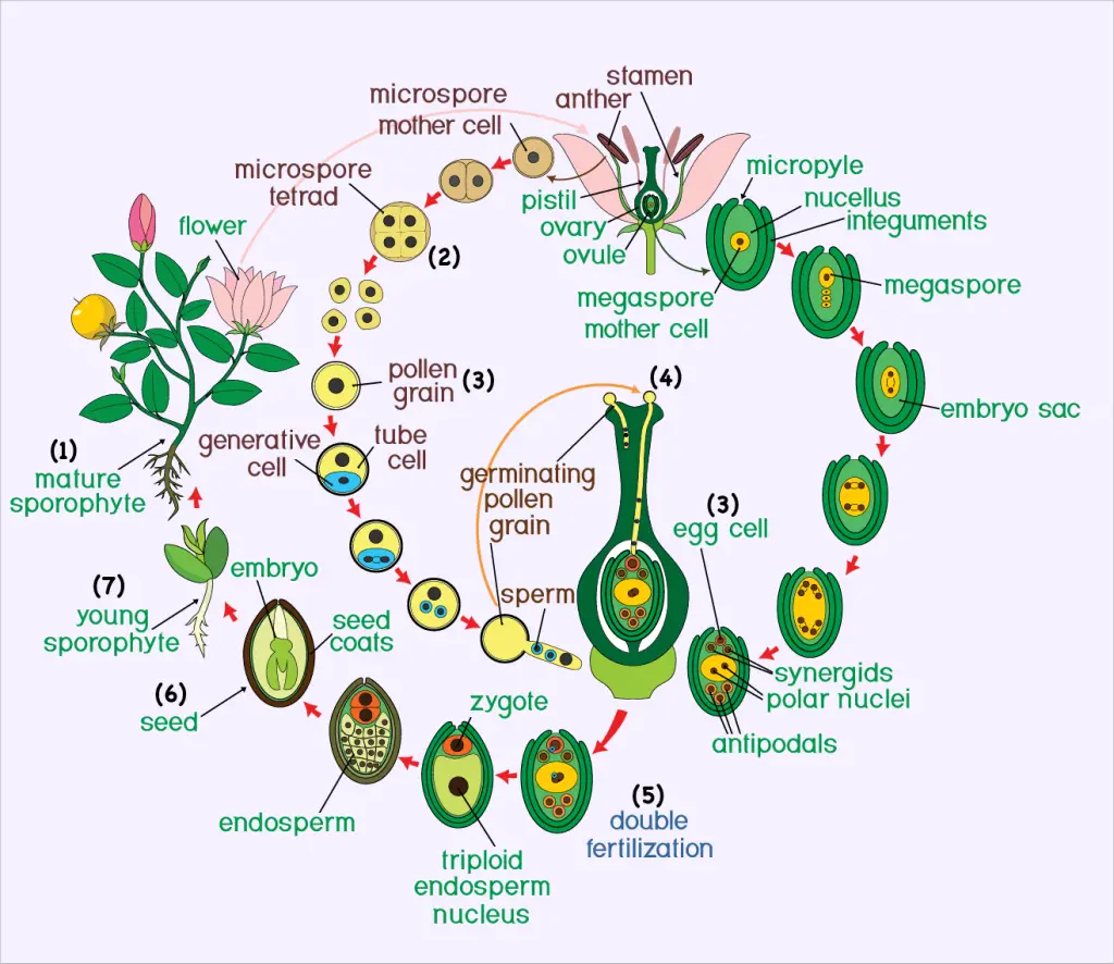
A plant that we usually think of, such as a tree or a flowering plant, is a sporophyte (usually diploid). A plant also has another stage in its life cycle, called the gametophyte stage (the sexual phase or haploids).
Pollen grains are the male gametophytes, and they are formed in anthers, the male parts of flowers. The function of pollen in plants is to transfer male genetic material from the anther of a single flower to the stigma of another flower. Pollen grains are produced by meiosis of microspore mother cells that are located along the inner edge of the anther sacs.
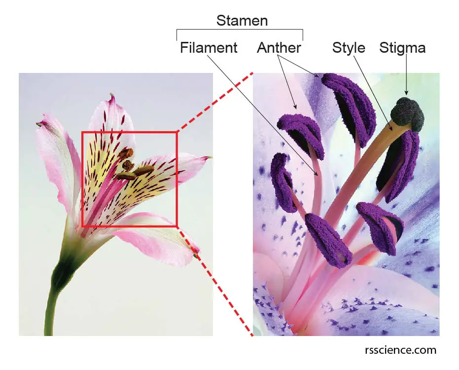
[In this figure] The anatomy of a flower.
The stamen is a male part of the flower. The stamen contains a filament and an anther where the pollens are produced. The stigma is the female part of the flower.
Life cycle of flowering plants
- The Adult Plant – This stage is called the sporophyte, and it has two sets of chromosomes in each of its cells.
- Inside the Flower – Special cells go through meiosis, a kind of cell division that cuts the chromosome number in half.
- Making Gametophytes –
▪ The male gametophyte is the pollen, which forms in the anther (the top part of the stamen).
▪ The female gametophyte is the egg, which forms inside the ovary at the base of the pistil. - Pollination – When pollen lands on the pistil of a flower of the same species, it begins to grow a pollen tube down into the ovary.
- Fertilization – The sperm travels down the tube and joins with the egg, creating a zygote.
- Seed Formation – The zygote develops into a seed, which contains the baby plant (embryo).
- New Plant Grows – When the seed finds the right place and conditions, it sprouts and grows into a new sporophyte, and the cycle begins again!
The size of pollen
Pollen comes in many sizes, from very tiny (less than 10 micrometers) to quite large (over 100 micrometers). How big a pollen grain looks can change depending on how wet it is and how it is prepared for study. Because of this and natural differences, scientists use size groups to describe pollen:
- Very small: less than 10 micrometers ( μm)
- Small: 10 to 25 μm
- Medium: 26 to 50 μm
- Large: 51 to 100 μm
- Very large: more than 100 μm
(For comparison, a human hair is about 70 micrometers thick!)
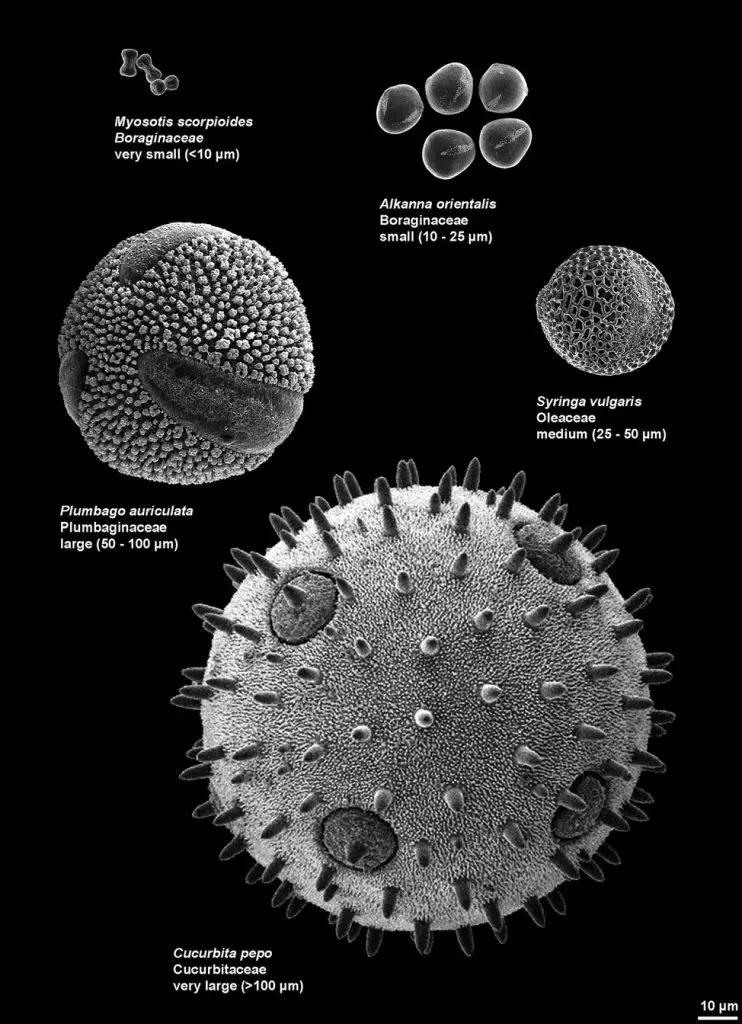

[In this figure] Different sizes of pollen grains, showing five size categories.
Myosotis scorpioides (Water Forget-Me-Not) – Very small pollen (<10 μm)
Alkanna orientalis – Small pollen (10–25 μm)
Syringa vulgaris (Common Lilac) – Medium pollen (25–50 μm)
Plumbago auriculata (Cape Plumbago) – Large pollen (51–100 μm)
Cucurbita pepo (Pumpkin or Squash) – Very large pollen (>100 μm)
Image source: Illustrated Pollen Terminology, Gardenia, Wikipedia, The University of Maine, The Spruce, My garden life.
The structure of pollen
The outer part of the pollen is the exine, which is composed of a complex polysaccharide called sporopollenin. Sporopollenin is one of the most chemically inert biological polymers and is usually well preserved in soils and sediments. The exine layer is often intricately sculptured in species-specific patterns, allowing material (as a kind of fossil) recovered from sediments to provide useful information to palynologists (the study of pollen) of plant populations in the past.
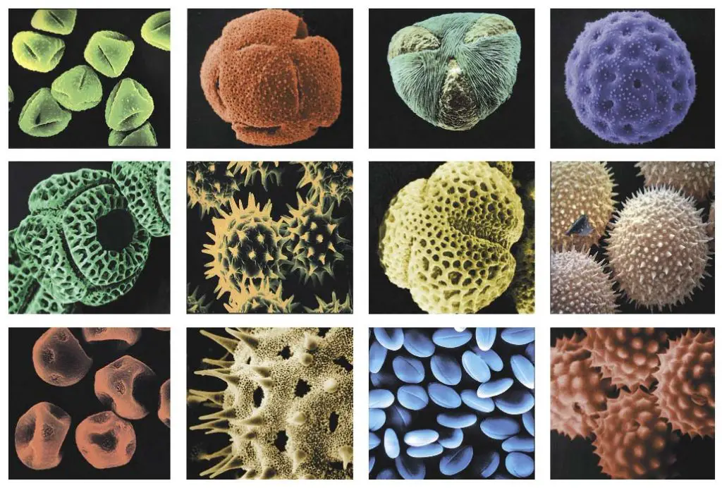
[In this figure] The appearance of pollens
False-colored scanning electron micrographs show the diverse ornamentation patterns on the surfaces of pollen from different species.
From Chapter 19 “Reproductive Development”, by J. Derek Bewley, Frederick D. Hempel, Sheila McCormick and Patricia Zambryski, in Biochemistry & Molecular Biology of Plants, edited by Bob Buchanan, Wilhelm Gruissem and Russell Jones, published by American Society of Plant Biologists (2000).
Each pollen grain contains two cells: the tube cell and the generative cell. The tube cell (also referred to as the tube nucleus) develops into the pollen tube. The generative cell divides into two sperm by mitosis.
Pollen can be spread by various means (wind, water, insects, etc.). Once the pollen lands on a compatible pistil, the process of pollination initiates. The tube cell produces a pollen tube that transfers the sperm to the ovule containing the female eggs.
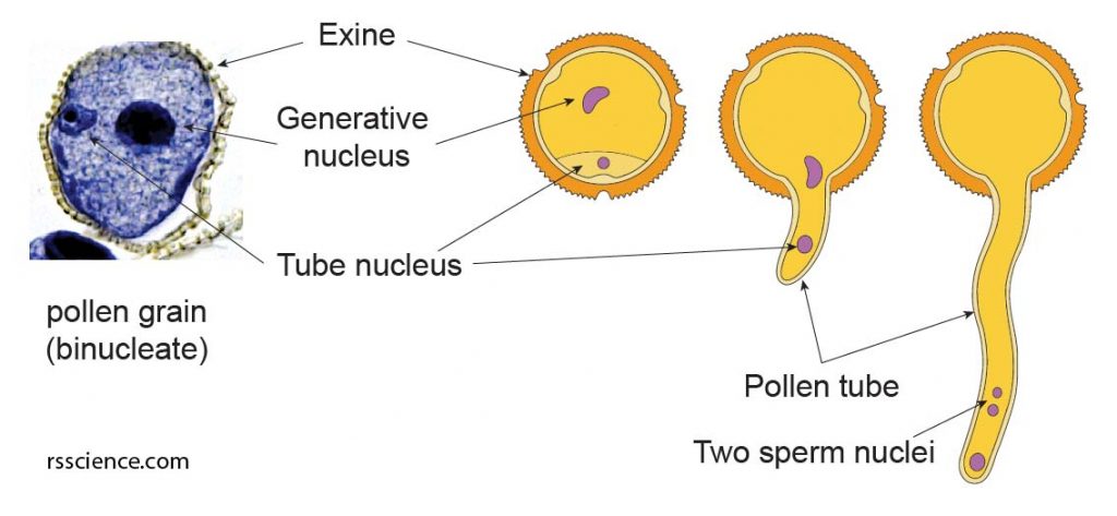
[In this figure] The structure of pollens
The pollen has a tough resistant outer wall made of sporopollenin, called exine. The exine exhibit protrusions and spines. This unique appearance is a character that can be used for species identification. The pollen itself is not a sperm; instead, it contains two nuclei. The tube nucleus develops into a pollen tube, while the generative nucleus develops into two sperm nuclei.
Pollen is one of the most common triggers of seasonal allergies. Each spring, summer, and fall, different plants release their tiny pollen grains for reproduction. However, these small particles can make our immune system confused. Most of the pollens that cause allergic reactions come from weeds and grasses, which are spread by wind. Certain species of trees, including pine, bitch, cedar, and oaks, also produce highly allergenic pollen.
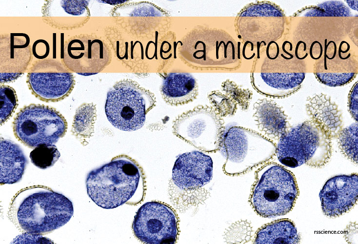
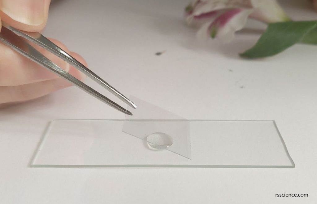
Pollen grains under a light microscope
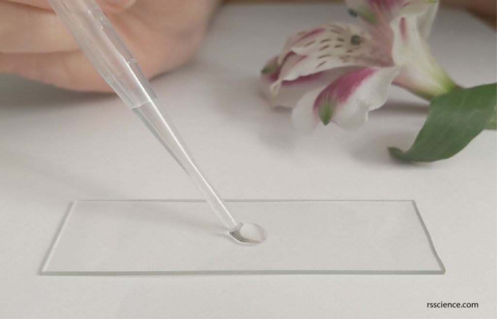
The material you need
- Blank microscope slides and coverslips
- Forceps
- A variety of flower pollens
- Droppers
- Compound microscope
- Water
Step 1: Use a dropper to take up a small amount of water and place it on a slide.
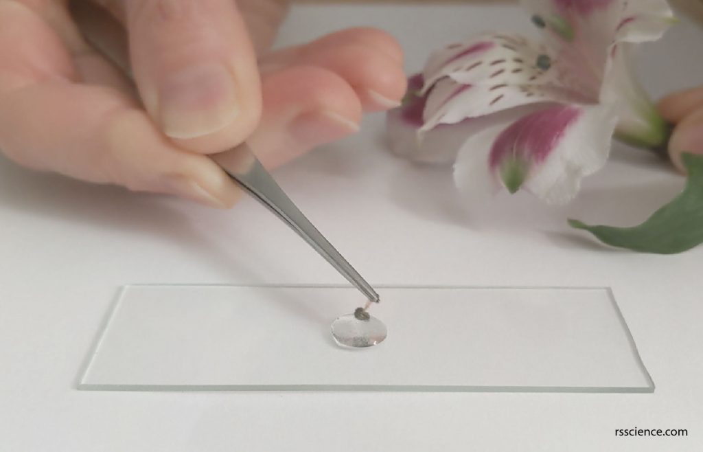
Step 2: Use forceps to grape a stamen and dip the anther that contains pollen grains to the water. This will help to release pollens.
Step 3: Place a coverslip on top. Use the forceps to lower the coverslip slowly with an angle. Allow one side of the liquid droplet touches the coverslip first. This permits air to escape from the other side. The slide is ready to view under a microscope.
What will you see?
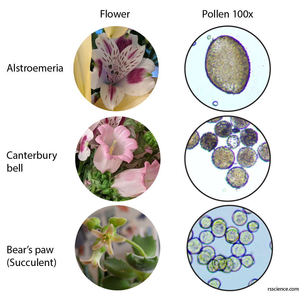
I collected pollens from three different flowers. They are various in size, shape, and texture. The pollens from alstroemeria are much bigger than that of canterbury bell flowers and bear’s paw succulent. The size of pollen grains is not correlated to their corresponding flowers.
You can also notice that there are several pores on the pollen surface. These are the place where the pollen tube grows out. 10% sucrose solution can be used for pollen germination. I will test it and report back in the near future.