
This article covers
What are centrosomes? A quick overview
Centrosomes are organelles that serve as the main microtubule organizing centers for animal cells. Microtubules are one type of filament protein in the cytoskeleton. Microtubule networks grow from the centrosomes and reach every inch of the cells. Microtubules can serve as a cell’s skeleton to change the cell shape.
Motor proteins that carry molecules and organelles can walk along the microtubule filaments like molecular trucks driving on an intracellular highway. Moreover, microtubule bundles form the cores of two special cellular structures, cilia, and flagella, which allow the cells to move and swim around.
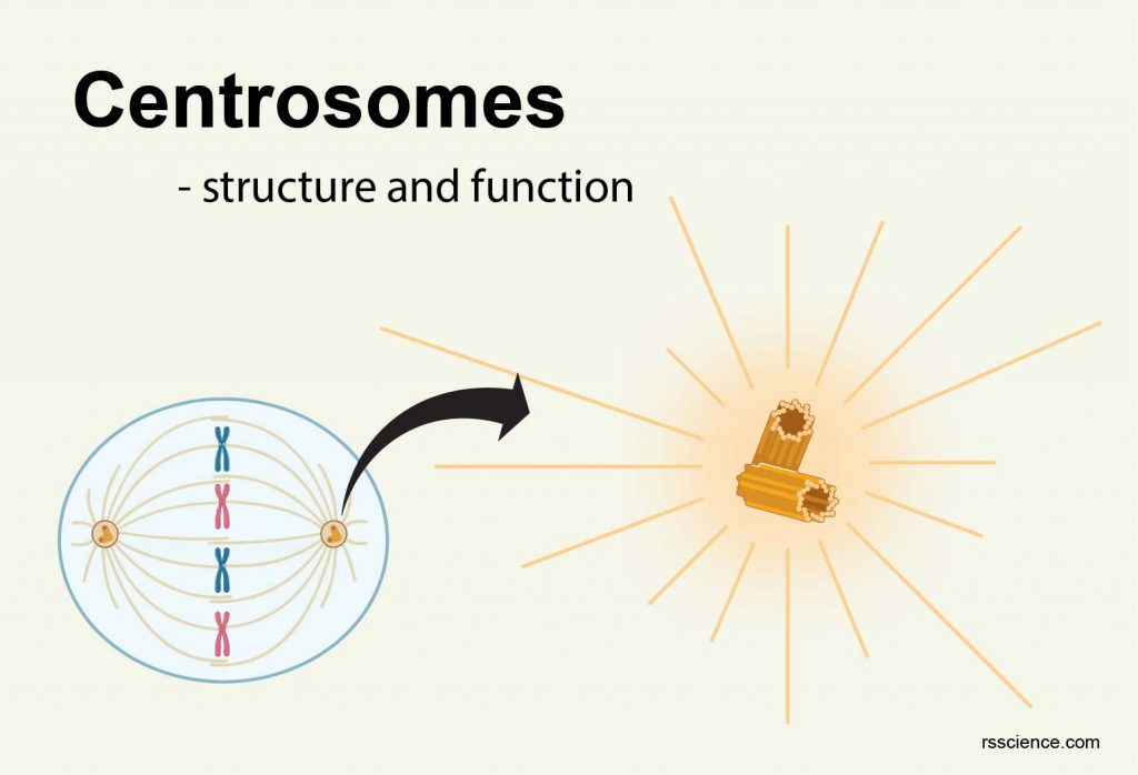
During cell division, centrosomes duplicate and move toward the opposite poles of dividing cells to help the precise separation of chromosomes (otherwise, the wrong chromosome numbers can cause cancer). All of these functions rely on the coordination of centrosomes!
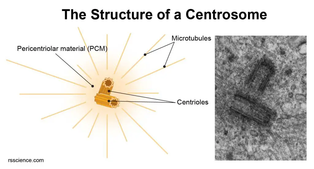
[In this figure] Illustration and electron micrography of the centrosome.
Left: A diagram showing the structure of a centrosome. A centrosome is composed of two centrioles arranged at right-angles to each other and surrounded by a protein mass called the pericentriolar material (PCM). The microtubules radiate from the centrosome to other parts of the cell. Right: Electron microscopic images of centrioles. (Image: johan-nygren)
The structure of centrosomes
Centrosomes are sometimes referred to as the “MTOC,” or “microtubule organizing center” of the cell. Centrosomes are made of two centrioles arranged at right angles to each other. Centrioles are barrel-shaped clusters of microtubule cores. Each centriole is based on a nine-triplet microtubule assembled in a cartwheel structure with several associate proteins.
In a centrosome, two centrioles are surrounded by a dense protein mass, termed the pericentriolar material (PCM), which is a complex of proteins that help additional microtubules to form. The PCM contains proteins that allow the centrosomes to start and stop the formation of microtubule filaments. This allows centrosomes to control the formation of mitotic spindle fibers and other structures that play important roles in the cells.
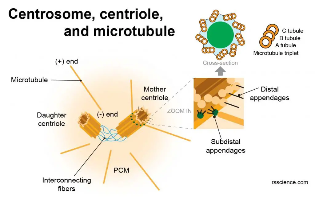
[In this figure] The detailed structure of centrioles and microtubules assembled in a centrosome.
A pair of centrioles are shown, each with ninefold symmetry due to the nine triplet microtubules (A, B, C tubules). Each centriole has pericentriolar material that nucleates microtubules around the ends closest to one another. Only the mother centriole has two sets of extra appendages, distal and subdistal; the latter seems to anchor microtubules. A series of interconnecting fibers link the closest ends of the two centrioles.
Before DNA replication, cells contain two centrioles, an older “mother” centriole, and a younger “daughter” centriole. The two centrioles are tied to one another at their proximal ends by interconnecting fibers. The mother centriole has radiating appendages (distal and subdistal) at the distal end of its long axis. Centrioles start duplicating when DNA replicates. After cell division, each daughter cell will inherit one of these pairs.
What are microtubules?
Microtubules are one of filament proteins of the cytoskeleton. They are hollow tubes made of tubulin protein units. Microtubules are anchored at the MTOC by their minus (-) ends, while their plus (+) ends continue to grow into the cell periphery. Microtubules have several functions in the cells. During cell division, microtubules form the mitotic spindle which separates sister chromatids equally apart so that one copy can go to each daughter cell. Microtubules also involved in transporting molecules within the cells and the formation of cellular structures like flagella and cilia.
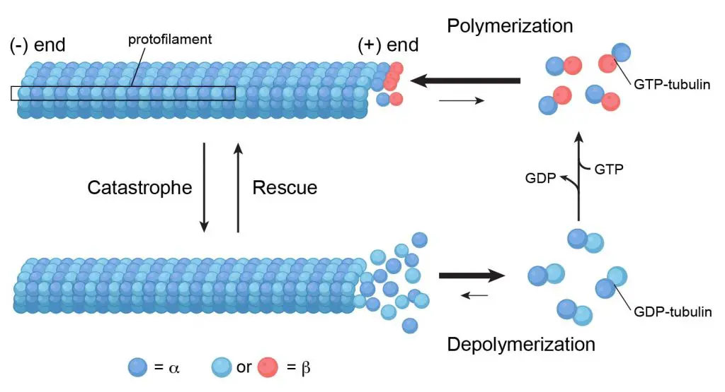
[In this figure] Microtubules are assembled from α‑tubulin and β‑tubulin. Soluble α‑tubulin/-β‑tubulin dimers polymerize into polar microtubules in the presence of GTP (energetic molecules similar to ATP).
To learn more about the microtubules and cytoskeleton, see our article of “Cytoskeleton”.
Centrosomes function
Centrosomes assist with several important functions, including:
I. Maintaining the chromosome number during cell division
In animal cells, centrosomes help the equal separation of sister chromatids in the cell division process.
What are sister chromatids?
When the cells prepare for the cell division, each DNA thread is organized into a much compact structure, called “chromosome”. After DNA replication, the chromosomes look like an X-shape (called sister chromatids) that remain attached at a centromeric region (centromere). Ideally, sister chromatids should be split into two identical chromosomes during mitosis. Otherwise, daughter cells may lose or duplicate certain genes.
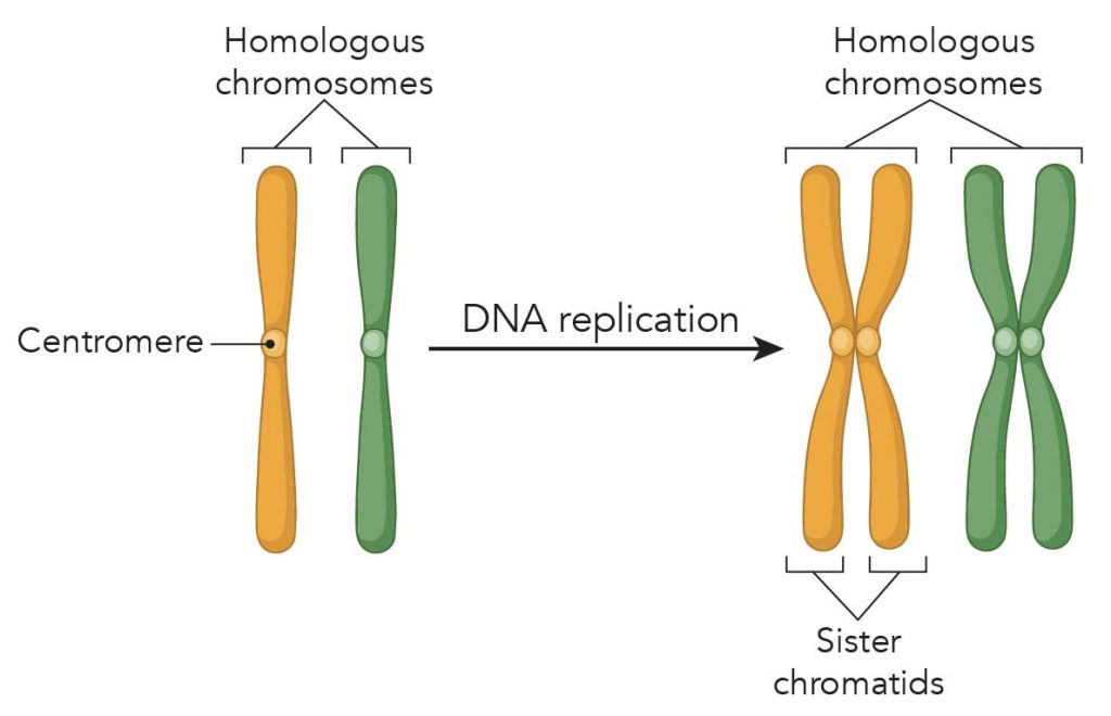
[In this figure] Chromosome replication forms sister chromatids. The X-shape sister chromatid has four arms attached at a centromere.
The phase of mitosis
Mitosis is a process of cell division in which one cell (the mother) divides and produces two new cells (the daughters) that are genetically identical. The goal of mitosis is to make sure that each daughter cell gets a perfect and fully equal set of chromosomes. If daughter cells have either more or missing genes due to uneven separation of chromosomes, the function of the cells will totally be messed up. These cells will very likely become cancer cells!
To ensure cells split up their duplicated chromosomes equally, our cells developed a Standard Operating Procedure (SOP). We call this carefully organized series of steps – the phases of mitosis. Centrosomes play a critical role to ensure this SOP done.
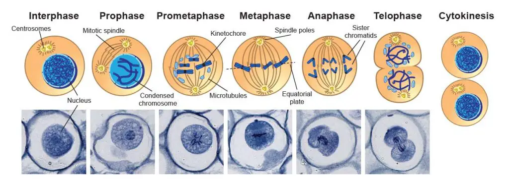
[In this figure] Mmitosis phases under the microscope.
Mitosis consists of five basic phases: prophase, prometaphase, metaphase, anaphase, and telophase. These phases occur in strictly sequential order. This specimen is Ascaris eggs (parasitic nematode worm) that contain active mitotic cells and can be purchased from Rs’ Science 25 Premade slide set.
The role of centrosomes during mitosis
As you can see in the phases of mitosis, the sister chromatids have to be pulled apart and separated equally into two new nuclei. Cells do this job exactly like how we pull a car out of mud using a cable and a winch. The “cable” is called “microtubule”. The centrosome works like a “winch” pulling the sister chromatids (the car) apart.
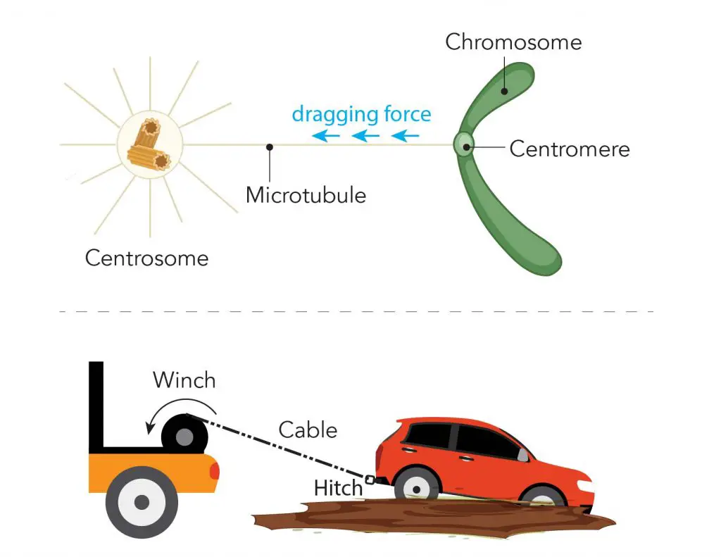
[In this figure] Illustration of microtubule, centromere, and centrosome during mitosis.
During mitosis, one end of the microtubule attached to the centromere regions of each chromosome, and the other ends are controlled by centrosomes. You can imagine that the centrosome like the winch pulls the sister chromatids (the car) apart by dragging the microtubule (the cable) anchored to the centromere (the tow hitch).
The action of centrosomes during mitosis
In a resting cell (not a cell cycle active cell), its centrosome is associated with the nuclear membrane. While mitosis begins, the nuclear membrane breaks down, releasing the centrosome to interact with the chromosomes.
The original centrosome duplicates once when DNA replicates. During the prophase, the centrosomes migrate to opposite poles of the cell. The mitotic spindle (or spindle apparatus) then forms between the two centrosomes.
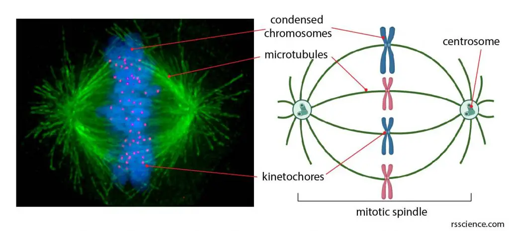
[In this figure] (Left) Fluorescent micrograph showing a mitotic spindle with condensed chromosomes in blue, kinetochores in pink, and microtubules in green during metaphase of mitosis. (Right) The illustration of the mitotic spindle, centrosome, kinetochores on condensed chromosomes, and microtubules.
Photo source: Afunguy at wiki (left), created with Biorender (right)
The mitotic spindle assembles by the attachment of microtubules to the centromeres of sister chromatids (forming an anchoring complex called kinetochore). Each sister chromatid attaches to both centrosomes from opposite cell poles. The coordinate action of both centrosomes allows all sister chromatids aligned in the middle of the cell (by opposing tension forces), forming a structure called an equatorial plate (in Metaphase).
Both centromeres retrieve their microtubule at the same time. The contraction force (generated by motor proteins, kinesin, and dynein) splits the sister chromatids apart from the attachment points of the centromere; then moving toward the opposite direction. Once all sister chromatids are equally separated into two new nuclei, the cytoplasm and cell membrane will also divide into two new cells. Upon division, each daughter cell receives one centrosome.
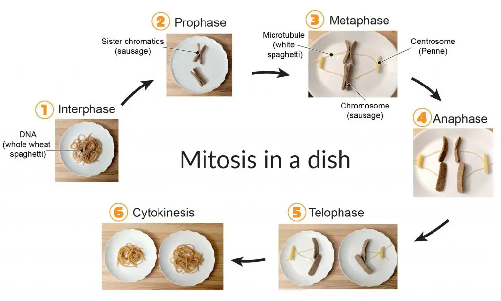
[In this figure] Food model of phases of mitosis.
This is a scientific project we published that uses food items in the kitchen to model the process of mitosis. Here, the whole wheat spaghetti represents DNA; sausage represents a condensed chromosome; Penne pasta represents the centrosome; and finally, white spaghetti represents the microtubule.
II. Organizing microtubule networks in the cells
Centrosomes as a “microtubule organizing center (MTOC)” control the distribution of microtubule networks inside the cells. Microtubule is a versatile cytoskeleton, and the formation of the mitotic spindle is just one of its functions. In non-dividing cells, microtubules also serve as an intracellular highway to transport molecules and organelles within the cells. There is a group of “motor proteins” that can carry cargos while walking along the cytoskeleton, which is just like many small trucks driving on an intracellular transportation system.

[In this figure] The microtubule organization varies according to the cell types and cell cycle. It determines the internal organization of organelles and vesicular trafficking that benefit the cell functions.
Photo source: Atlas of plant and animal histology
Centrosomes can also orchestrate large changes to cell membrane shape under other circumstances, such as cell movement or phagocytosis.
Extended read:
III. Forming the cilia and flagella
Microtubules support special cellular structures that allow cells to move.
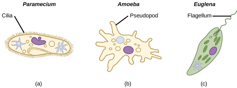
[In this figure] Three ways of locomotion for protozoa.
Cilia – coordinately beat to swim. Pseudopod – crawl on the surface by changing the cell shape. Flagellum – swim by rotating like a propeller.
Photo credit: Lumen.
One example is the “flagellum” (plural: flagella), which is a “tail” that can propel a cell forward. A centrosome is sitting near the root of a flagellum. A long microtubule bundle (called axoneme) extends from the mother centriole and forms a lash-like appendage that protrudes from the cell body. Usually, a cell has only one or two long flagella.
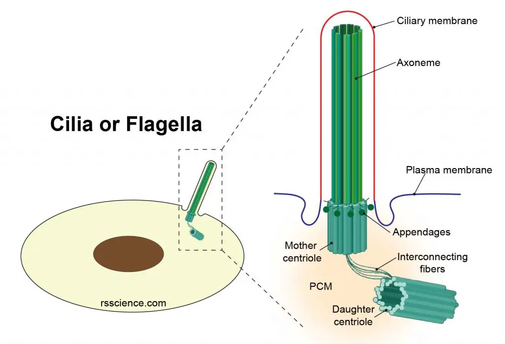
[In this figure] Anatomy of the centrosome-cilium/flagellum complex.
Microtubules are also found inside the cilia (singular: cilium), which are hair-like small protrusions. These cilia are in constant motion and help single-celled microorganisms like paramecium move around. Cilia also cover the surface of our bronchial epithelial cells to move microbes and debris up and out of the airways. The microtubules function in a similar way in both flagella and cilia. The difference is that a cilium is much shorter than a flagellum, and a cell could have hundreds of cilia.
In order to generate numerous cilia, centrosomes will replicate many times (a process called ciliogenesis) in cells. Once the cells start preparing for cell division, the cilium/flagellum complexes disassemble. The centrosomes fuse and return to guide the mitosis.
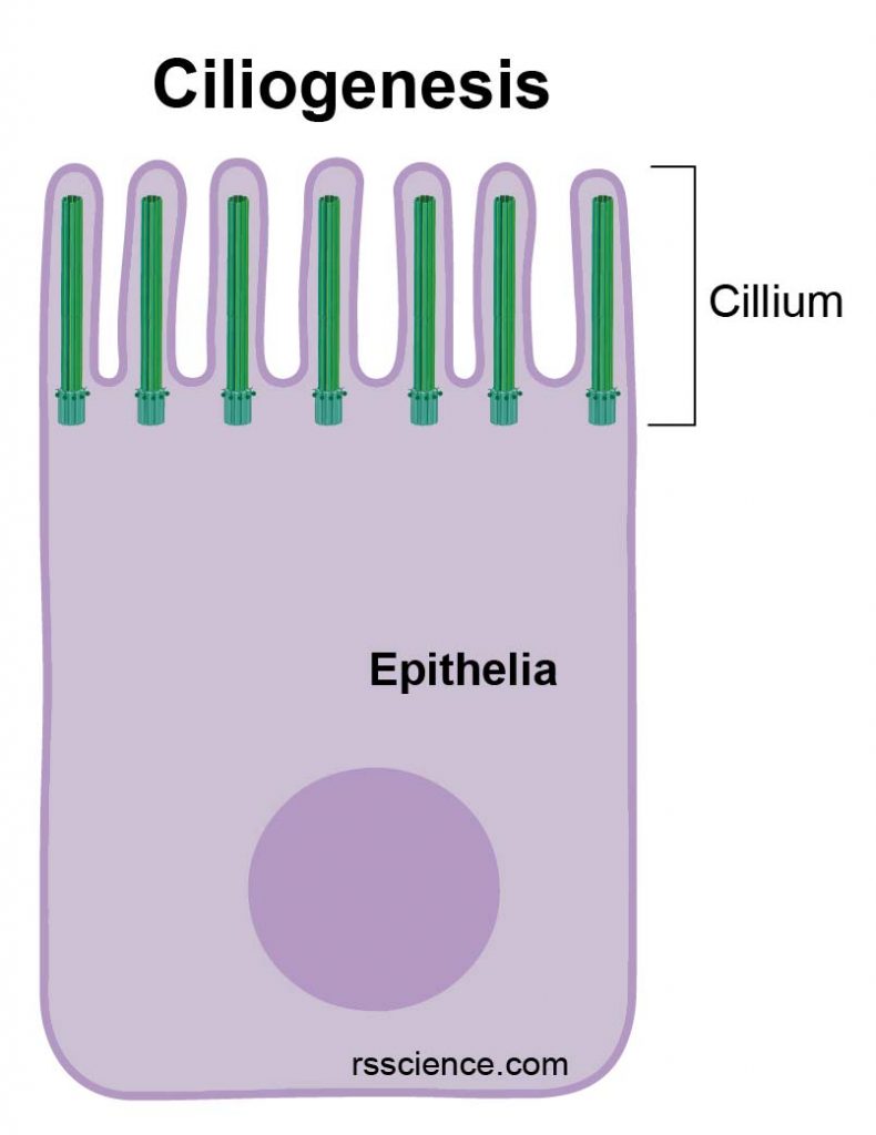
[In this figure] An airway epithelial cell possessing many cilia.
The base of each cilium has a centrosome as the root, which generated by ciliogenesis.
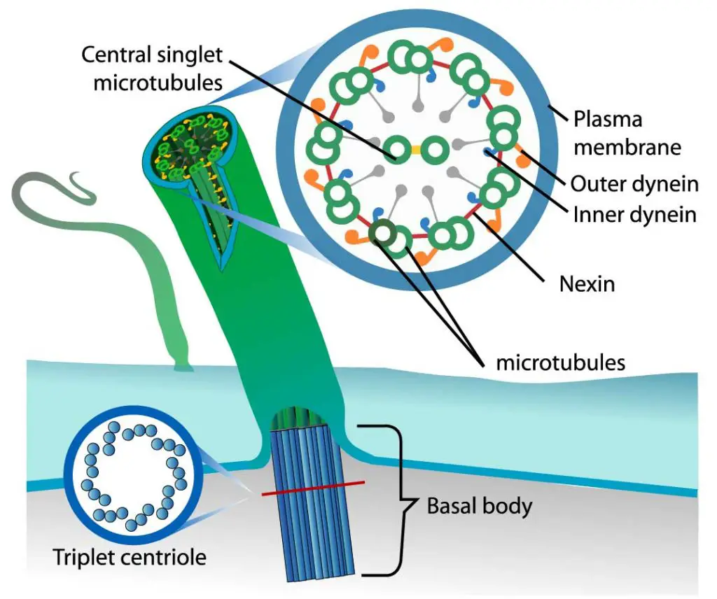
[In this figure] The organization of cilium.
Each cilium contains nine pairs of microtubules forming the outside ring and two central microtubules (9+2). This structure is known as an axoneme. Microtubules are held together by cross-linking proteins. There are motor proteins, called dynein, setting across each paired microtubule fiber.
Photo credit: LadyofHats on wiki.
In the microtubule bundles of cilia and flagella, there are many “motor proteins” in-between microtubule filaments. These motor proteins (dynein) use ATP as energy to crawl along the microtubules. When dynein proteins move upward on one side but downward on the other side, the cilium bends. The repeat of bending-relaxing cycles makes cilia act like oars, beating back and forth to create movement.
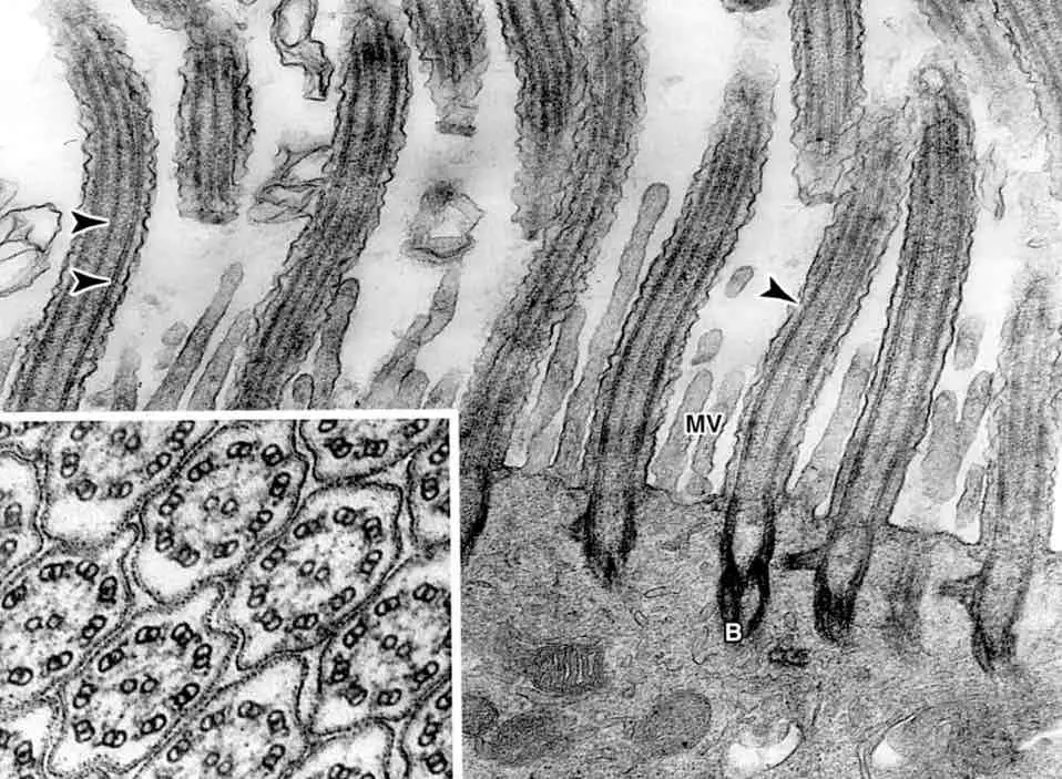
[In this figure] Electron micrograph of cilia on the surface of epithelial cells.
The inserted image shows the 9+2 microtubule arrangement inside the cilia from a cross-section view.
Photo source: Columbia University
Note: The “flagella” we mention here are eukaryotic cells’ flagella (can be found in euglenas and some green/brown algae). Bacteria also have a cellular structure called “flagella”. However, bacterial flagella are very different in structures and compositions. Some scientists attempted to replace the name of eukaryotic flagella with “undulipodia” or “cilia”. However, the usage of “flagella” in eukaryotic cells is still very common.
Centrosome alterations in cancer cells
Aberrant structures and numbers of centrosomes in a cell have been associated with tumors. The presence of defective centrosomes is often linked to the appearance of genome instability, failure in cell division, and the loss of cell differentiation. Centrosome amplification (more than two centrosomes during mitosis) in human cells can form irregular mitotic spindles, resulting in aneuploidy (an abnormal number of chromosomes), which very likely cause a cancer.
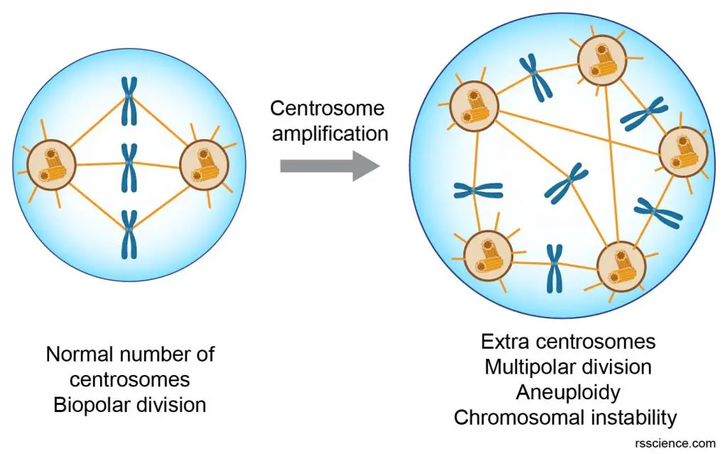
[In this figure] A schematic diagram showing the consequence of centrosome amplification.
The discovery of centrosomes
The centrosome was discovered by Edouard Van Beneden in 1883, and later described and named in 1888 by Theodor Boveri as the “special organ of cell division”. The name came from Latin centrum “center” + Greek sōma “body”. Theodor Boveri also noticed centrosome aberrations in cancer cells.
Frequently Asked Questions:
Do plants and fungi have centrosomes?
No, plants and fungi don’t have centrosomes. The centrosome is thought to have evolved only in the animal lineage of eukaryotic cells. Fungi and plants lack centrosomes and therefore use other structures to organize their microtubules and mitosis.
Are centrosomes essential for all animal cells?
Maybe not. Although centrosome has a key role in efficient mitosis in animal cells, the centrosome is not essential for mitosis in certain animal species like fly and flatworm. In an experiment done in the fruit fly (Drosophila), even the centrioles were destroyed by a laser, the mitosis proceeds normally with a morphologically normal spindle. Moreover, the fruit fly grows normally. This suggests the cells should have other ways to organize their microtubules.
How is a centrosome different from a centromere?
The centrosome is the organelle that contains two centrioles. Whereas centromere is a highly constricted region on the chromosome. A centrosome is a microtubule-organizing center, whereas centromere holds together the sister chromatids in a replicated chromosome.
Summary
- Centrosomes are organelles that serve as the main microtubule organizing center (MTOC).
- During cell division, each cell has two centrosomes. They move toward the opposite positions of the cells and form a mitotic spindle.
- The microtubules extend from the centrosome and attach to the centromeres of sister chromatids. Both centromeres retrieve their microtubule at the same time to split the sister chromatids apart and move into new cells.
- Centrosomes also form the core of two structures involving in cell mobility: cilia and flagella.
References
“Centrosome”
“Controling centrosome numbers”
“Centrioles, Centrosomes, and Cilia in Health and Disease”
“Mechanism limiting centrosome duplication to once per cell cycle”