
This article covers
What is euglena
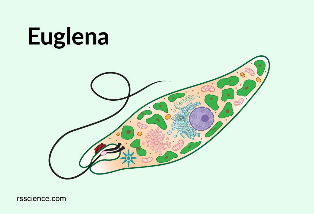
Euglena (Greek: eu = true, glene = eye-ball) is a genus of single cell eukaryotes with flagella, and they can be found in freshwater pond and ditches. Euglena gracillis is one of the species that has been used as a model organism for studying cell biology in the lab. Other species, such as Euglena viridis and Euglena sanguinea, can thrive in a short time; subsequently, their abundance can change the surface color of the pond to green and red, respectively.
Euglena shares some characteristics of both plants and animals. For example, euglena contains chloroplasts; as a result, they can make their own food, a characteristic of plants. In contrast, euglena can also move using its flagella and consume food through phagocytosis, which are characteristics of animals. Euglena also lacks a cell wall.
[In this video] An euglena under a microscope.
While I examined the pond life under a microscope, I came across this slow-moving euglena. Although the flagella are not obvious in this video, you can appreciate many chloroplasts and one red eyespot in a tear-drop shape organism. The red eyespot is located in the anterior of the euglena. Notice the way of euglena’s movement; it moves forward and also rotates its body axis.
Structure of euglena
The euglena cells are tear-drop shaped with a blunt end (head) and a pointed end. The common features of euglena cells are a nucleus, endoplasmic reticulum, Golgi apparatus, mitochondria, ribosomes, lysosomes, and a contractile vacuole. The unique features of euglena include pellicle, flagella, an eyespot, a paraflagellar body, and paramylon. Let’s discuss the unique characteristics one by one below.
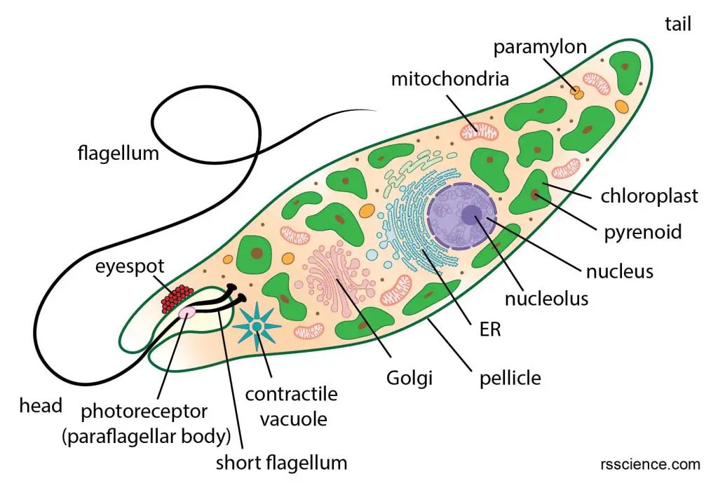
[In this figure] Euglena anatomy and its organelles.
Pellicle
Unlike plant cells, euglena does not have a rigid cellulose cell wall. Instead, they have a flexible and tough pellicle that facilitates their flexible and contractible movement. The pellicle is tough enough to maintain its shape but also flexible enough to allow changes in the body shape, known as metaboly movement or euglenoid movement.
The euglena body is covered by a pellicle which lies under the plasma membrane. The pellicle is made up of a layer of fibrous elastic proteins and microtubules. The microtubules arrange in strips spiraling around the cell. These pellicle strips slide over one another, giving euglena its remarkable flexibility and contractility to change its shape.
[In this video] Metaboly movement.
The metaboly movement is characterized by elegantly concerted, large-amplitude
distortions of the entire cell.
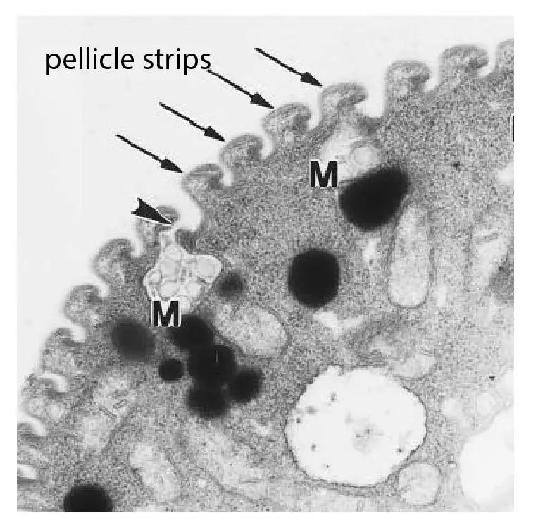
[In this figure] TEM image of Euglena showing the pellicle strips in cross-section.
The pellicle strips (arrows) look wavy, with ridges and grooves; as a result, giving pellicle striated appearance under a light microscope. The arrowhead points toward a pellicle pore where the biogenic lubricant, the mucus (M), is secreted.
Photo credit: Gruenberger C.
Flagella
Flagellum (plural: flagella) is a long whip-like structure at the front of the euglena cells. Typically, euglena has two flagella. One is long and can be seen under a light microscope, but the other is very short without protruding from the cells. The function of flagella is to help euglena swim.
Structurally, cilia and flagella are indistinguishable. They both possess a central bundle of microtubules, called axoneme. Each axoneme contains nine pairs of microtubules (doublet) forming the outside of a ring and two central microtubules, known as 9+2. There are motor proteins, called dynein, attached to Tubule A, one of the doublet. Microtubules are held together by cross-linking proteins. Each doublet is connected by Nexin protein.
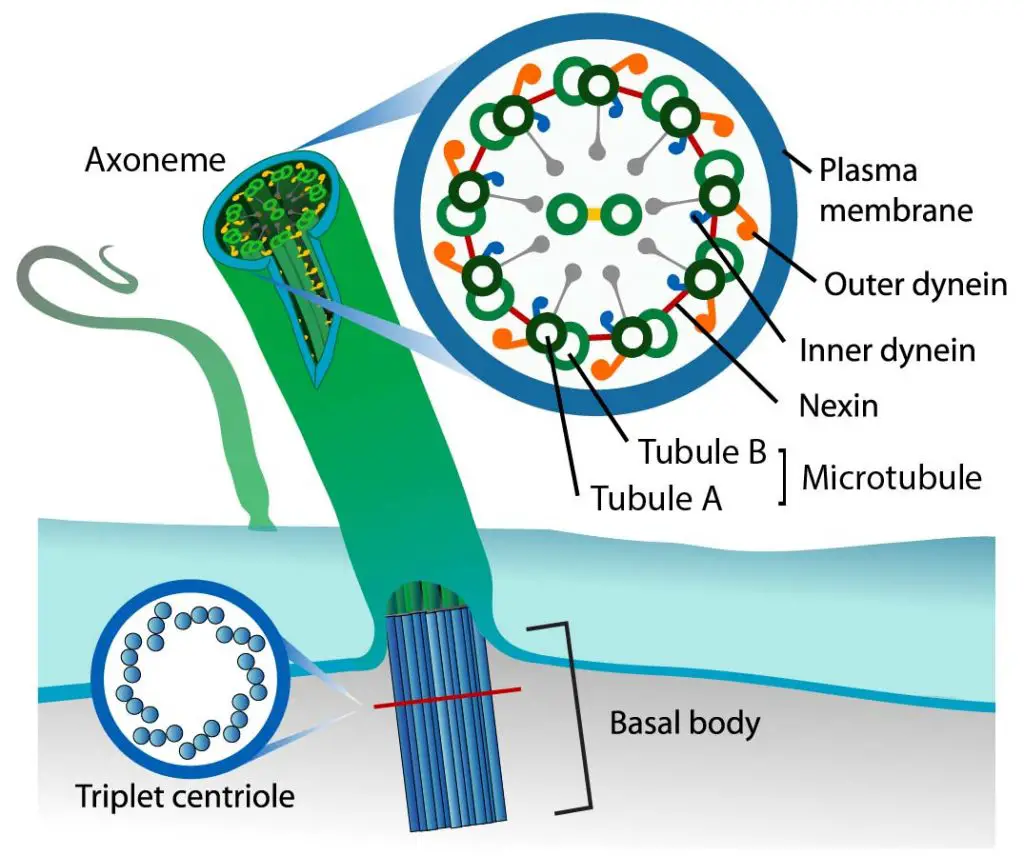
[In this figure] A diagram of flagella.
Photo credit: modified from LadyofHats on wiki.
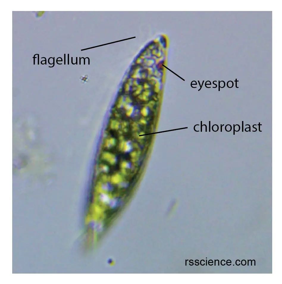
Eyespot
Euglena has a bright red eyespot, also called stigma. It is made up of carotenoid pigment granules. The eyespot is not an actual eye; instead, it is more like a sunglass for a photoreceptor. The eyespot filters the sunlight and allows certain wavelengths of light to reach photoreceptors (also called paraflagellar body). Therefore, the eyespot can tell the euglena where the light source comes from.
Paraflagellar body or photoreceptor
Paraflagellar body (also called photoreceptor) is a swelling structure at the base of the flagellum that is photosensitive. It is the photoreceptor that senses light. Paraflagellar body, together with an eyespot, is located close to the flagella; thus their proximity promotes light-guided directional movement.
Chlorophyll containing chloroplast
Euglena also has chloroplasts throughout its body. Its chloroplasts contain chlorophyll a and b to produce sugar by photosynthesis; therefore, euglena can survive with light without eating.
Euglena chloroplasts contain pyrenoids, a subcellular compartment inside chloroplasts. Pyrenoids’ main function is to generate a CO2 rich environment for ribulose diphosphate carboxylase, one of the enzymes for carbon fixation in photosynthesis.
The photosynthesis produces paramylon, a starch-like carbohydrate. It serves as food storage and enables euglena to survive when light is not available.
How does euglena eat
Although euglena is able to make its own food by photosynthesis, it can also consume food via phagocytosis, a process to engulf food particles in a vacuole. A lysosome then fuses with a food vacuole, releasing enzymes to digest food. Euglena also has a contractile vacuole to collect and remove excess fluid from the cell. Without the contractile vacuoles, the euglena may burst.
How does euglena move
Flagellar movement – use fragella to turn and twist
Euglena moves by whipping and turning its flagella in a way like a propeller. The beating of the flagella created two motions. One is moving euglena forward (transitional motion), and the other one is rotating the euglena body (rotational motion). You can see how scientists study the euglena movement below.
[In this video] Reconstructed swimming kinematics of E. gracilis.
The resulting trajectory of the cell can be seen as a smooth circular helix (the “backbone” trajectory), perturbed by periodic “swirls” at the flagellar beating time scale. The cell completes one turn of the helix while undergoing a full rotation around the axis of the helix. The Euglena’s body is not to scale with the displacements for visualization purposes.
Movie credit: Rossi M. et al., PNAS 2017
Euglenoid movement – use pellicle for peristaltic movement
Euglena is able to alter its shape and then return to its initial shape like an elastic rubber band, a process called euglenoid movement (metaboly).
The movement is created by peristaltic waves. When peristaltic waves travel through the body, they trigger the body to become much shorter and wider first at the anterior end, and then in the middle, and finally at the posterior end.
This smooth movement is due to a unique structure on the euglena, called pellicle.
[In this video] Metaboly movement.
Metaboly movement allows euglena to change its shape and return to its initial shape coupled with movement.
Euglena reproduction
Euglena reproduces asexually through binary fission on its longitudinal axis. When environmental conditions become unfavorable and too difficult for them to survive, such as low moisture or scarce food supply, euglena forms a protective cyst around itself and becomes dormant.
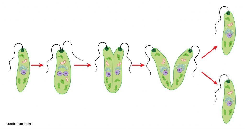
[In this figure] A diagram of euglena reproduction.
Euglena under a microscope
The material you need
- Pond water
- Microscopic slides and coverslips
- Droppers
- Compound microscope
Steps
- Use a dropper to obtain some pond water, and place a drop on a microscope slide.
- Place a coverslip gently on the sample. You can check how to mount a bubble-free slide.
- Place the slide on the microscope stage and start viewing.
What will you see
[In this video] Euglena under a light microscope.
The euglena whips its flagella for directional movement, and it also rotates its body. In addition, green chloroplasts and red eyespot are present. Magnification 100x.
Euglena fun facts
As a food and biofuel source
Euglena gracilis is an outstanding resource of dietary protein, vitamins, lipids, and also the β-1,3-glucan paramylon, which is only found in euglenoids.
Paramylon is marketed as an immunostimulatory agent in nutraceuticals.
Interestingly, a Tokyo-based Euglena Company marketed Euglena-based food and beverage products in 2005. Now they shift their business model to biofuels using euglena.
Reference
Euglena Viridis: Habitat, Structure and Locomotion | Protozoa
Bioproducts From Euglena gracilis: Synthesis and Applications