
This article covers
What are ferns?
Ferns are an ancient group of plants. They pre-date the evolution of humans, mammals, reptiles, and birds based on the fossil record. Their origins can be traced to the late Devonian period, approximately 360 million years ago – far older than dinosaurs!
They were thriving on Earth for two hundred million years before the flowering plants evolved. Even though they are so ancient, ferns already have true roots, stems, and complex leaves similar to current plants. Ferns also have vascular systems that can efficiently transport water and nutrients. Thus, we classify ferns as vascular plants. This is why some giant ferns can grow up to 20 feet tall compared to tiny nonvascular plants, such as mosses and liverworts.
Ferns are classified as the lower vascular plant division – Pteridophyta. Unlike the other vascular plants such as the flowering plants and conifers where the adult plant grows immediately from the seed, ferns reproduce via spores, and ferns do not have seeds and flowers.
The life cycle of ferns requires an intermediate plant stage called a gametophyte (the sexual phase in plants) between fern spores and maturity. Ferns are restricted and can only propagate in a suitably moist condition. As we know them now, most ferns grow in wet areas under a forest canopy, along creeks, streams, and other sources of permanent moisture. They cannot grow in hot, dry areas like a cactus or cold frozen places like conifers.
The structure of fern
Ferns come in various shapes and sizes, but they all have 3 major parts – the fronds, the rhizome, and the reproductive structures called sporangia.
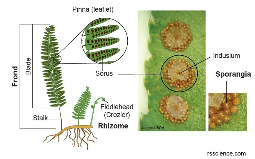
[In this figure] The structure of the fern.
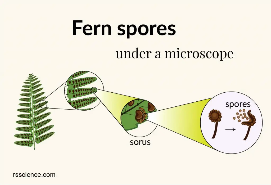
The fronds, the rhizome, and the sporangia are 3 major parts of the fern. The fronds are the leaves of the fern. The rhizome is the stem of the fern plant. The sporangia are reproductive structures and are found in clusters (called sori; singular sorus) underneath the fronds.
Fronds
The fronds are the leaves of the fern and consist of a blade and a stalk. The flat blade is the expanded, leafy part of the frond and the stalk connects the frond to the rhizome.
New fronds are produced from the rhizome, and they are tightly coiled into a spiral (called a fiddlehead or crozier). As they grow and mature, these fiddleheads slowly unroll. Fronds have a dual function – photosynthesis and reproduction.
Ferns’ blades grow in a massive variety of forms. The frond may be simple and undivided, or it may be divided into many divisions (called pinnae). Pinnae of some species can further divide into sub-leaflets, called pinnules. Pinnules can even be split into lobes and generate more complicated frond shapes such as beech fern, northern wood fern, and Maidenhair fern (my favorite fern).
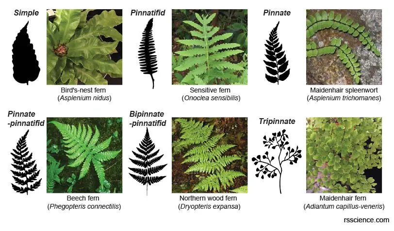
[In this figure] Fern leaf divisions.
Fern leaves display a variety of divisions depending on the species. Various degrees of leaf divisions are shown in the series of frond silhouettes.
• Simple – The fronds are undivided (bird’s nest fern).
• Pinnatifid – The fronds are divided into segments (sensitive fern).
• Pinnate – The fronds are divided into segments completely separated from each other maidenhair spleenwort).
Rhizome
The rhizome is the stem and root in combination and looks like a branching vein system. Laterally growing rhizomes can creep along or under the ground to expand their territory. Sometimes, rhizomes may even climb up a tree. At some points, the rhizomes can grow vertically into a short trunk. The young fronds sprout directly from the rhizomes. Some rhizomes in tree ferns, such as a king fern or a crown fern, can grow into a tree-like solid stem that gives rise to a tuft of fronds.
Sporangia
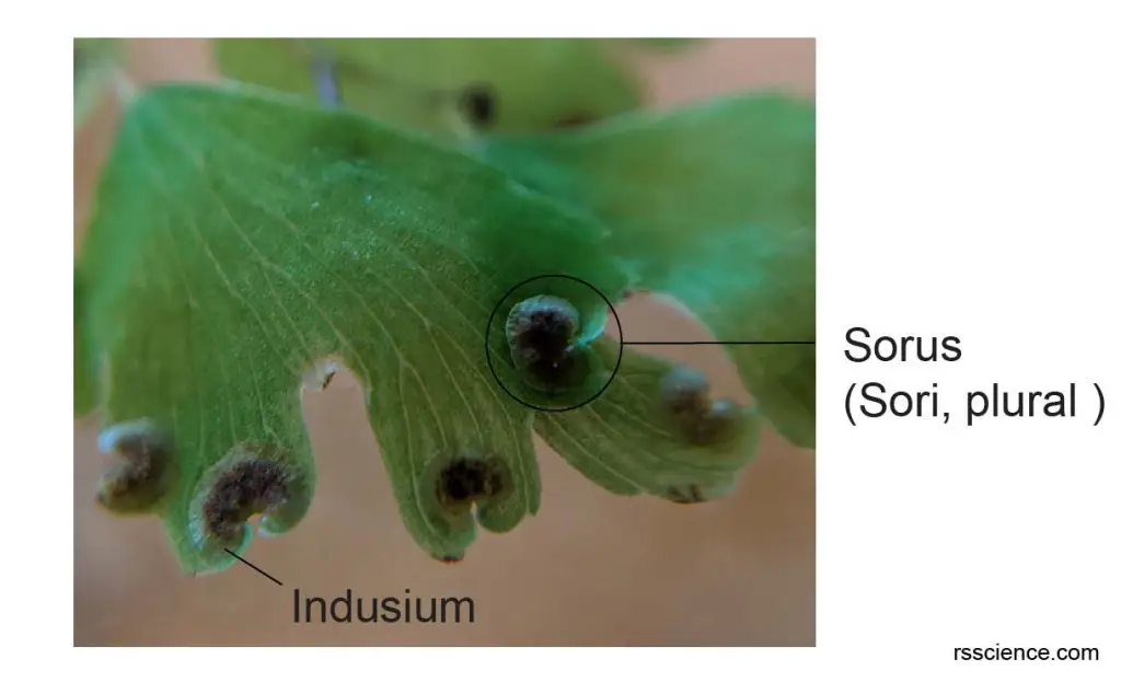
The sporangia (singular sporangium) are found in clusters called sori (singular sorus) underneath the fronds. They are often brown, black, and orange. The fern spores are inside the sporangia. In some species, there is a piece of protective tissue covering the sporangia, called indusium. Not every frond has sporangia underneath it. Fronds that have sporangia are called fertile fronds.
When the sporangia open, they release the spores. If the fern spores land in a habitable environment, they can grow into a tiny plant called the gametophyte. This intermediate, short-lived plant has 2 sets of reproductive organs – the antheridia (male) and the archegonia (female). In suitably moist conditions, fertilization takes place either on the same gametophyte or an adjacent one. Fertilization gives rise to a new sporophyte – the actual fern plant.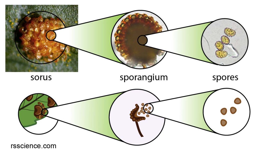
[In this figure] The microscopic images and the Illustration of sorus, sporangium, and spores.
Sorus looks like a little dot found on the back of the fronds. Many sporangia make up one sorus. The sporangia are not always visible in some ferns because they are covered by a structure called an indusium. The spores are inside sporangium. The upper panel is microscopic images.
Photo credit: modified from Botany Blog.
Fern reproduction and life cycle
The life cycle of the fern is an alternation of diploid sporophytic and haploid gametophytic phases. Diploid means that the cells contain two complete sets of chromosomes, one from each parent. The leafy “fern” plants are their sporophyte. Mature sporophytes can produce spores.
However, a fern’s spores don’t grow into a leafy sporophyte. They aren’t like seeds of flowering plants. Instead, they produce a haploid generation. In a haploid stage, each cell contains one set of chromosomes or half of the genetic complement (like a human sperm or egg cell). This version of the pant looks like a little heart-shaped plantlet. It is called the gametophyte.
Within the gametophyte, sperm is produced within a structure called an antheridium. The egg is produced within a similar structure called an archegonium. Eggs are sometimes produced on the same gametophyte as the sperm, but some species may occur on a separate gametophyte. When water is present, sperm uses its flagella to swim to an egg and fertilize it (this is why ferns can only flourish in a moist environment). The fertilized egg becomes a diploid zygote formed by the combination of DNA from the egg and sperm. The zygote grows via cell division into a new sporophyte. 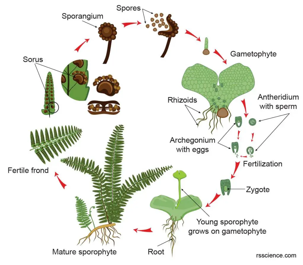
[In this figure] The life cycle of the fern.
The life cycle consists of diploid sporophytic and haploid gametophytic phases. The sporophytes are mature plants that produce spores, but the spore cannot grow into a little plant. Instead, spores are haploid, and they grow into a heart-shaped plant, called gametophyte. It produces sperms and eggs. After sperms seek out eggs to fertilize, the zygote develops into a sporophyte.
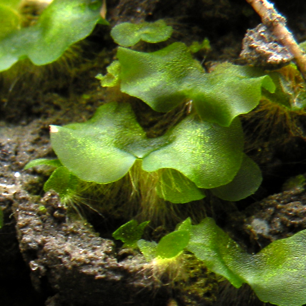
[In this figure] The heart-shaped gametophyte.
Observe fern spore under a microscope
The material you need
- Fern frond specimens
- Microscopic slides and coverslips
- Droppers
- Tweezers and dissecting needles
- Compound microscope
Steps
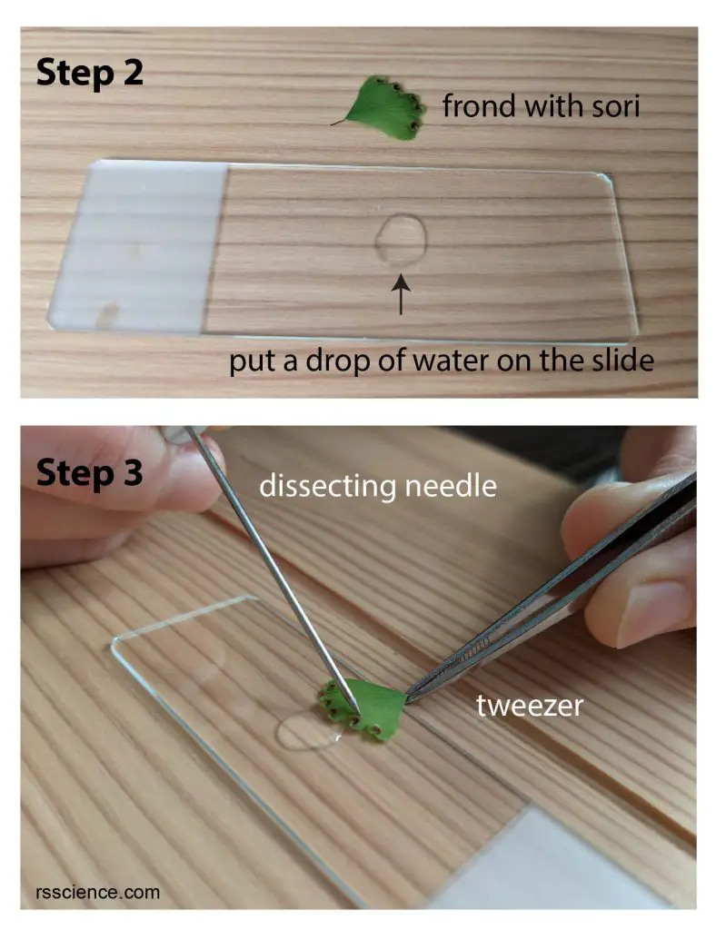
- Obtain a fern frond, and make sure there are sori underneath it. Pay attention to the color of the sori. The black sorus is riper than the white one.
- Put a drop of water on the microscope slides.
- Use tweezers to hold the frond, and use a dissecting needle to open sorus. Do this step next to a drop of water, so the spores can be released into water.
- Put on a coverslip, and ready to see.
What will you see
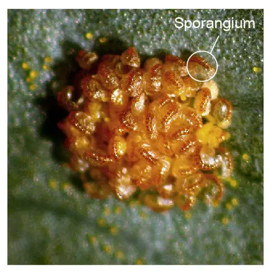
[In this figure] Sori.
The sori are underneath the frond (leave). The position of the frond is upside down. This is Maidenhair fern.
[In this figure] Sori under a dissecting microscope.
The sporangia become visible with 3x magnification.
Photo credit: Botany Blog
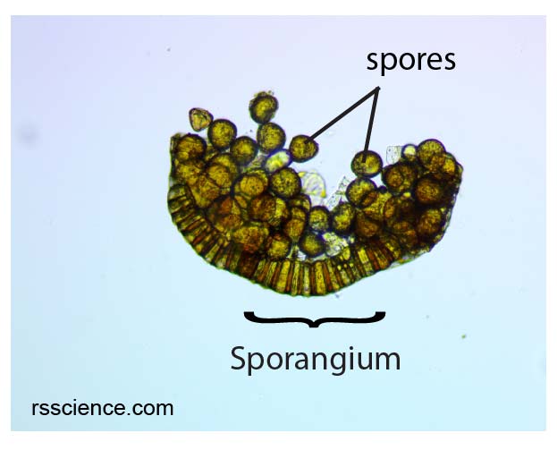
[In this figure] Sporangium from Maidenhair fern under a compound microscope.
A sporangium was mounted on a slide. There are many spores inside a sporangium. 100x magnification.
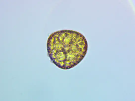
[In this figure] A spore from Maidenhair fern under a compound microscope.
A spore was mounted on a slide and viewed at 400x magnification.
[In this video] Fern leaf under the microscope catapulting its spores.
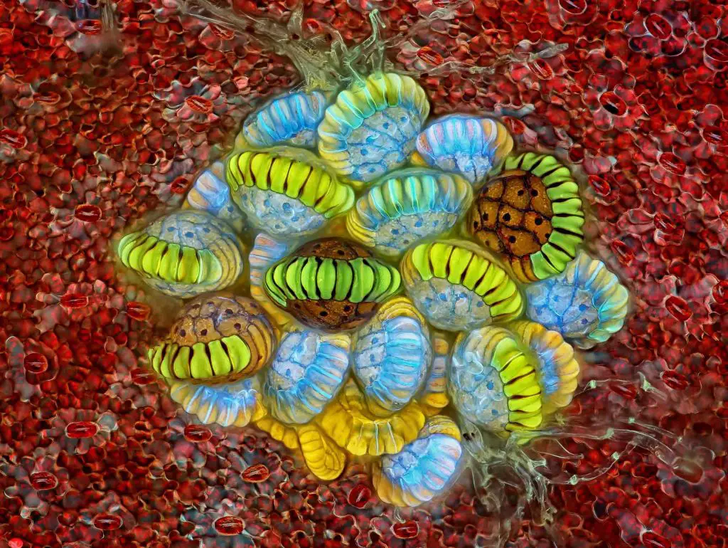
[In this figure] Fluorescence image of fern sorus.
Magnification, 10x.
Photo credit: Rogelio Moreno, 2018 photomicrograph competition.