
This article covers
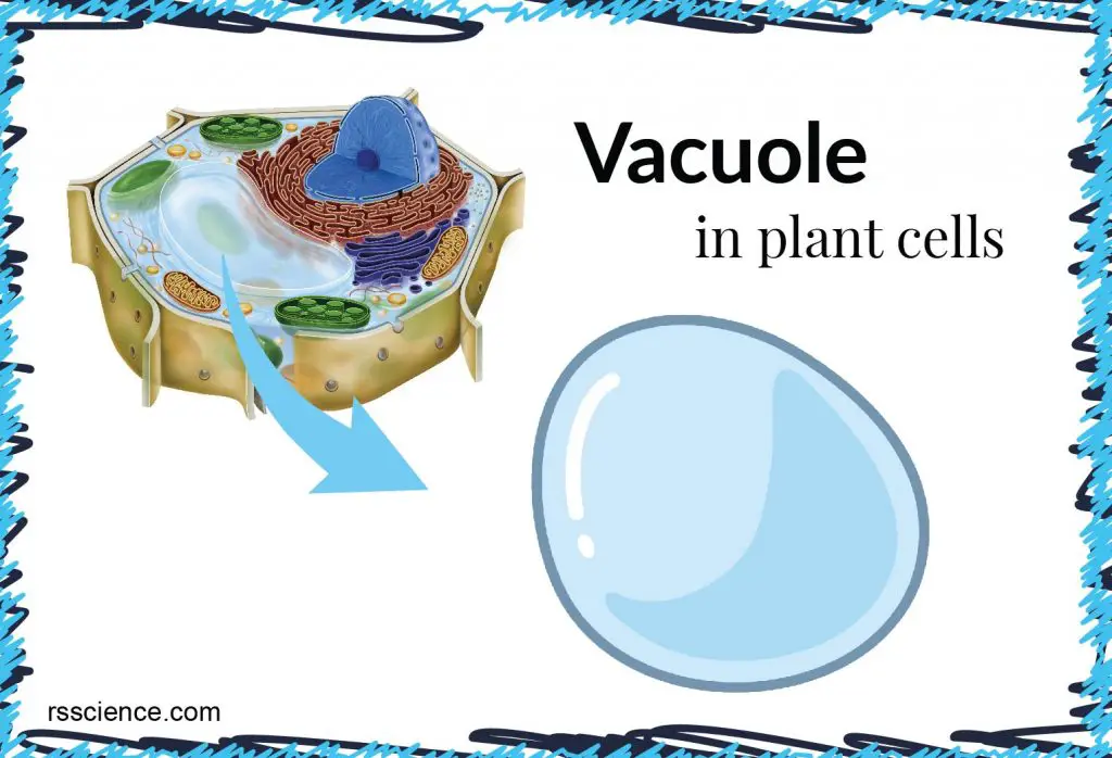
What is a vacuole?
A vacuole is a membrane-bound organelle (like a bubble) that is present in all plant cells. Some animal and fungal cells also have vacuoles, but they are much smaller. Most mature plant cells have one large central vacuole that can occupy as much as 80% of the cell volume, making the vacuole the most prominent organelle in plant cells.
By storing various materials, the central vacuole keeps its water potential as low as the cytoplasm’s potential and maintains force (turgor pressure) against the cell wall. Turgor pressure is essential for supporting plants in an upright position.
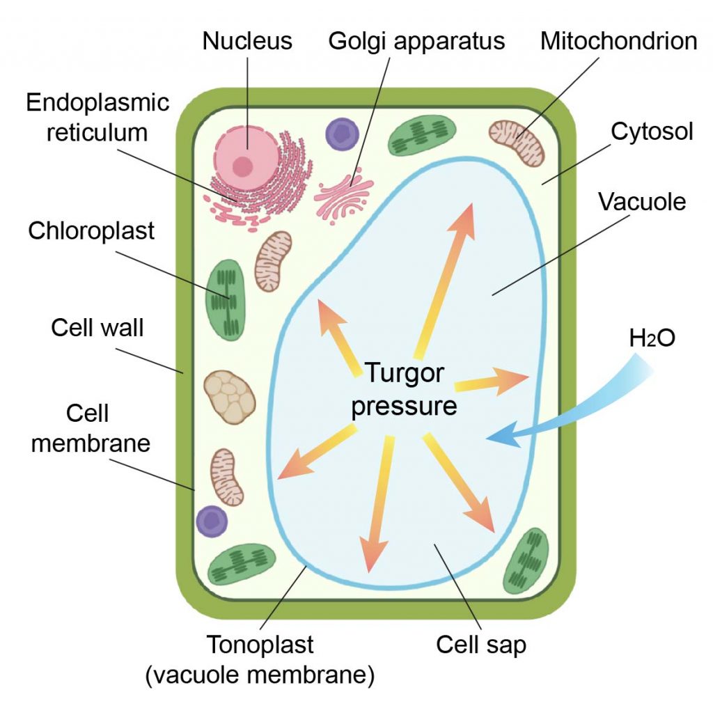
[in this figure] The anatomy of a plant cell.
A plant cell contains a large vacuole that occupied most of the plant cells; therefore, it helps to position other organelles in the cells. The central vacuole stores water and nutrients and creates turgor pressure to support plants in the upright position.
The structure of vacuoles
The vacuole is a membrane-bound, water-filled organelle which contains inorganic ions and organic compounds. The vacuolar membrane, called the tonoplast, contains various transporters. These transporters function as pumps or valves that control the import and export of substances across the vacuolar membrane, including
(1) Proton pump – move H+ ions to adjust the pH value of cells, which stabilize the cytoplasmic pH environment.
(2) Aquaporins – control water permeability and regulate the turgor pressure of cells.
(3) Ion transporters – control the flow of specific ions, like calcium, potassium, and sodium ions that maintain cytoplasm homeostasis.
Plant cell’s vacuole does not have a defined shape or size; its structure varies according to the cell’s need.
The biogenesis of vacuoles
Vacuoles are complex organelles, and their biogenesis remains unknown. However, studies have suggested that vacuoles assemble from smaller vesicles that are derived from the Golgi apparatus. These small vesicles fuse together to form pre-vacuoles, which are vacuole precursors. The continuous fusion of these pre-vacuoles ultimately results in the formation of a large vacuole.
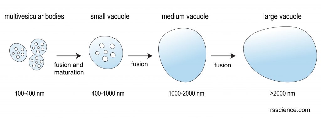
[In this figure] The working model of vacuole formation in plant cells.
Vacuoles are mainly derived from small vesicle fusion and maturation.
Photo source: modified from CHHK researchers.
Where is the vacuole located in a cell?
Most mature plant cells have one large central vacuole that typically occupies more than 30% of the cell’s volume. It can occupy as much as 80% of the volume for specific cell types and conditions.
Due to the large size of the vacuole, it pushes all contents of the cell’s cytoplasm and organelles against the cell wall. A good example is cytoplasmic streaming.
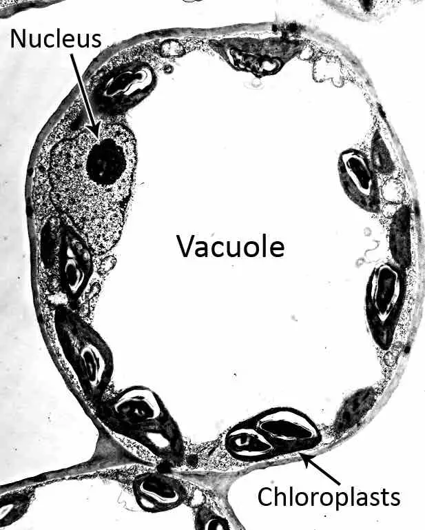
[in this figure] Electron microscopic (EM) image of a plant cell.
A large vacuole occupied most of the space of the cell and pushed all contents of the cell’s cytoplasm against the cell wall.
Photo credit: UF
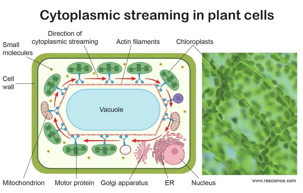
[In this figure] Cytoplasmic streaming in plant cells.
Cytoplasmic streaming circulates the chloroplasts around the central vacuoles in plant cells. This optimizes the exposure of light on every single chloroplast evenly, maximizing the efficiency of photosynthesis. The right image is the actual cytoplasmic streaming of chloroplasts in Elodea cells.
Created with BioRender.com
What is the biological function of the vacuole?
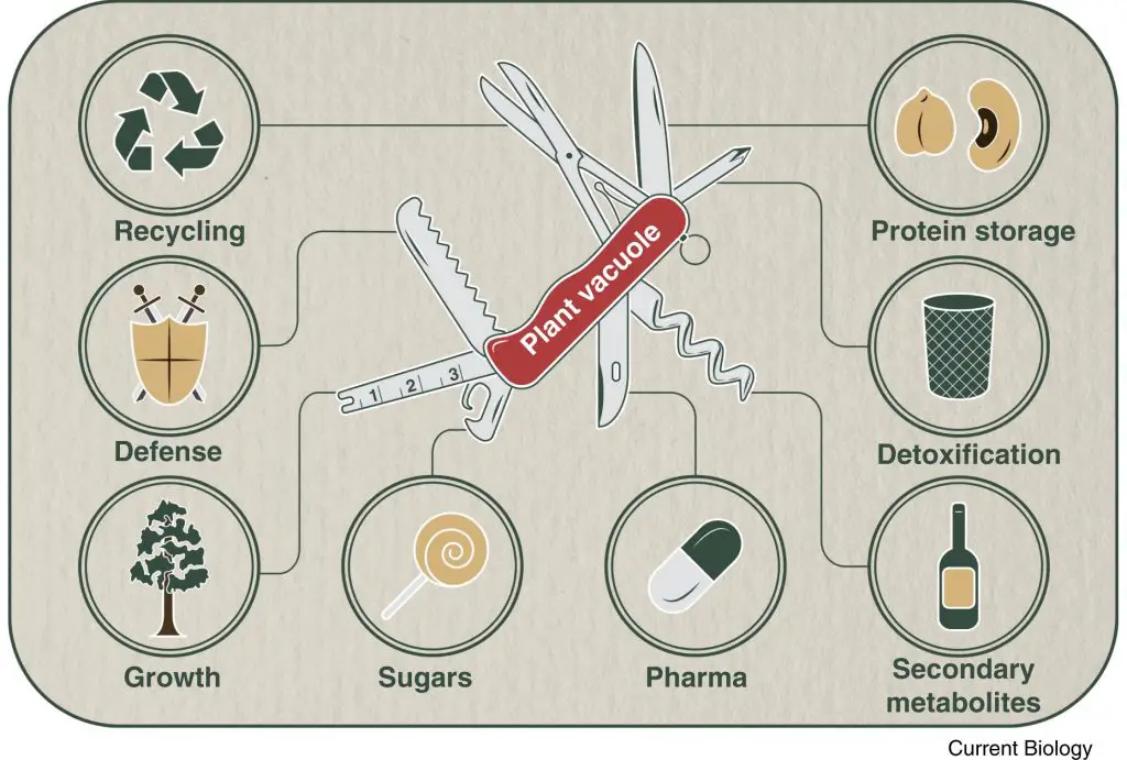
[In this figure] The multifaceted role of plant vacuoles.
Photo credit: Current Biology
Storage
The vacuoles serve as storage spaces for plant cells. The fluid (called cell sap) is enclosed by a membrane called tonoplast. In the fluid, there are food and various nutrients, including sugars, minerals, amino acids, nucleic acids, ions, and special chemicals. Vacuole also functions as a reservoir for the cell to store excess water. The regulation of water content helps maintain the balance of osmotic pressure and internal pH value inside the cells.
In seeds that contain a lot of proteins such as soybeans, vacuoles store proteins as protein bodies that can be used in germination. In oil-seeds such as sunflower seeds, lipids stored in vacuoles (oil bodies) are transported in peroxisomes and metabolized to produce energy for germination.
Defense
Plants lack an immune system, but each plant cell has its own defense weapons. The defense proteins and enzymes that can kill bacteria and viruses are stored in the vacuoles. There are two defense mechanisms that vacuoles can perform, depending on the targets.
Viral infections lead to vacuole membrane breakage and release enzymes into the cytosol, where they can attack viruses. For bacteria outside of the cells, the vacuole membrane fuses with the cell membrane; the vacuole enzymes then release to the extracellular space where they can kill pathogens like bacteria.
Compartment
The large size of the vacuole pushes all contents of the cell’s cytoplasm against the cell wall, thus keeping the chloroplasts closer to light. Sometimes, the vacuole can even be used as a compartment to store waste products, so the rest of the cell is protected from contamination. We can fairly say that vacuole is a versatile organelle.
Vacuole and turgor pressure
Most of the plant cell’s volume depends on the water level in its vacuole. Inflated vacuoles allow plants to support structures such as leaves and flowers due to the turgor pressure. Cell turgor is the level of hydrostatic pressure against the cell wall of the plant cell. The gain and loss of water in vacuoles depend on how much water is available to the plant. A drying plant has lost much of its water, and the vacuoles are shrinking. It still maintains its basic structure due to the cell walls. However, the entire plant looks depressed with drooping leaves and limping stems. When the plant finds a new water source, the vacuoles are refilled, and the plant regains its structure.
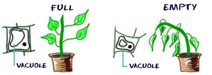
[In this figure] The vacuole, turgor pressure, and the appearance of the plant.
When a plant receives adequate amounts of water, the central vacuoles of its cells swell as the liquid collects within them, creating a high level of turgor pressure, which helps maintain the plants’ structural integrity. In the absence of enough water, however, central vacuoles shrink, and turgor pressure is reduced, compromising the plant’s rigidity, so that wilting takes place.
Stomata opening and closure
Vacuole also plays a unique role in the leaves. During photosynthesis, leaves take in CO2 and release O2 through stomata. Stomata (singular: stoma) are microscopic pore structures on the underside of the leaf epidermis. A pair of guard cells surround each stoma, and these cells control the opening and closing of the stomatal pore between them.
Guard cells regulate the opening and closing in response to various environmental signals, such as day/night rhythms, CO2 availability, and temperature. The stomata also regulate the passage of water molecules. If the stomata were always open, plants would lose too much water via evaporation from the leaf surface; a process called transpiration.
How do guard cells control the opening and closing of stomata? To open the stomata, the vacuoles enlarge by taking up water, which in turn changes their osmotic pressure. To close the stomata, the vacuoles shrink due to the loss of water. Guard cells have cell walls that are thicker on the inner side than the outer side. This unequal thickening of the paired guard cells causes the stomata to open when they take up water (inflated vacuoles) and close when they lose water (shrunken vacuoles).
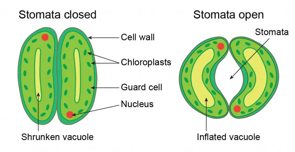
[In this figure] The illustration of vacuole controlling the opening and closing of stomata.
Stomata are on the underside of the leaf. The gas exchange occurs when stomata are open. Stomata closed due to shrunken vacuole, and stomata open due to inflated vacuole.
How to see the vacuole under a microscope
Although the vacuole does not take as much dye as other organelles of the cell (the vacuole does not contain many stainable constituents), you can still see and study the structure of vacuoles under a compound microscope. The trick is to use dyes that can stain the cell sap inside the vacuole.
The material you need
- Dyes: Neutral red is the most common dye for vacuole staining. Alternatively, we can pick dyes that are pH-sensitive. The pH level of vacuole depends on different types of plants and conditions.
You can purchase Neutral red at Amazon. - Plant specimens (roots, leaves, onion skin, etc.)
- Microscopic slides and coverslips
- Petri dishes and droppers
- Tweezers, blades, or microtomes
- Compound microscope
Steps
- Obtain a thin specimen of the plant tissues. You can try to peel a thin layer of skin cells using tweezers from leaves or onion skin. If you have a handhold microtome, try to section a piece of the plant’s stem.
- Stain the plant specimens with a Neutral red solution. You may need to try different dye concentrations and staining time.
- Rinse the specimens with water.
- Prepare a mounted slide for observation.
What will you see
With the right staining condition, the Neutral red will only stain the vacuole of live plant cells without staining any other organelle. The vacuoles appear deep red in color.
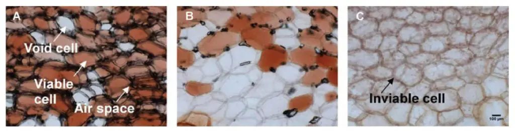
[In this figure] An example of using Neutral red to stain fresh onion cells.
(A) Neutral red stains vacuoles only in viable cells. (B,C) When cells are damaged by high pressure, cell integrity loses, and vacuoles leak. You won’t see Neutral red staining in inviable cells.
Photo credit: Gonzalez ME et. al.
In addition, the availability of fluorescent probes allows studying the morphology and physiology of the vacuole. Let’s look at some fluorescence images below.
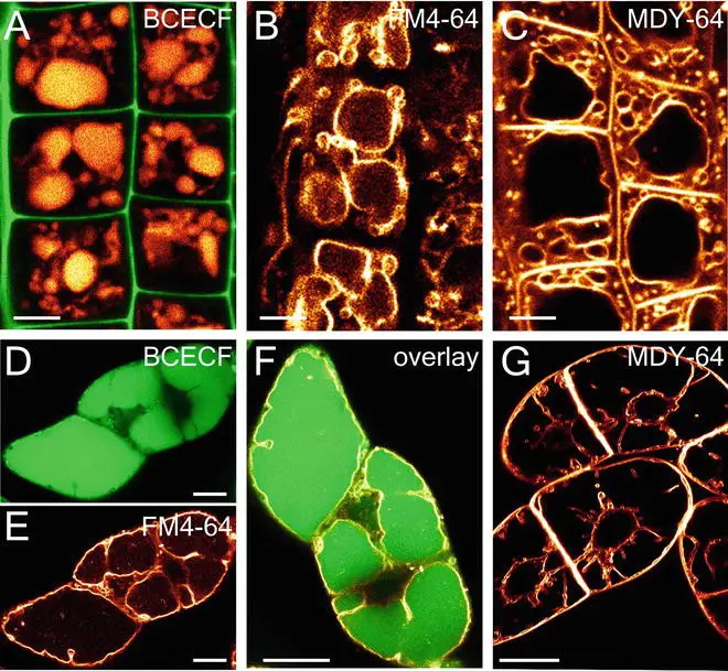
[In this figure] Fluorescence microscope images of vacuoles.
BCECF is a chemical that labels the acidic lumen of the vacuole. FM4-64 and MDY-64 can label lipid molecules of the tonoplast membrane. If both BCECF and FM4-64/MDY-64 are used for staining, the lumen and membrane of vacuoles can be visualized.
Photo credit: Scheuring D. et. al.
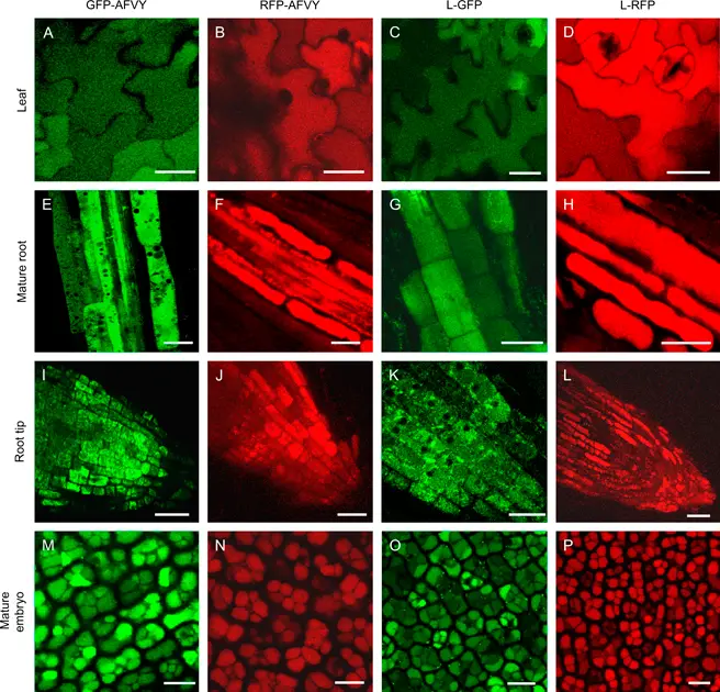
[in this figure] Fluorescent proteins are very useful to study vacuoles under a fluorescent microscope.
Generically engineering vacuole proteins fused with green (GFP) or red fluorescent proteins (RFP) allows scientists to study the size, number, and function of vacuoles in different tissues of plants.
Photo credit: Frigerio L. et. al.
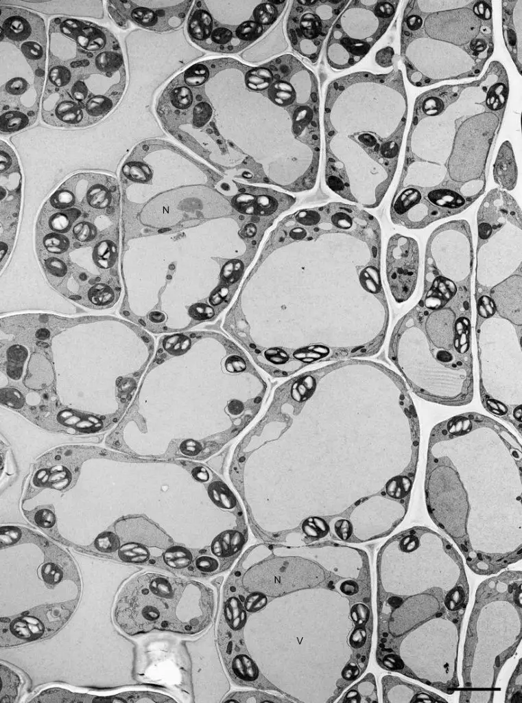
[in this figure] The transmission electron microscopic image of vacuoles.
N is the nucleus and V is the vacuole.
Photo credit: Noguchi T. et. al.
Summary
- Vacuoles are multi-functional organelles, which provide storage, defense, nutrient storage (protein or lipid), compartment (separate toxic waste from the rest cells), and control of the opening and closure of stomata.
- A mature plant contains one large central vacuole that takes up 30-80% of the cell volume.
Reference
- “Plant vacuoles” Current Biology
- “Vacuoles and Storage Organelles” Atlas of Plant Cell Structure, pp 89-106
- Microscopic Quantification of Cell Integrity in Raw and Processed Onion Parenchyma Cells
- Neutral red staining for plant vacuoles
Related posts
Cell Biology on the Dining Table – Plant Cell Model