
This article covers
What is chloroplast?
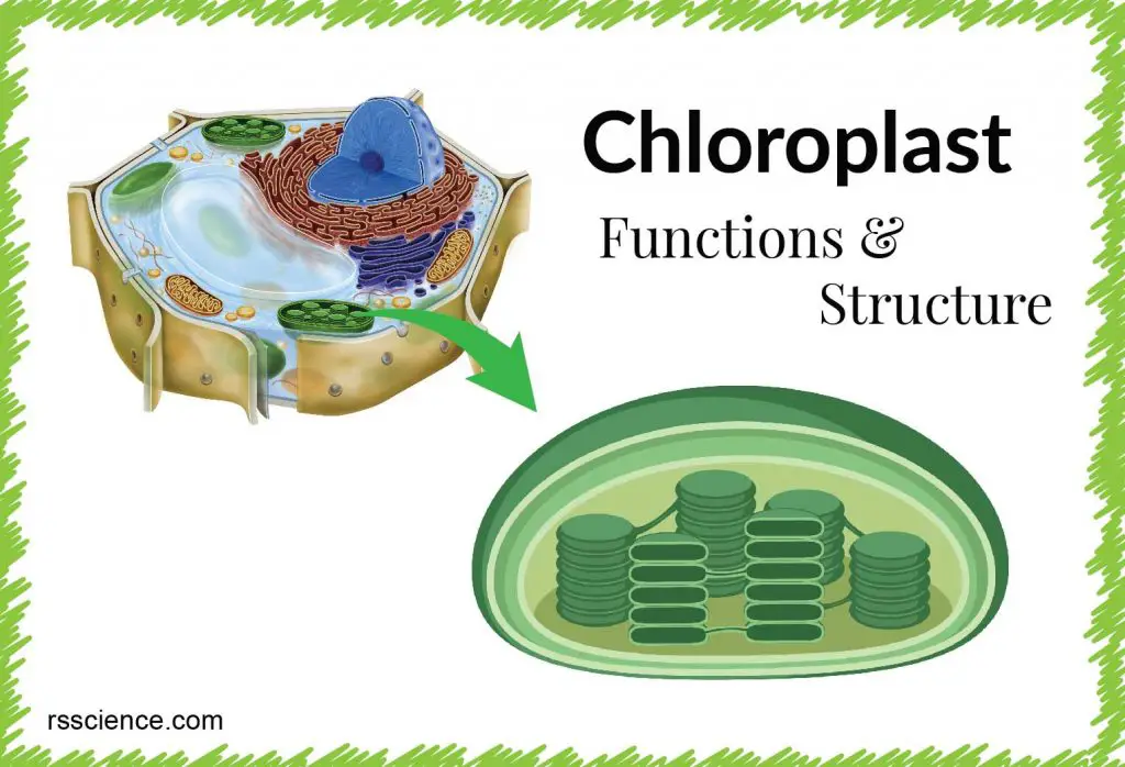
The chloroplast is a double membrane organelle found in plants and certain algae cells. The main function of the chloroplast is to convert energy from the Sun into chemical energy (glucose) for growth, a process called photosynthesis. This is why we call chloroplasts are like solar panels inside the cells.

[In this figure] The chloroplast and its relative location inside the plant cells.
The structure of chloroplasts
Inside the chloroplast space enclosed by inner membranes, there is a protein-rich fluid called the stroma, which corresponds to the cytosol of the original cyanobacterium. Chloroplast DNA, ribosomes, starch granules, and many proteins can be found floating in the stroma.
Suspended within the chloroplast stroma is the thylakoid system. Thylakoids are membranous sacks containing chlorophyll molecules and are where the light reactions of photosynthesis happen. In most vascular plants, the thylakoids are arranged in stacks called grana (singular: granum). However, in certain C4 plants (like rice) and some algae, the thylakoids are free-floating.
The stacks of thylakoid sacs are connected by stroma lamellae. The lamellae act like the skeleton of the chloroplast, keeping all of the sacs at a safe distance from each other and maximizing the efficiency of the organelle. If all of the thylakoids were overlapping and bunched together, there would not be an efficient way to capture the Sun’s energy.
The size of chloroplasts is roughly 1–2 μm thick and 4–6 μm in diameter[1].
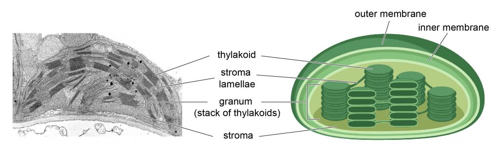
[In this figure] Transmission electron micrographic image of chloroplast and its structure.
[Left] The thylakoid (dark region) is the area where photosynthesis happened. They are connected by stroma lamellae. Some dark spots around the stroma are lipid.
Photo source: modified from University of Wisconsin-Madison Libraries
Where is the chloroplast located in a cell
The chloroplast is located throughout the cytoplasm of the cells. They can often be found in the plant leave cells, guard cells (specialized cells to control gas exchange), and cells of the green tissue of the plants. In fact, the reason we see plant green is due to the chloroplasts.
Chloroplast movement
Chloroplasts are motile: they move depending on the availability of sunlight.
Under the shade conditions, chloroplasts move to the area where they can absorb as much light as possible. When the light is strong such as the full sun, chloroplasts move from the cell surface to a side wall where they can minimize photodamage. In the dark, chloroplasts migrate to the bottom of cells, although the purpose of this movement in the dark is not clear[5].
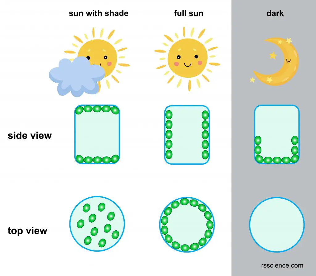
[In this figure] Chloroplast movement in different conditions.
The position of chloroplasts in the side view and top view are shown. In the shade condition, chloroplasts move to the position where it can absorb as much light as possible. In the full sun condition, the chloroplasts avoid the sun, minimize the photodamage. In the dark, the chloroplasts stay at the bottom of the cells.
Cytoplasmic streaming
Cytoplasmic streaming is a movement of the fluid cytoplasm within a cell. You can often see chloroplasts move along the cell walls.
Find out more about cytoplasmic streaming, please check “what is cytoplasmic streaming“.
[In this video] Looking at chloroplasts moving by cytoplasmic streaming in plant cells (Elodea) – DIC microscope/ 1250x.
How many chloroplasts can be found in one cell?
The number of chloroplasts per cell ranges from 1 to 100 per cell, depending on organisms. Chlamydomonas reinhardtii is a single-cell green alga that contains a single large cup-shaped chloroplast.
Chlamydomonas is an important model organism because we can manipulate its genetics and culture them easily. Interestingly, we can modify not only the nuclear genome of chlamydomonas, but also its chloroplast genome. This provides a great tool to understand the role of nuclear genes in chloroplast gene expression and chloroplast biogenesis.
Other plants, such as Arabidopsis thaliana, contains ~ 100 chloroplasts per epidermal cell. Coffea arabica has 13-20 chloroplasts per guard cell, so it really depends on the species[2-4].
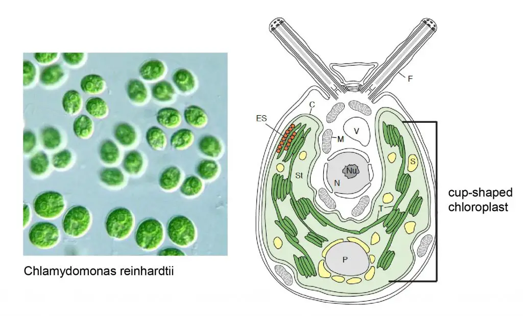
[In this figure] Chlamydomonas reinhardtii and its cup-shaped chloroplast.
Left is the image of Chlamydomonas, and right is the illustration of organelles in Chlamydomonas. Thylakoid membranes (T) and starch grains (S) within the stroma (St).
Photo source: modified from Alchetron and Trends in Plant Science.
What is the biological function of chloroplast?
Chloroplasts convert the light energy of the Sun into sugars (a process called photosynthesis) that can be used by cells. At the same time, photosynthesis produces oxygen (O2) and consumes carbon dioxide (CO2). For this reason, plants are the basis of all life on Earth. They are classified as the producers of the world.
Chloroplasts also provide diverse functions for plant cells, including fatty acids synthesis, amino acid synthesis, and plant innate immunity.
Photosynthesis
Chlorophyll is a little green molecule sitting on the surface of each thylakoid sacs and is the core of photosynthetic reaction. When the energy from the Sun hits a chloroplast and its chlorophyll molecules, light energy is converted into the chemical energy found in compounds such as ATP and NADPH. This part is called light-dependent reactions. It happens in a series of electron transportation between protons across the membrane, which is similar to what we previously described in mitochondria.
Later, these energy-rich compounds move into the stroma where enzymes fix the carbon atoms from carbon dioxide (CO2) to synthesize sugar molecules. This part is called the Calvin cycle or dark reactions. Oxygen (O2) is a byproduct of this chemical reaction. Plants and animals then use the sugars (glucose) for food and energy (plant cells have mitochondria, too).
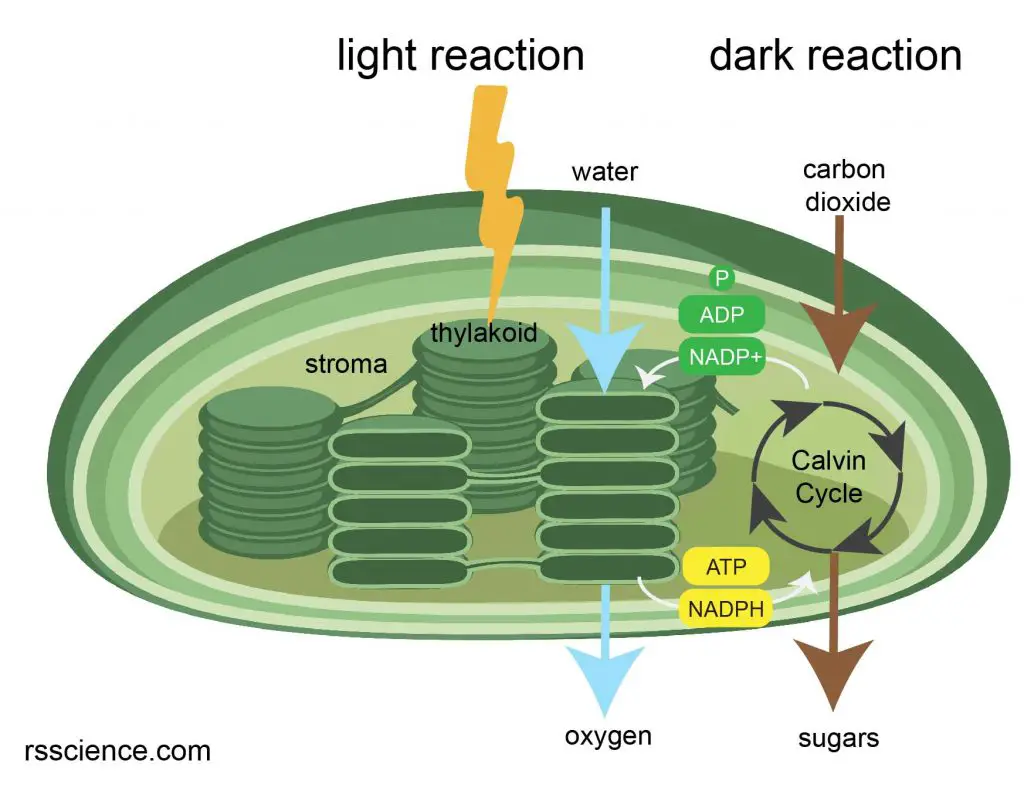
[In this figure] Illustration of photosynthesis.
Chlorophylls in the thylakoid absorb the energy from the Sun and transfer the energy to ATP and NADPH. In the dark reaction, the enzymes and proteins in the stroma use high energy molecules such as ATP and NADPH to convert carbon dioxide to sugars.
Innate immunity
Unlike animals, plants do not have immune cells (white blood cells). Each cell has to defend itself. Plants have two immune response, hypersensitive response and systemic acquire resistance.
Hypersensitive response
When pathogens are present in a cell, the cell seals itself off and undergoes apoptosis (a programmed cell death). This can prevent the pathogen from spreading to other cells.
Systemic acquire resistance
The infected cells can also release signals to inform the rest of the plant cells that the pathogens are present. Therefore, the rest of the plant cells can prepare for the defense.
The chloroplast is a key player for two immune responses. Chloroplast can purposely damage their photosynthetic system to produce reactive oxygen species (ROS), which can directly kill pathogens (bacteria) within the cells. Pretty much like the mechanism by which macrophages use ROS to kill bacteria in humans. The residual (low level) of ROS serves as an “SOS” signal and trigger defense molecules production in the rest of the plant cells.
How does the chloroplast divide?
In single-cell algae, they often have one chloroplast; therefore, the chloroplast replicates and divides before the cytokinesis completion.
Land plant cells generally have many chloroplasts. Their chloroplasts divide according to the cell’s need, not always couple with cell division. Different environmental stimuli affect the chloroplast division rate. For example, the chloroplasts divide and increase their number when the cell grows bigger[6].
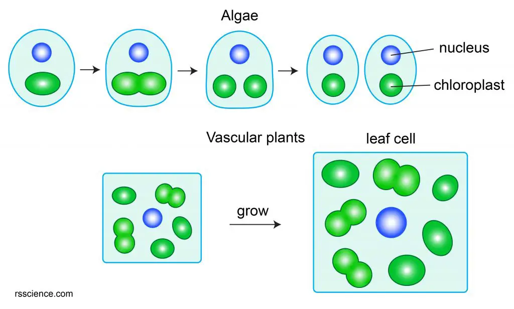
[In this figure] Chloroplast division in algae and vascular.
Single-cell algae often have one or a few chloroplasts. Chloroplast division occurs every cell cycle right before the separation of two daughter cells (cytokinesis). In vascular plants, during the leaf development, the cell division stops, but the nuclear DNA replication still continues, resulting in the bigger nuclei and cells. During this time, the chloroplast division continues without cell division, resulting in an increase of the chloroplasts in size and number.
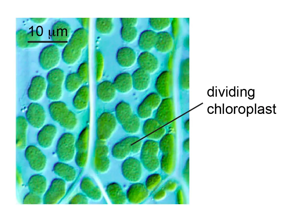
[In this figure] Dividing chloroplasts in the moss P. patens.
Photo source: Plant physiology
Chloroplast division process
The division process starts when the division proteins assemble into a ring structure (Z-ring) within the chloroplast stroma. Next, the two plastid-dividing rings (PD rings) form along the z-ring. Then the chloroplast constriction begins.
Next, dynamin proteins (motor protein) assemble around the PD ring, providing force to squeeze the chloroplast. As the constriction site becomes narrower and narrower, two chloroplasts separate[7].
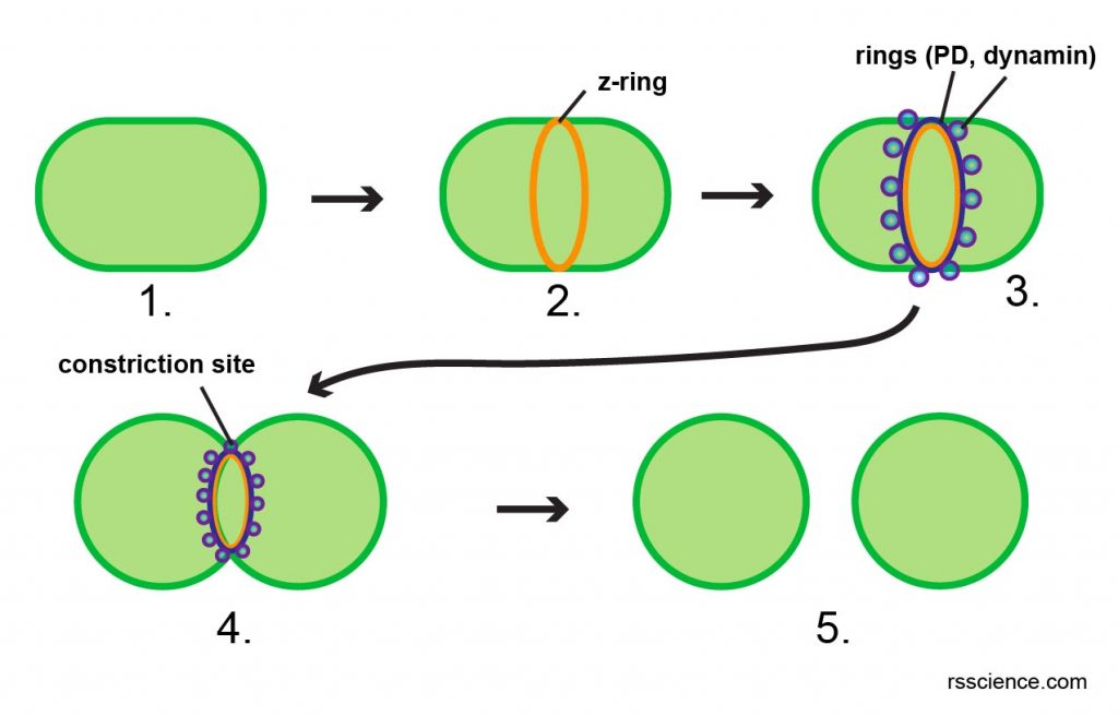
[In this figure] Cartoon diagram showing the chloroplast division steps.
Division machinery such as protein complex assembles into a ring structure (2 and 3). These ring structures apply pressure to narrow the constriction site (4). Then, the chloroplast completes the division, and two daughter cells form (5).
The origin of chloroplasts – the endosymbiotic theory
Chloroplasts and mitochondria share many in common. Both organelles have two layers of membranes – called outer and inner membranes. Chloroplasts and mitochondria also have their own copies of DNA, which are independent of the cell nuclei. There are specialized ribosomes inside chloroplasts and mitochondria to make proteins only for these organelles. Scientists believe chloroplasts and mitochondria are derived from the bacteria that were engulfed by the early ancestors of today’s eukaryotic cells. This theory is called the endosymbiotic theory.
Around 1.5 billion years ago, some prokaryotes incorporated other prokaryotes into their cells. These incorporated prokaryotes then lost their ability to live independently and become integrated as part of the hosts. They later became specialized in specific functions, such as energy production in both mitochondria and chloroplasts.
Mitochondria appear to be related to Rickettsiales proteobacteria, and chloroplasts appear to be related to nitrogen-fixing filamentous cyanobacteria. This endosymbiotic theory can explain why chloroplasts and mitochondria have two layers of membrane, their own DNA and ribosomes.
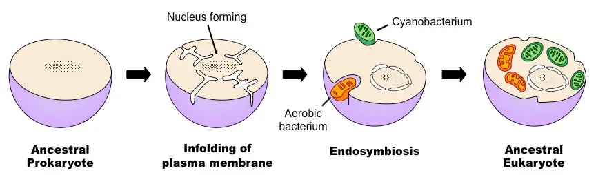
[in this figure] Overview of the process of endosymbiosis.
Photo credit: BioNinjia
Can animals live like plants?
Elysia chlorotica (common name the eastern emerald elysia) is one of the “solar-powered sea slugs”, utilizing solar energy to generate energy. The sea slug eats and steals chloroplasts from the alga Vaucheria litorea. The sea slugs then incorporate the chloroplasts into their own digestive cells, where the chloroplasts continue to photosynthesize for up to nine months – that’s even longer than they would perform in algae.
The sea slugs stay nourished thanks to the sugars produced by photosynthesis. In the past, how this animal managed to do this was a complete mystery.
In a new study appearing in the Biological Bulletin[9], researchers reveal that the sea slug has incorporated genes from the algae that it eats. These genes, which encode for both chloroplast proteins and chlorophyll synthesis, are transferred from algae to the sea slug’s chromosomes so that the stolen chloroplasts can be well maintained for a long time.
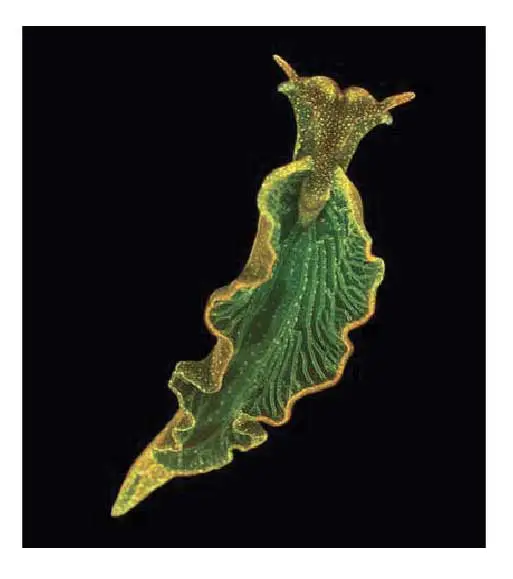
[In this figure] Elysia cholorotica, a sea slug found off the U.S. East Coast, can steal photosynthetic chloroplasts from algae.
Photo source: Mary S. Tyler/PNAS
How to see the chloroplast under a microscope
The material you need
- Blank microscope slides and coverslips
- Forceps
- Elodea or leaves (the thinner, the better)
Steps
- Clean a slide with a piece of Kimwips paper.
- Add a drop of water on the slide.
- Cut a small piece of Elodea leaves and put on the slide.
- Place a coverslip on top. Lower the coverslip slowly with an angle. Allow one side of the liquid droplet touches the coverslip first. This permits air to escape from the other side. You can use the forceps to help you control the coverslip.
- The slides are ready for viewing.
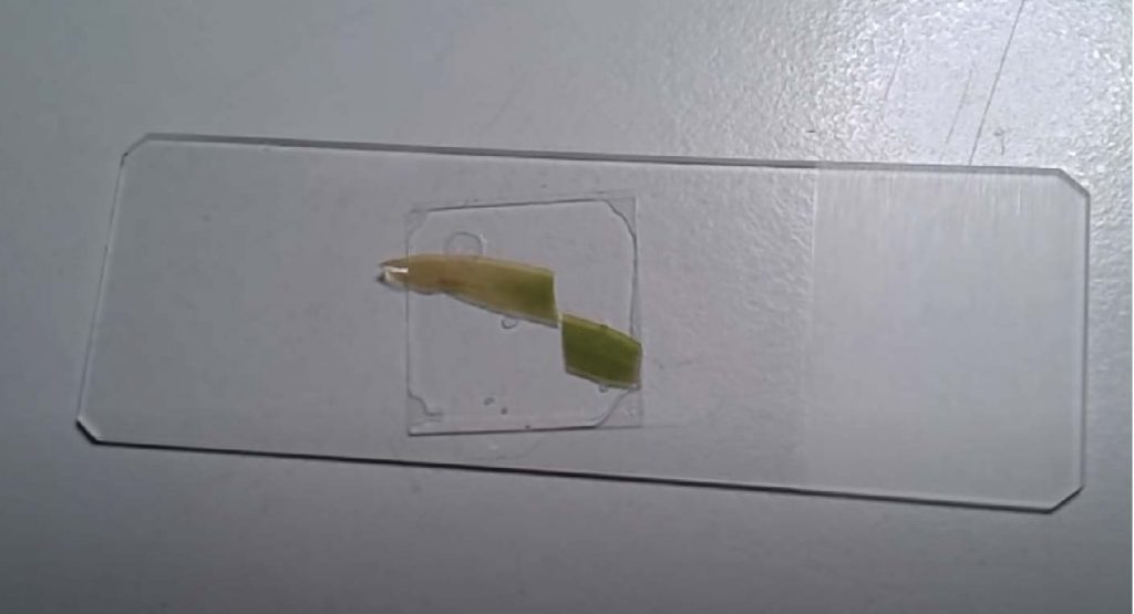
[In this figure] Place a piece of leaf or elodea on the slide.
Cover with a coverslip. The slide is ready for viewing.
What will you see
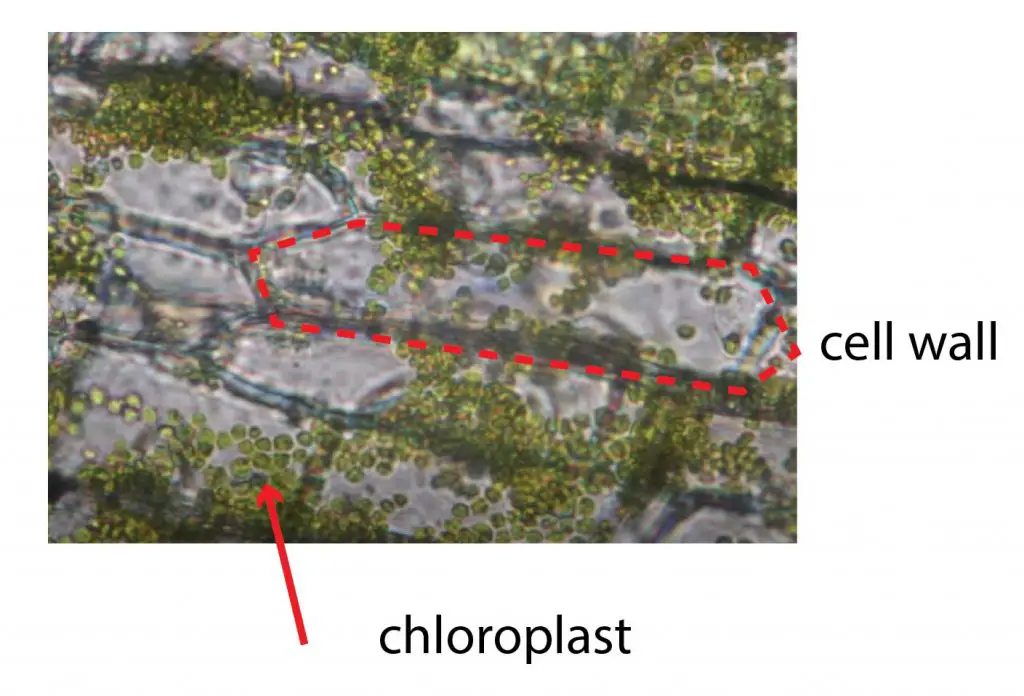
[In this figure] The chloroplasts under a light microscope.
You can see individual cell walls and many green chloroplasts. These chloroplasts move!! You may need to use higher magnification to see these movements.
Summary
- Chloroplasts are organelles that conduct photosynthesis and produce energy for the plant cells.
- Chloroplasts consist of many stacks of sac structures, called thylakoid system. Chlorophyll in thylakoid absorbs the energy of the Sun and through a series of reactions to produce sugar. At the same time, the reaction produces oxygen (O2) and consumes carbon dioxide (CO2).
- Chloroplast plays an important role in plant innate immunity.
- Chloroplasts are motile; they move according to the light condition.
- Chloroplasts and mitochondria share many in common. They both have two layers of membranes, their own DNA and ribosomes. They are believed to be derived from endosymbiotic bacteria engulfed by the early ancestors of today’s eukaryotic cells.
Reference
- http://book.bionumbers.org/how-large-are-chloroplasts/
- https://bionumbers.hms.harvard.edu/search.aspx?trm=chloroplast
- Königer M, Delamaide JA, Marlow ED, Harris GC. Arabidopsis thaliana leaves with altered chloroplast numbers and chloroplast movement exhibit impaired adjustments to both low and high light. J Exp Bot. 2008 59(9):2285-97. p.2288
- Etienne H, Bertrand B. Trueness-to-type and agronomic characteristics of Coffea arabica trees micropropagated by the embryogenic cell suspension technique. Tree Physiol. 2001 Sep21(14):1031-8. p.1034 table 2
- Wada M, Kong SG. Actin-mediated movement of chloroplasts. Journal of Cell Science 2018 131: jcs210310
- Miyagishima SY. Mechanism of Plastid Division: From a Bacterium to an Organelle. Plant Physiol. 2011 Apr; 155(4): 1533–1544.
- wiki-chloroplast
- Bozeman Science
- Rumpho ME et al. Horizontal gene transfer of the algal nuclear gene psbO to the photosynthetic sea slug Elysia chlorotica. Proc Natl Acad Sci USA. 2008 Nov 18;105(46):17867-71
Pingback: Which plant cell organelle contains its own DNA and ribosomes? – Dr. Biology Questions and Answers
Pingback: Which organelle produces sugars while releasing oxygen, and in which cell could it be found? – Dr. Biology Questions and Answers