
We have a series of posts about microscope’s function, history, and types.
I. Background and Introduction
- What is a Microscope? – Function and Magnification
- Who Invented the Microscope? – History of Microscopy
- Are Microscopes the Same? – A Introduction of Current Microscope Family
II. Components of a Microscope
III. Specialized Microscopes
IV. Practical Guides to Work with Your Microscopes
This article covers
The early history of microscopes – from simple glass magnifiers to compound microscopes
A convex lens that can bend and focus light
The key for a microscope to magnify the image is the convex lens.
The oldest objects resembling lenses can date back 3,000 years. This “Nimrud lens” is a crystal disk with a convex shape that belonged to an ancient Near East empire, Assyria. It has been used as a magnifying glass or as a burning-glass to start fires by concentrating sunlight. There were also records showing that ancient Greek know the optical properties of water-filled spheres.
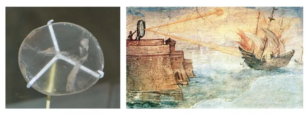
[In this figure] Left: The “Nimrud lens” of Assyrians is a 3000-year-old piece of crystal unearthed in modern-day Iraq. It is displayed in the British Museum, London. Right: A wall painting from the Uffizi Gallery in Florence, Italy, shows the Greek mathematician Archimedes’ mirror burning Roman military ships. Painted in 1600 by Giulio Parigi. Could the mirror really get it hot enough to burst into flame is unknown. However, it proved that ancient Greek may already learn the power of a focused light beam.
First simple microscopes (or magnifying glasses)
The earliest known use of simple microscopes or magnifying glasses (consisted of only one convex lens) dates back to the 13th century. At that time, the use of lenses in eyeglasses became popular in Italy and may lead to the widespread use of simple microscopes. However, these single lens magnifying glasses had only minimal magnification.
Van Leeuwenhoek’s microscopes
Antonie van Leeuwenhoek (1632-1723) was a Dutch businessman and scientist. He is commonly known as “the Father of Microbiology” and one of the first microscopists and microbiologists.
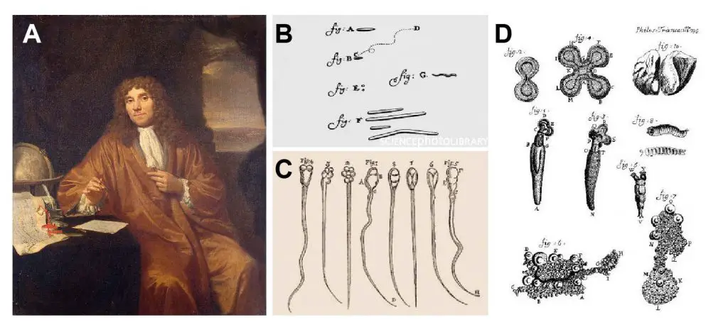
[In this figure] (A) Antony van Leeuwenhoek painted by Jan Verkolje. Van Leeuwenhoek is best known for his pioneering work in microscopy and his contributions toward establishing microbiology as a scientific discipline. (B) Drawings made by Leeuwenhoek of what could be the first bacteria observed. (C) Sperms from rabbit and dog drawn by Leeuwenhoek. (D) Rotifers observed by Leeuwenhoek. He named these tiny creatures “wheel animalcules.”
Antonie van Leeuwenhoek made more than 500 optical lenses. He also created at least 25 single-lens microscopes of different types. Leeuwenhoek modified his microscopes to be capable of magnifying up to 275 times. Using his microscopes, Leeuwenhoek reported the first discovery of protists (he called infusoria) in 1674 and bacteria (he described as “little animals” or animalcules) in 1783. He also observed the vacuole inside the cells, mobility of sperms, and the banded pattern on muscular fibers.
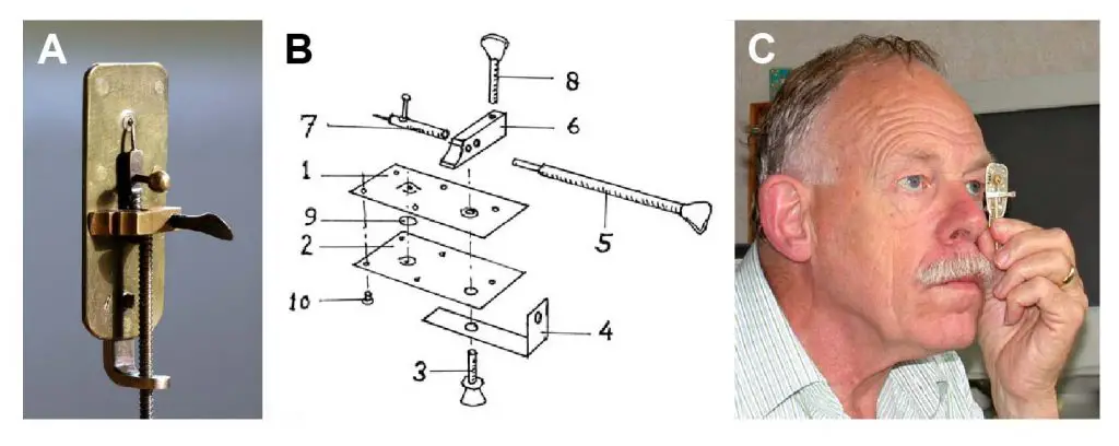
[In this figure] (A) A replica of Van Leeuwenhoek’s microscope. It is only 5-cm long. The lens is in a small hole. The other side of the microscope had a pin, where the sample was attached to stay close to the lens. (B) Exploded design of a van Leeuwenhoek’s microscope. If you want to learn how to make one, check out this great article. (C) The way of using a Van Leeuwenhoek’s microscope. You have to place the lens very close in front of the eye while looking in the direction of the sun. (Don’t look at the sun, it will hurt your eyes.)
[In this video] A cool video showing how to make a paper Leeuwenhoek microscope.
First compound microscopes
The compound microscope’s actual inventor is unknown, although many claims have been made over the years. Among these claims, the Janssen brothers were believed to develop the first compound microscope in the early 1600s.
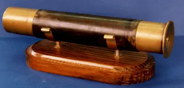
[In this figure] Janssen’s compound microscope consisted of two convex lenses aligned in series: an object-glass (objective) closer to the object or specimen; and an eyepiece (ocular) closer to the observer’s eye.
Galileo Galilei (Italian astronomer) is also sometimes cited as a compound microscope inventor. He found that he could magnify small objects by viewing through the wrong end of his telescope. After seeing the compound microscope built by others, Galileo built his own improved version.
The name of “microscope”
In 1625, Giovanni Faber coined the name “microscope.” This name came from the Greek words “micro,” meaning “small,” and “scope,” meaning “to look at.”
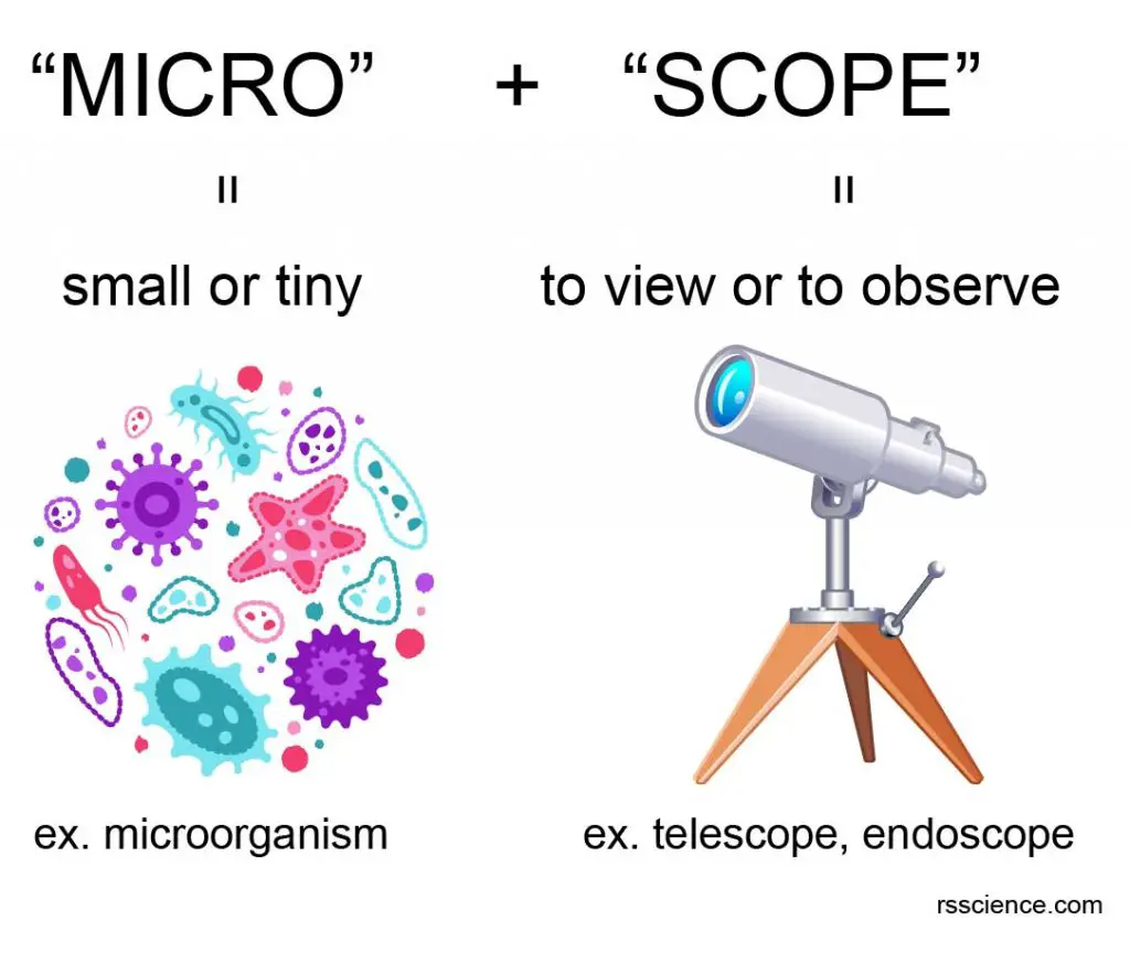
[In this figure] The name “microscope” came from two words – “micro” and “scope”.
Robert Hooke and cell theory
The power of “compound microscopes” allowed Robert Hooke (1635-1703) to establish the cell theory, which states that all organisms are made of cells, all life functions occur in cells, and all cells come from other cells.
While observing cork through his microscope, Hooke saw tiny empty cavities, which he illustrated and described as cells. Hooke’s discovery led to the understanding of cells as the smallest units of life—the foundation of cell theory.
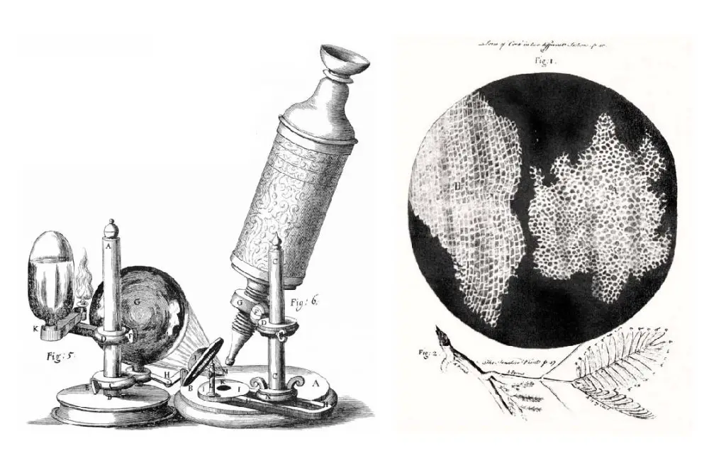
[In this figure] Left: The compound microscope used by Robert Hooke to discover “cells.” Right: Cell structure of cork illuminated by Robert Hooke in Micrographia, 1665.
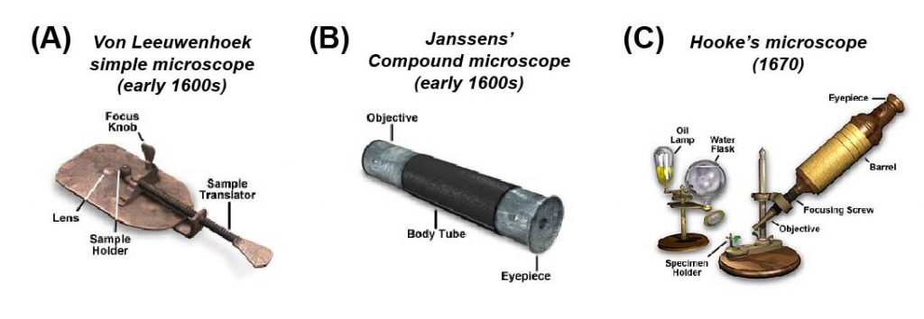
[In this figure] A collection of “antique” microscope dated to the early development of microscopy. (A) Anton von Leeuwenhoek’s microscope was a simple glass magnifier with only one convex lens. He discovered many microorganisms, such as Paramecium, using this simple microscope. (B) Janssen brothers developed the first compound microscope with two aligned convex lenses. It looked very similar to a tubular telescope. (C) Robert Hooke observed “cells” using his modified compound microscope. He used an oil lamp as a light source and a water flask to focus the light beam.
Photo credit: Olympus.
Compound microscopes became a popular and essential scientific instrument
The 18th and 19th centuries witnessed a great improvement in the mechanical and optical quality of compound microscopes. Advances in machine tools allowed more sophisticated optical parts to be fabricated. Newly-invented lenses corrected the optical aberration (both spherical and chromatic) that hampered the quality of microscopic images. Many British and German microscope manufacturers flourished during this time period.
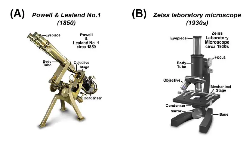
[In this figure] (A) In the 18th and 19th centuries, advances in machine tools allowed more sophisticated microscope parts to be fabricated. Many British and German manufacturers started selling commercial microscopes. The microscope illustrated was manufactured by Hugh Powell and Peter Lealand in London about 1850. (B) The classical design of the Zeiss Laboratory microscope was produced in the early 20th century. This type of microscope is very functional, and many are still in use today.
Photo credit: Olympus.
Light microscopes become a very powerful tool
Today, the family of microscopes was significantly expanded. Microscopes had become an essential instrument in modem technology, research, and industry. In the family tree of an optical (or light) microscope, the principle is still the same: “generating the magnified images through the interaction of visible lights and the subjects.” However, scientists had made tremendous progress to see more specific (such as fluorescence light), deeper (such as the 3D image of cells and tissues), faster (such as automated system), and even smaller (such as beyond the diffraction limit of light). Click the type of microscope to know more.
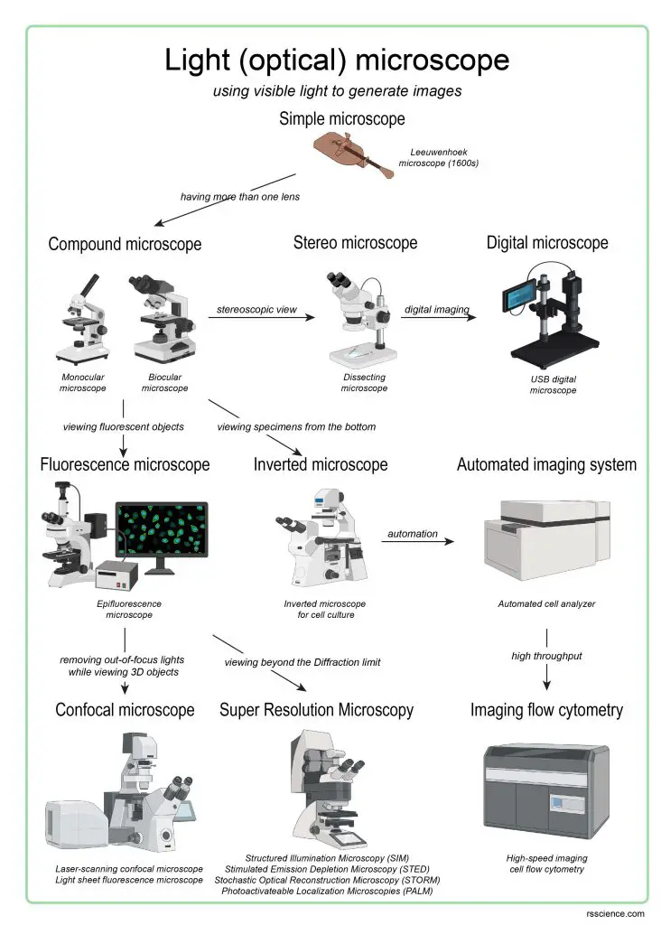
[In this figure] The overview of current light microscopies.
The family tree shows the evolution of light (optical) microscopes.
Electron microscopes change the game
In order to see smaller things, the scientists have to find an illumination with a wavelength much smaller than the visible light. In the early 1930s, they realized that electrons could do so and thus developed the transmission electron microscope (TEM).
According to modern physics, an electron has both properties as a particle and a wave. As a result, an electron has a wavelength (called De Broglie wavelength) depending on its energy (or speed). For example, if an electron is speeded up in a vacuum tube to 1,000,000 meters per second (equal to 2.2 million miles per hour!), the De Broglie wavelength we get is around one-tenth of a nanometer, which is about the size of an atom.
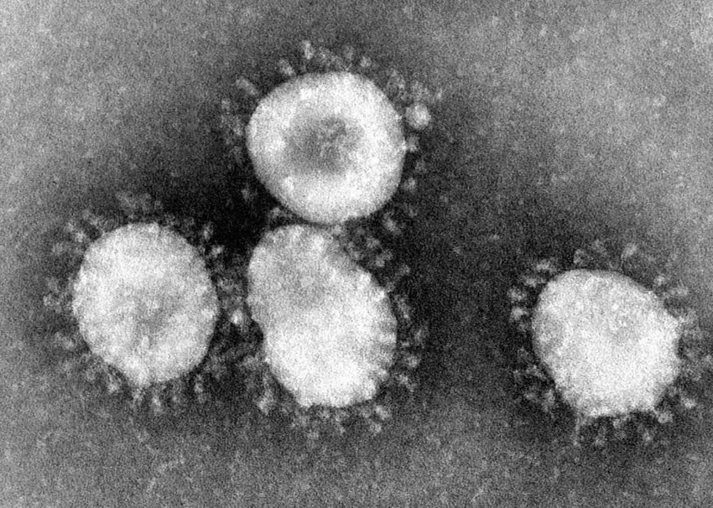
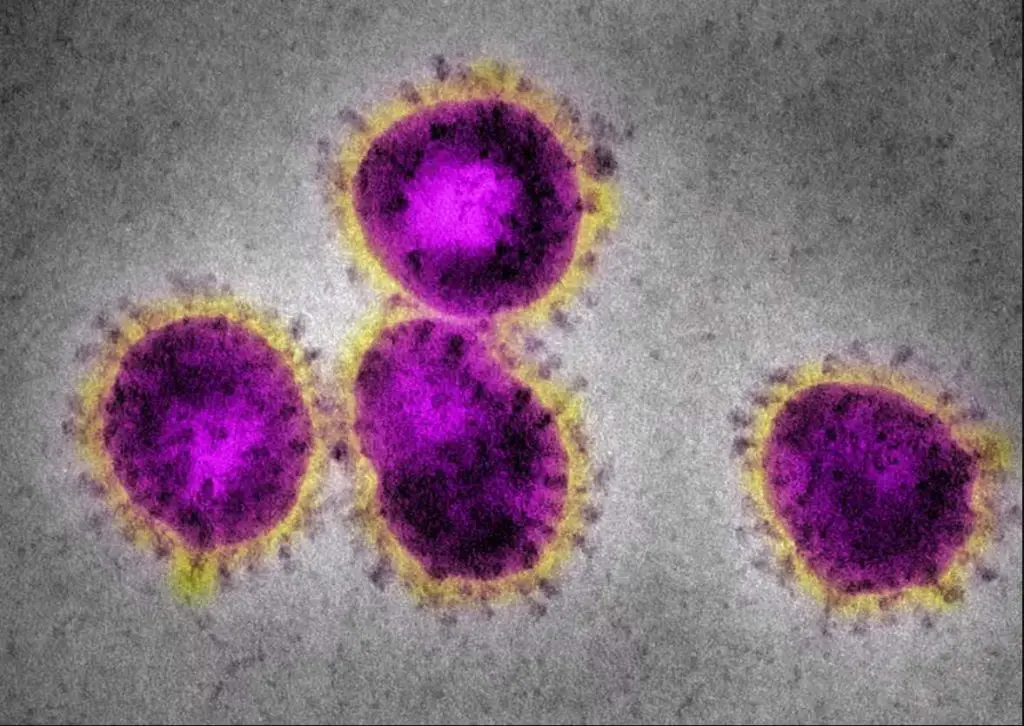
[In this figure] Coronaviruses have a halo or crown-like (corona) appearance.
The new virus, COVID-19, is one kind of coronaviruses. Original electron microscopic images are always black-and-white. The colors are artificially painted for better visualization (right). Photo credit: CDC.
The advance of microscopy never stops
Scientists never give up inventing new types of microscopes and push our limit to see invisible things. For more detail, you can read “the advanced microscopies.”
3D printing techniques also help the development of smaller and cheaper microscopes. You may DIY microscopes (open source) at home and have similar power to professional microscopes in a research laboratory.
Summary
In this article, we learned:
- A simple microscope is the earliest type of microscope. It has only one lens and functions as a magnifying glass.
- Anton von Leeuwenhoek (1632-1723) discovered many microorganisms like protists and bacteria with his simple microscope. He is considered the pioneer of microbiology.
- Compound microscopes use two convex lenses to obtain higher magnification. The first compound microscope was invented in the early 1600s.
- Robert Hooke (1635-1703) observed “cells” using his modified compound microscope and established the cell theory that cells are the basic units of life.
- In the 20th century, microscopes had become the essential instrument and driving force of new technology. Many new microscopes, such as electron microscopy, continue to advance.
References
“Making an Antoni van Leeuwenhoek microscope replica” by Hans Loncke“