
We have a series of posts covering all the aspects of microscopes.
I. Background and Introduction
What is a Microscope? – Function and Magnification
Who Invented the Microscope? – History of Microscopy
Are Microscopes the Same? – Types of Microscopes
II. Components of a Microscope
III. Specialized Microscopes
IV. Practical Guides to Work with Your Microscopes
This article covers
Microscopes open up the door to the tiny wonderland!
Cells are building blocks for all living organisms, but they are too small to be seen with our naked eyes. With the aid of microscopes, we can amplify the power to see these tiny cells. There are also many tiny creatures that we can not see with our naked eyes, such as plankton and bacteria.
Below is a length scale of biological objects and the types of microscopes and approaches to study them.
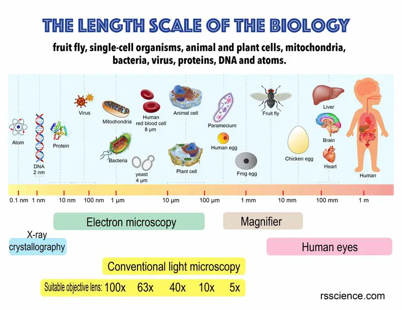
[In this figure] A biological scale from a single atom to the human body.
An overview of microscopes
What is a microscope? A microscope is an instrument used to see objects that are too small to be seen by the naked eye. Microscopes are commonly used in science laboratories and classrooms to visualize all kinds of tiny objects, such as cells, microorganisms, tissue structures, materials, and electronics. Microscopes provide magnification (enlarging the image) and give the images a contrast (making them stand out of the background). To do so, microscopes are made up of a few lenses for magnification, each lens with its own magnification powers and focusing strength.
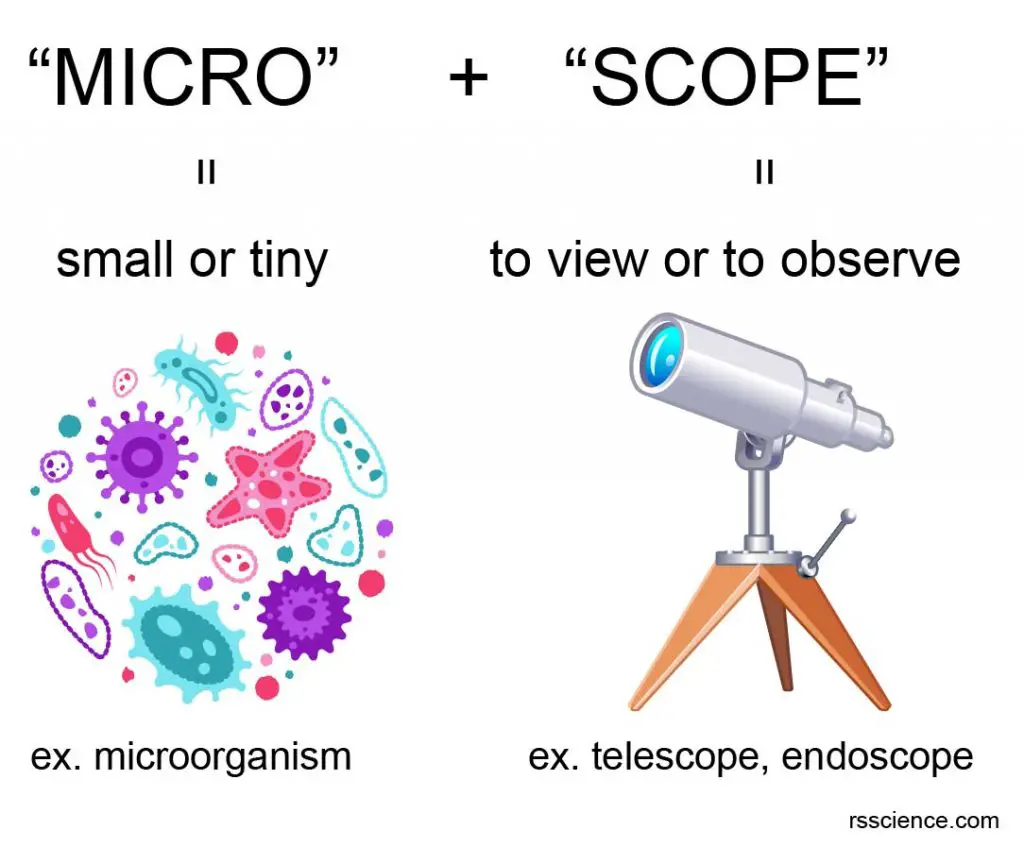
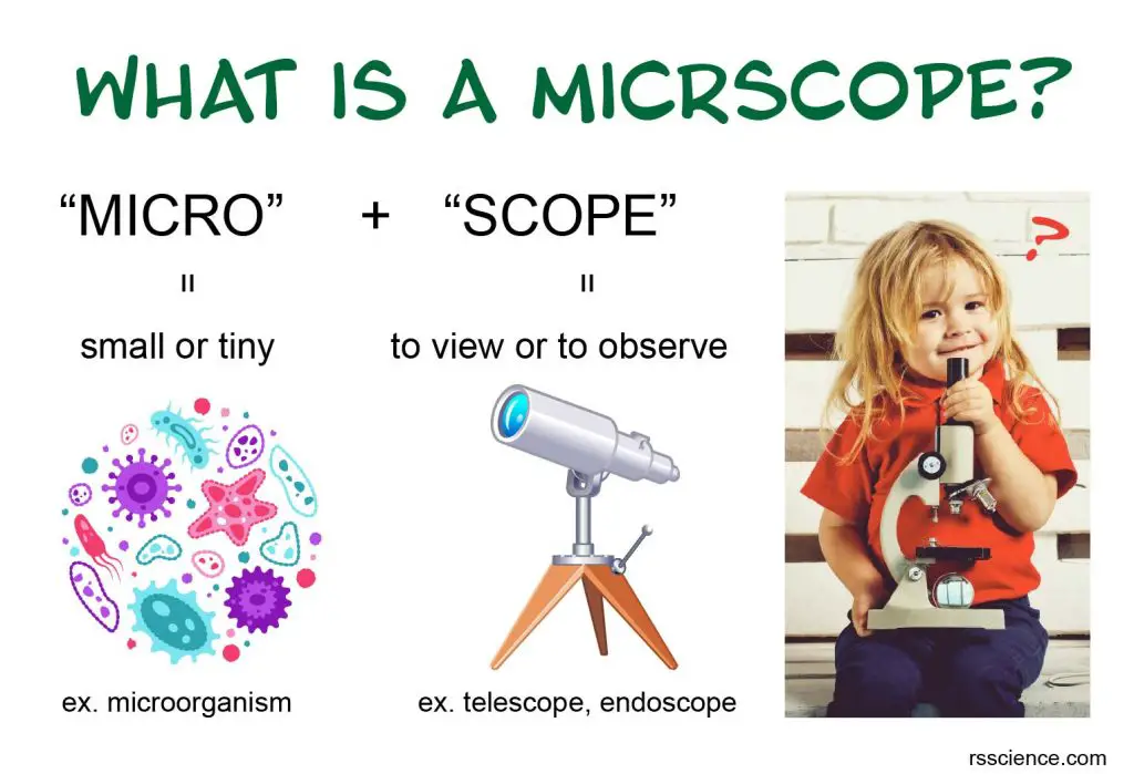
[In this figure] The name “microscope” came from two words – “micro” and “scope”.
“Micro” means small or tiny. “Scope” means to view or to observe. Therefore, a microscope can be understood as an instrument to see tiny things.
What microscopes can do?
Microscopes’ power of magnification can help us to see many things that are too small to observe with our naked eyes. The main application of microscopes is scientific research. In fact, microscopes can do more than that. Here are some examples:
Microbiology – Microscopes allow us to see things we could never see before. This is exactly what Antonie van Leeuwenhoek did in the late 1600s to make the first observations of bacteria and protozoa. The microscopy with special stain techniques, such as Gram stains, allows us to identify pathogens that cause diseases.
Cell Biology – Microscopy is the major tool to study how organelles and cytoskeleton function in the cells. Many researches provide insight into the mechanism of diseases.
Human Physiology and Medicine – Histological analysis of tissue organ sections and blood smear helps diagnose many diseases.
Reproductive medicine – Without the precise injection under the microscope, In vitro fertilization (IVF) is impossible.

[In this figure] Applications of microscopes: (A) Gram stains of bacteria; (B) Fluorescent image of cytoskeleton; (C) Histology of brain tissue; (D) Blood smear; (E) Microinjection for In vitro fertilization (IVF).
Forensic science – Microscope is frequently used to identify tiny criminal evidence like hairs.
Environmental monitoring – To monitor the aquatic ecosystem, several key planktons, such as algae, euglena, and rotifers, are important indicators that can be observed under a field microscope.
Agriculture – Microscope is useful for several aspects of agriculture like soil validation and pest control.
Art and jewelry appraisal – Portable microscopes are essential tools to evaluate the value of artworks.
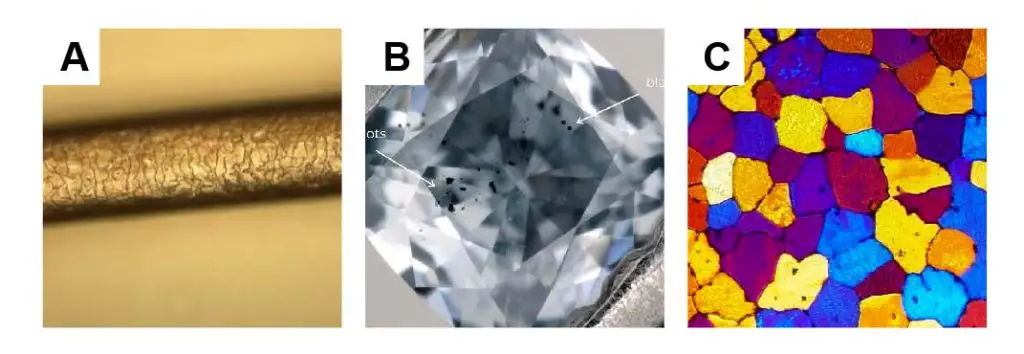
[In this figure] Applications of microscopes: (A) Human hair; (B) Diamond inspection; (C) Metallography showing metal grain microstructures.
Metallurgy and Manufacture – Metallographic microscopes are used to identify defects in metal surfaces, to determine the quality of metal alloys, and to study rocks, ceramics, and minerals. Flight crashes due to metal fatigue relies on microscopy to find tiny evidence.
Semiconductor – Microelectronics and semiconductor manufacturing heavily rely on microscopes to ensure quality control.
Nanotechnology – The development of nanomaterials like carbon nanotubes and graphene can not advance without electron microscopes.
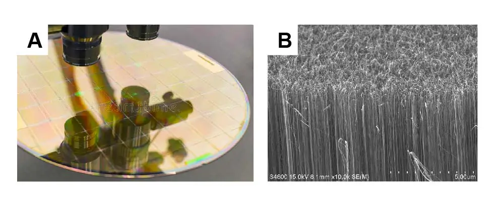
[In this figure] Applications of microscopes: (A) Inspection of a silicon wafer; (B) Forest of carbon nanotubes under a scanning electron microscope (SEM).
Thus, microscopes can be considered one of the most important modern technology instruments, which cannot be overstated.
Magnified observation and instruments
A microscope is an optical instrument used to view small objects by enlarging them with convex lenses. Depending on the design, a light microscope usually has a magnification ranging from 10x to 1000x. Higher magnification requires the usage of electron microscopes.
| Magnification | Instrument | Example |
| 1x | Naked eye | Hair (approx. 0.1 mm) |
| 2x – 5x | Magnifying glass | Plant and insect in detail |
| 10x – 20x | Stereo microscope | Insect’s compound eyes |
| 50x | Compound microscope | Daphnia, Rotifers, Water bears |
| 100x | Compound microscope | Paramecium, Amoeba |
| 200x | Compound microscope | Pollen, Euglena |
| 400x | Compound microscope | Cheek cells, Onion skin cells |
| 800x – 1,500x | Compound microscope | Red blood cells (8µm), bacteria (1µm) |
| 2,000x – 1,000,000x | Electron microscope | Objects smaller than 1μm, such as a virus (100 nm) and DNA (2 nm) |
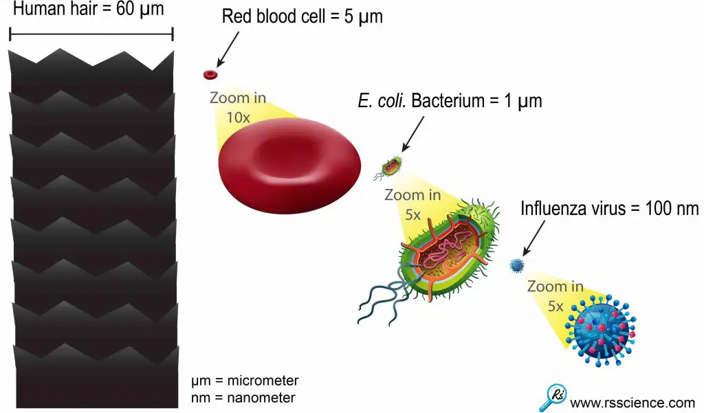
[In this figure] This relative size chart can give you an idea of how small the viruses are.
One influenza or “flu” virus particle is 600 times smaller than the diameter of human hair. 1 meter = 1,000 millimeters = 1,000,000 micrometers = 1,000,000,000 nanometer.
The structure of a microscope
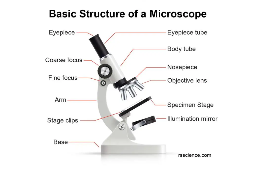
A general light microscope mainly consists of an objective lens, ocular lens, lens tube, stage, and light source. An object placed on the stage is magnified through the objective lens. When the target is focused, a magnified image can be observed through the ocular lens.
Check Compound Microscope Parts – labeled diagram and their functions for more details.
The principle of magnification using a microscope
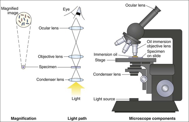
[In this figure] Principles of bright-field (light) microscopy. (Modified from Atlas RM: Principles of microbiology, St Louis, 2006, Mosby.)
Photo credit: Role of Microscopy
For a light microscope, visible light passes through the specimen and then a series of lenses. Lenses bend the light, which results in magnification.
Simple microscope
In order to better understand how the magnification is generated, let’s start with only one convex lens. This kind of instrument is called a simple microscope or magnifier.
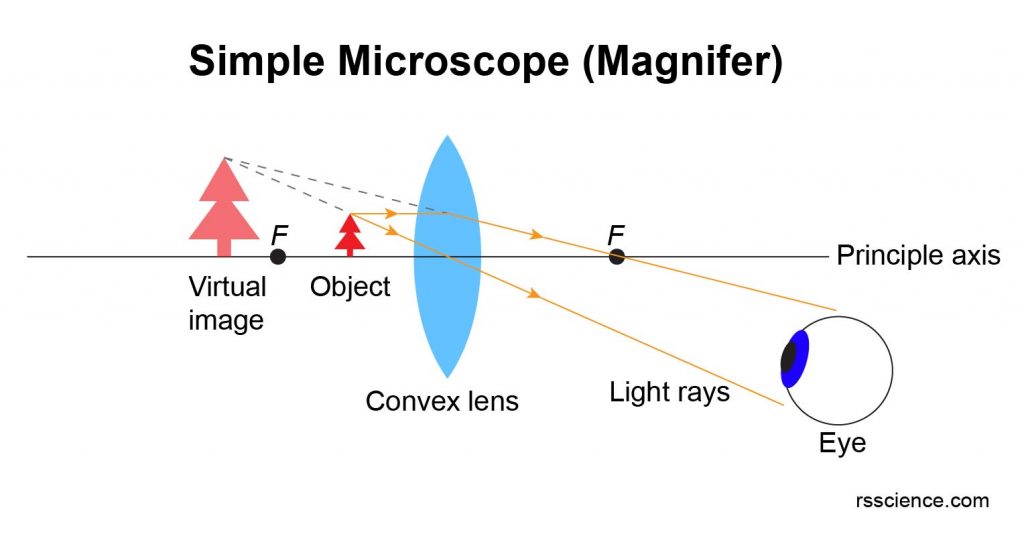
The image above shows how to draw a ray diagram for an object nearer than the lens F. “F” is the focal point of this convex lens.
The bottom of the object is placed on the principal axis. Two rays of light are drawn from the top of the object. The first ray of light is parallel to the principal axis. Any light ray parallel to the principal axis will be refracted, change direction, and cross the principal axis at the focal point.
The second ray of light goes from the top of the object and passes straight through the center of the lens. This ray does not refract.
As you can see, the rays are diverging (moving apart) on the right side of the lens. The eye looks back along with the rays that seem to have come from a point behind the object where the two rays of the light cross. This is where you draw the top of the virtual image. The bottom of the image is still on the principal axis.
The image made by a magnifying glass is virtual, upright, and bigger than the object. The image is called virtual because the light rays never really go there. The virtual light rays are drawn as dotted lines.
You can experiment with the DIY magnifying glasses with a recycled plastic bottle or water-filled balloon.
Compound microscope
Now we move forward to a compound microscope of two convex lenses (called Objective and Ocular lenses).
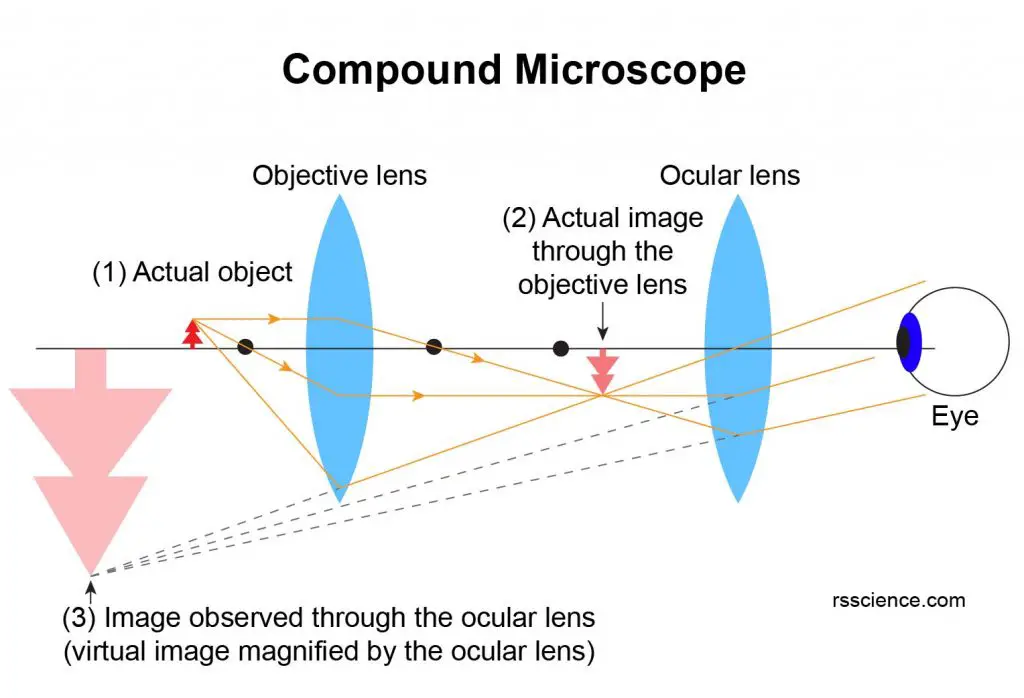
As you can see in the above image, the actual object (1) will be placed near but outside the focus point of the Objective lens. This generates an inverted and bigger image (2) between two lenses. This image is formed by real light rays and setting within the focus range of the Ocular lens. Now, if you observe this image behind the Ocular lens, the image will be magnified again. The final image (3) is virtual and inverted with the combined magnification contributed by both Objective and Ocular lenses.
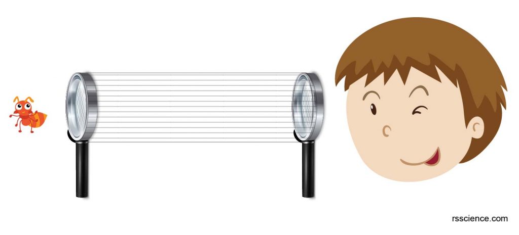
[In this figure] If you have two magnifying glasses, try to install them at each end of a paper roll to make your DIY microscope.

[In the figure] The low to high magnifications of vicia root tip and human blood cells.
The total magnification of a compound microscope is determined by the combination of Objective and Ocular Lenses. Assuming you have 10x eyepieces and 100x objective, the total magnification of this combination is 1,000x (10×100 = 1000).
Scientists modified the optical components and invented many special microscopes for different purposes, including dark-field, phase contrast, and differential interference contrast (DIC) microscopy. On the other hand, modern microscopes no longer consist of only two convex lenses. In fact, a better quality of imaging, more lenses, condensers, and filters are added to adjust the optical performance.
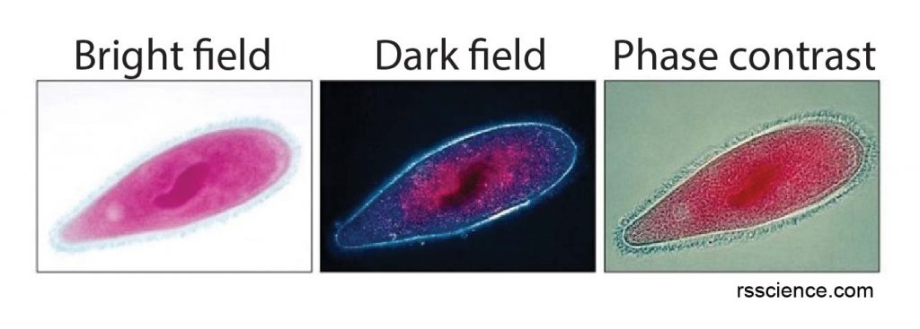
[In this figure] The same specimen (a stained paramecium) is observed with different settings: Bright-field, dark-field, and phase contrast.
Summary
Microscopes are amazing tools if you want to explore the microscopic wonderland. In this article, we learned:
- A microscope is an instrument used to see objects that are too small to be seen by the naked eye.
- Microscopes are essential tools for modern technology. We need microscopes to study, manufacture, and inspect all the tiny objects, including microorganisms, cells/tissues, materials, and electronics.
- Usually, an optical microscope has the power of magnification, ranging from 10x – 1500x. It is enough to see things as small as red blood cells (8 µm) and bacteria (1 µm).
- The magnification of microscopes comes from the ability of convex lenses to bend and focus light rays.
- Compound microscopes consist of two aligned convex lenses to multiplex the power of magnification.
In the next article of this microscope series, we will review the history of microscopes.
References
“CHAPTER 6 Role of Microscopy”
“Microscope Types & Principles”
“What is the Ray Diagram for a Magnifying Glass?”