
This article covers
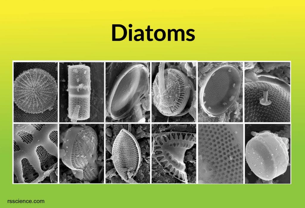
Diatoms
Diatoms, a big group of microalgae, are free-floating unicellular algae found in both the oceans and freshwater.
A unique feature of diatom cells is that they are enclosed within a cell wall made of silica (hydrated silicon dioxide, like glass) called a frustule. The photonic structures in the frustules such as pores and chambers on the micro to nanoscale interact with the visible light spectrum, creating colorful, shining, and opal-like appearances. Therefore, they are described as “jewels of the sea.”
The abundance of diatoms has a huge impact on the earth because one-fifth of the photosynthesis on the earth is done by diatoms. Is it a great deal?
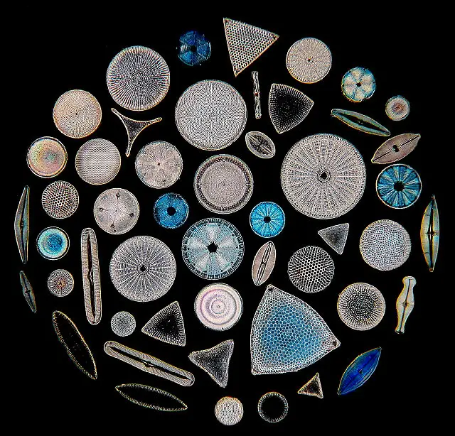
[In this figure] The beauty of diatoms on a microscopic slide.
Photo credit: micromagus.net
The discovery of diatoms
In 1703, an anonymous Englishman wrote to the Royal Society of London to report his observation he made using a simple microscope. He has been looking at the roots of pondweeds. As he looked closer, he found “many pretty branches, composed of rectangular oblongs and exact squares.” These tiny beautiful shapes, seem kind of plant-like, are what we know today as diatoms.
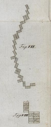
[In this figure] The observation of rectangular oblongs in 1703.
Photo credit: The Royal Society.
Does the image below look like the “an anonymous Englishman” saw in 1703? I found this diatom (Tabellaria) in the pond water in the spring near my home. I feel like I am sitting on a time machine.
Tabellaria forms colonies of cells connected by their corners. Tabellaria grows by division. The daughter cells remain attached to the mother cells, forming the zig-zag chains.
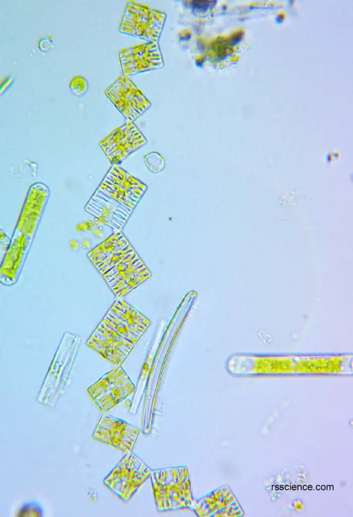
[In this figure] Tabellaria under a light microscope.
Tabellaria are square and rectangle, are often formed a zig-zag chain.
Diatom structure
Diatoms can be divided into two groups by their shapes, the pennate diatoms and the centric diatoms. Most diatoms are pennate diatoms (bilaterally symmetric), while a few diatoms are centric diatoms (radical symmetric).
Unlike the plants’ cell walls that are made of cellulose, the diatom cell walls are made of silica (i.e. glass), called frustule. The frustule morphology looks like a perti dish, consists of two halves. The part acting like a container is called hypotheca (slightly smaller than the other half), and the bigger part acting like a lid is called epitheca.
The frustule has a narrow and long opening called raphes, and their shells are typically elongated parallel to these raphes. Diatoms can secrete mucus through raphes, enable them to move along solid surfaces.
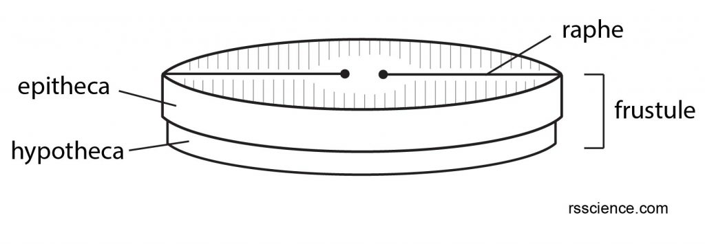
[In this figure] Illustration of the structure of a diatom frustule.
The upper half is called epitheca, slightly bigger than the lower half part, hypotheca. There are one or two slits, called raphe.
Like plants and algae, diatoms contain chlorophyll A in the chloroplast for photosynthesis. Chlorophyll A absorbs energy from the wavelength on the red-orange and blue-violet spectrum and reflects green light; therefore, most plants look green. In addition to chlorophyll A, diatoms also have chlorophyll C, fucoxanthin, and carotene, giving them golden-brown color.
Diatoms store energy as unique polysaccharide
chrysolaminarin or different lipid molecules. Some groups of
diatoms are capable of producing lipids almost up to 70% of their
body volume; therefore, they are considered to be explored as a source for biofuels.
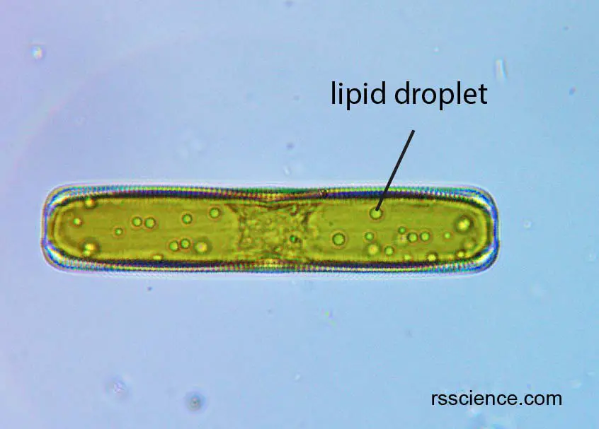
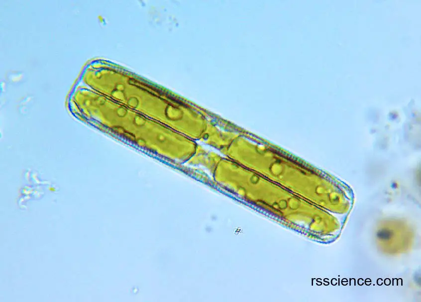
[In this figure] Lipid droplet in a diatom.
Diatoms can store energy as lipid. The round and shiny circles inside a diatom are lipid droplets.
How does diatom move
Most diatoms are nonmotile, but some species are capable of gliding over the surface due to their ability to secrete mucus. In addition, sperms in some species (centric diatoms) have flagella and can swim to meet female gametes (mature female germ cells such as an egg).
Diatom reproduction
Asexual reproduction
Diatoms mostly reproduce asexually by binary fission (vegetative reproduction), producing two daughter cells with identical genes. Because the cell wall is made of silica, it is very rigid. It cannot accommodate to increase sizes as the diatom grow bigger.
When diatoms divide, each daughter cell receives one of the frustules from parents, acting as an epitheca, the bigger frustule. The epitheca in daughter cells is used to construct a second smaller frustule, hypotheca (remember the petri dish idea). The daughter diatom that receives the bigger frustules becomes the same size as its parents. However, the one that gets the smaller frustule becomes smaller than its parents. This causes a reduction in the size of the diatom population.
You might wonder, the daughter cell will eventually become too small to survive. Yes. Therefore, they will need sexual reproduction, acting like a refresh button for bringing back to the normal size.
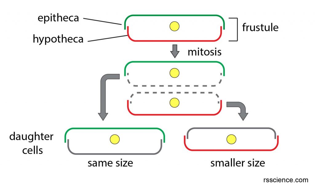
[In this figure] Illustration of diatoms asexual reproduction and size reduction in daughter cells.
The asexual reproduction generates one daughter cell with the same size as its parents and another daughter cells with a smaller size.
Sexual reproduction
In sexual reproduction, the diatoms undergo meiosis to produce male and female gametes (1N, equivalent to humans’ sperms and eggs), which then fuse to form a zygote (2N). The zygote then develops an auxospore, which has a soft membrane; therefore, can be expanded. A new diatom cell is formed inside the auxospore, beginning a new generation.
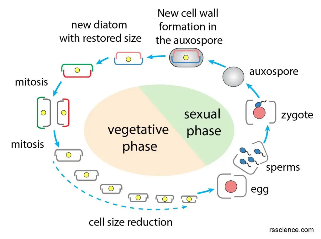
[In this figure] Life cycle of a diatom.
When the diatom size is too small to survive, diatoms will undergo meiosis to generate eggs and sperms. The sperm fertilizes an egg and form a zygote, which then develops an auxospore. The auxospore is a soft stage, which allows expansion. A new diatom forms inside the auxospore. Once it reaches the normal size, it releases.
More diatom images
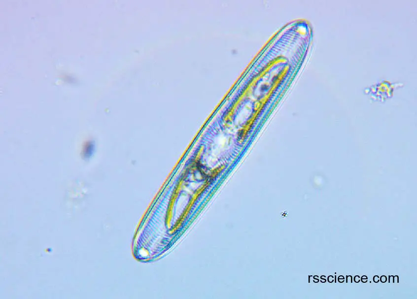
[In this figure] A diatom under a light microscope.
[In this figure] A diatom under a light microscope.
The lipid droplets are inside the diatom. Notice the yellow-brownish color of diatom.
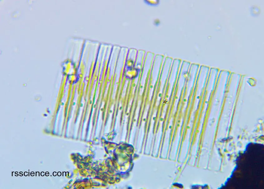
[In this figure] Diatoms under a light microscope.
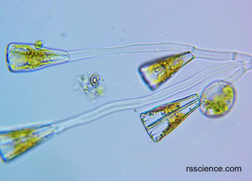
[In this figure] Diatoms under a light microscope.
Reference
https://diatoms.org/what-are-diatoms
https://diatoms.de/en/diatoms/what-are-diatoms
https://www.micromagus.net/microscopes/pondlife_plants01.html
https://websites.rbge.org.uk/algae/diatoms_introduction1.html