
You may wonder how the world looks like on a microscopic scale. Are cells cute? How about an ant look like an elephant? Welcome to the microscopic wonder world!
Looking at ordinary things under a microscope can change your perspective and the way you look at the world. If you have no idea where to start, we have listed 10 everyday things you should look at under a microscope that will blow your mind.
Before we start – we are talking about “compound microscope”
The same specimen may look totally different under different types of microscopes. For example, a stereo microscope will be perfect for the surface texture of a rock. However, if you put the same rock under a compound microscope, you will see nothing (because the light cannot pass through solid rock). The image below is a perfect example.
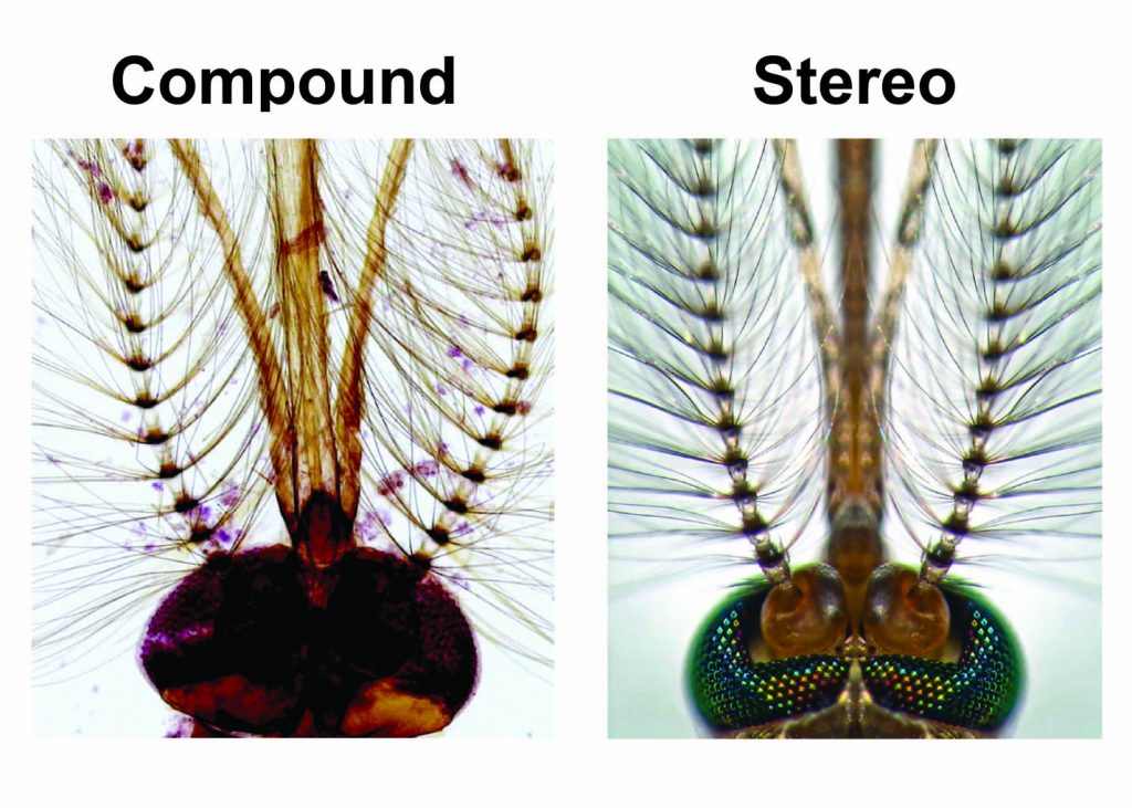
[In this figure] Looking mosquito head under compound and stereo microscopes.
A stereo microscope allows you to see the surface of specimens with a 3-dimensional view. Under a stereo microscope, you can see the metallic texture and colors of the mosquito’s compound eyes. In contrast, the light has to pass through the specimen to form the image under a compound microscope. In this case, the region of compound eyes is too thick to form a clear image. Right Image credit: Dr. Gareth Paul Jones, 2013 Photomicrography Competition, Technique: Stereomicroscopy, Fiber Optic Illumination. Magnification: 70x.
In this article, we selected the specimens ideal for compound microscopes. This is because compound microscopes are the most common ones and probably the first microscope most people have. However, if you have other types of microscopes such as stereo, pocket, and USB digital microscopes, please also try these tips and I guarantee you will see something fantastic!
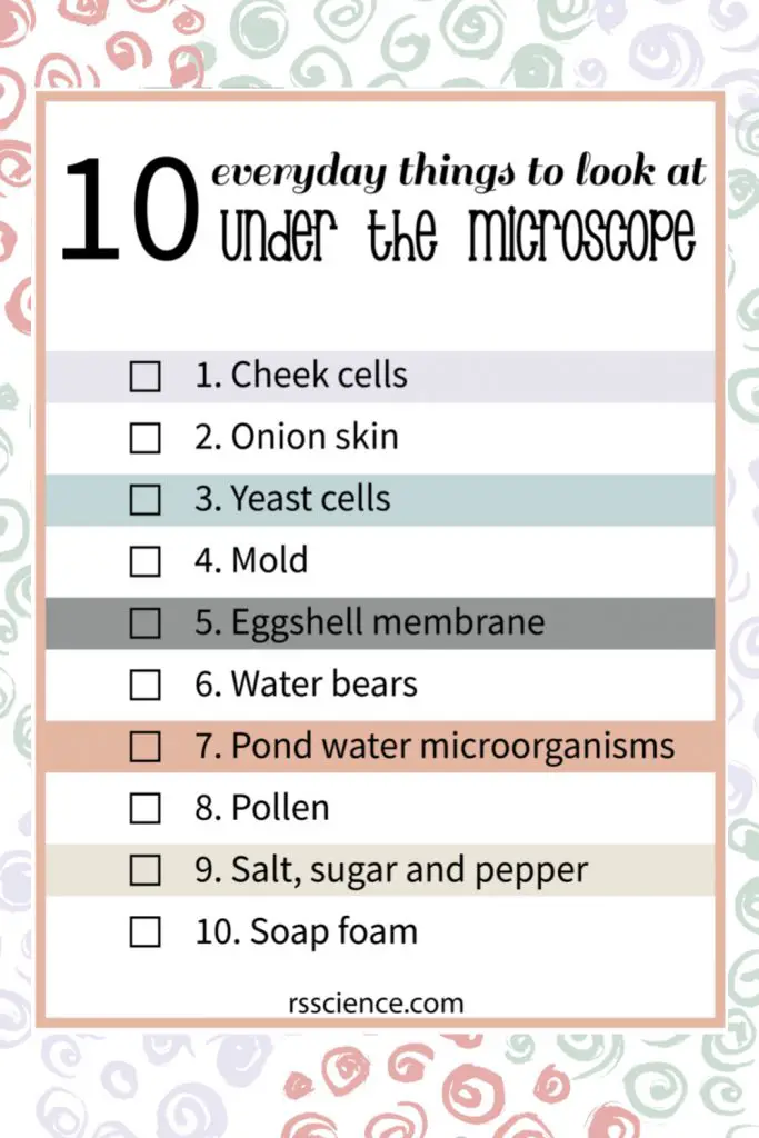
This article covers
1. Cheek cells
Cheek cells (more specifically, epithelial cells) form a protective barrier lining your mouth. Due to their heavy workload, many cheek cells retire everyday and new cells replace their positions. Old cheek cells are easily shed from the mouth lining; therefore, it is easy to obtain them for observation.
All you need to do is to gently scrape the inside of the mouth using a clean, sterile cotton swab and then smear the swab on a microscopic slide to get the cells onto the slide. You can see our step-by-step guide “Look at your cheek cells“.
In addition, you can learn the cell structure by looking at your own cheek cells through identifying cell membrane, cytoplasm, organelles, and nucleus.
Click “Cell Organelles and their Functions” to learn more about the function of each cell organelle.
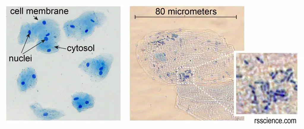
[In this figure] Cheek cells stained with Methylene Blue.
The left image is a low magnification. You can see the nuclei stained with a dark blue (because Methylene Blue stains DNA strongly). The cell membrane acts like a balloon and holds all the parts of a cell inside, including a nucleus, cytosol, and organelles.
The right image is a high magnification. This check cell is about 80 micrometers in diameter. We measured the cell size by using a microscopic meter slide.
You can also see some small rod-shaped bacteria on the right image. Don’t worry, they are normal oral microbes.
2. Onion skin
The onion skin is a layer of protective epidermal cells against viruses and fungi that may harm the sensitive plant tissues. This layer of skin is transparent and easy to peel, making it an ideal subject to study plant cell structure. You can follow our step-by-step guide “Look at the Plant Cells” to prepare your own onion skin slide.
Onion skin cells are well-organized and uniform in shape because the plant cells have something, we (animal cells) do not have – the cell walls. The cell walls are made up of cellulose, which protects the cell and maintains its shape.

[In this figure] Microscopic view of onion skin.
Without stains, you can only see the cell walls of onion cells. By staining Eosin Y, now you can see a nucleus inside an onion cell.
3. Yeast cells
Yeasts are single-celled fungi with an oval shape that play extremely important roles in our daily life. Without these tiny microorganisms, human beings can not make bread, beer, wine, or all kinds of fermented food products. You can easily grow your yeast culture by mixing active dried yeast (for bakery use), sugar, and warm water. You can follow our step-by-step guide “Grow your own yeasts“.
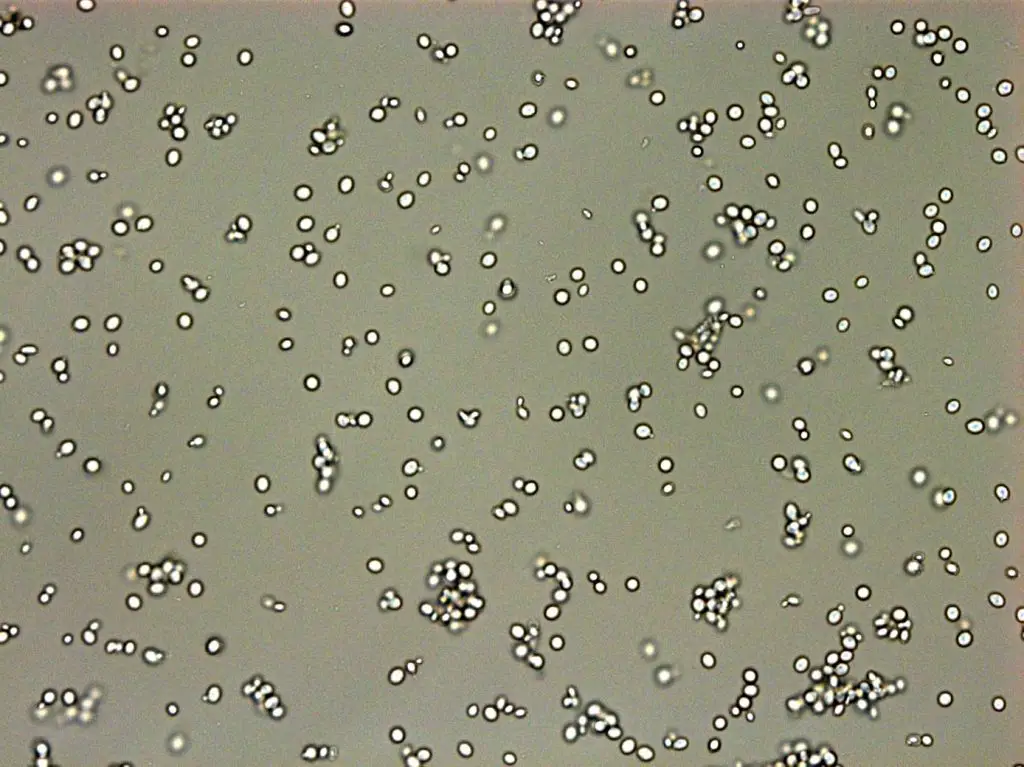
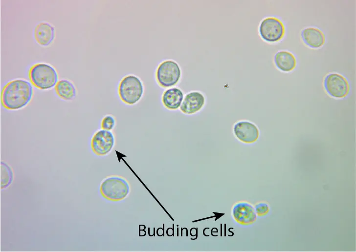
[In this figure] Microscopic view of yeast cells.
Yeast cells with an oval shape and some of them are dividing (budding yeasts).
4. Mold
Mold is a type of fungi that is different from yeasts and mushrooms. Mold grows in the form of filaments or fibers that may look like a lawn and produce spores. You can find them growing on rot fruits with white, green, or black colors.
You can directly observe the mold under a stereo microscope or prepare a spore specimen for a compound microscope. We suggest wearing protective glasses and masks while you collect the mold spores. Some of them are harmful when you breathe in. For safety, we recommend buying the prepared slide of mold.
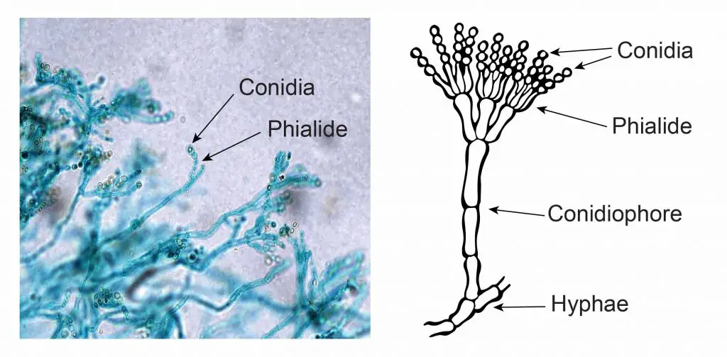
[In this figure] Penicillium is a common green mold that grows on rot fruits. However, Penicillium is also essential to our human beings because it can produce the first antibiotics in the world – Penicillin. Under the microscope, Penicillium molds grow like a lawn with many fiber-like structures, called hyphae. Some hyphae will branch and produce spores on their tips.
5. Eggshell membrane
Have you ever notice a very thin film attached to the inside wall of an eggshell? This is an eggshell membrane. In fact, it contains two membranes that defend against bacterial invasion. If you give these layers a tug, you will find they are surprisingly strong! They are made partly of keratin, a protein that is also present in human hair. What do they look like? see the image below!
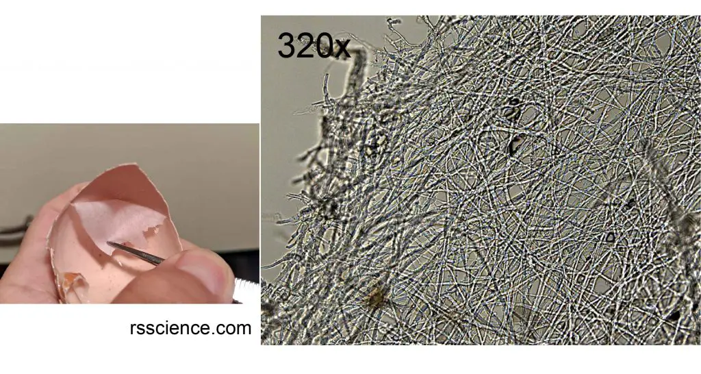
[In this figure] Looking at the edge of an eggshell membrane, you will see a web of many keratin fibers.
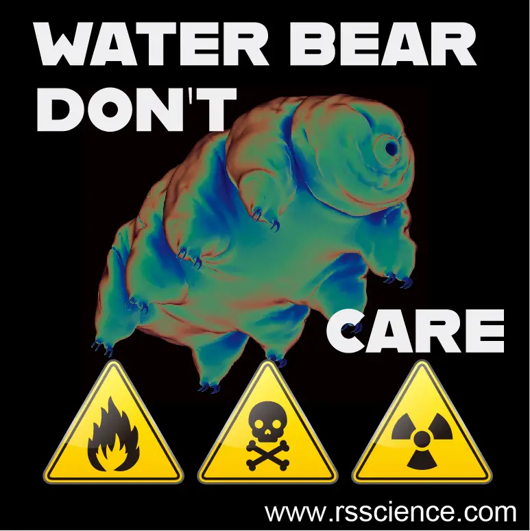
6. Water bear
Water bears (official name, Tardigrades) are the superstars in the science classroom. They can survive in many extreme environments such as extremely high and low temperatures, low pressure, dehydration, and high radiation.
These microscopic animals have eight legs with four to eight claws on each. You can see them swim or crawl with their eight legs slowly under a microscope. Scientists found that they are almost indestructible and can even survive in outer space.
Below is a cute sticker we created for the water bear. Is it funny?
Water bears can be found almost everywhere – such as moss, ferns, lichens, soil, beaches, dunes, and other damp habitats. Just bring some simple tools when you have a hike next time and go hunting for your water bears. You can see our field research story “How to find water bears“.
[In this video] A water bear is recovering from a piece of frozen moss collected from suburban Boston.
7. Pond water microorganisms
There are so many interesting things to look at from a drop of pond water. It is totally a “jungle” out there!
We can divide all the pond water microorganisms into two groups: Protozoa (animal-like) and Protophyta (plant-like). Algae (singular, alga) are the majority of plant-like pond lives and exhibit a wide range of shapes. Some algae grow like green silks attached to rocks. Some single-celled algae float freely. Some big algae, such as the giant kelp, can grow up to 100 feet in length. Many algae have chloroplasts meaning they can absorb energy from sunlight.
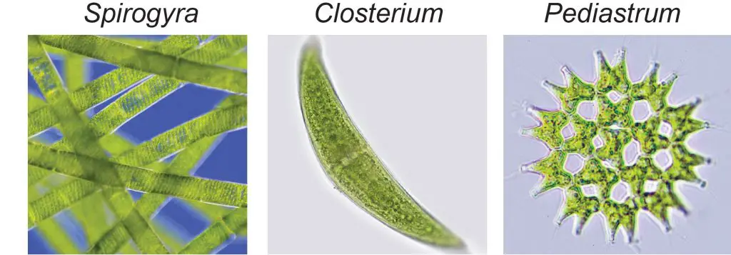
[In this figure] You can collect filamentous Spirogyra (or water silks) from rocks. Chlorella (Sphere-shaped), Closterium (crescent-shaped), or Pediastrum (cluster of 8-32 cells) can be found floating in green pond water.
Many microscopic animals live in pond water that you can not see without a microscope. They can be single-celled microorganisms like Paramecium, micro-predators Rotifers, vorticella, stentors, or tiny Crustacean Daphnia.
[In this video] Within every drop of pond water lurks an invisible world, alive with an amazing variety of microscopic creatures. You can find simple life forms such as bacteria, great oxygen-producers like algae, all kinds of alien-like protozoans, and cute microscopic animals like water bears.
[In this video] Rotifer’s cilia can move like a rotating wheel to create a vortex and suck in food. Rotifer anchors itself while feeding.
[In this video] Seeing several kinds of pond life microorganisms under the microscope.
You can see many small ciliates swim around a piece of food using their hair-like cilia. Two ciliates play around behind a piece of aquatic plant. A twisting nematode, a planarian, a water bear, and rotifers.
Be careful when you approach any ponds, creeks, rivers, or lakes. Please follow our step-by-step guide “microlife hunter” to catch your microorganisms. A microscopic slide with a single concave that can hold some water will be helpful.
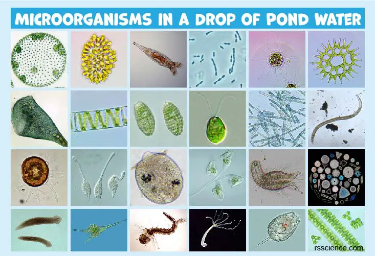
Please also check out our post “34 Microscopic Organisms in a Drop of Pond Water“. How many microscopic organisms do you see in your sample?
8. Pollen
I personally have a pretty nasty symptom every year during the allergy season, but this can not stop me from exploring the beauty of pollen under the microscope!
Grains of pollen is produced by the stamens (male) of flowering plants. They are carried by animals (like bees), wind, or water to the pistils (female). Each pollen grain contains two cells in a hard shell. Observing pollen grains is easy. You just need to use a toothpick or Q-tip to collect some pollen grains onto a microscopic slide. Add a drop of water and cover it with a coverslip.
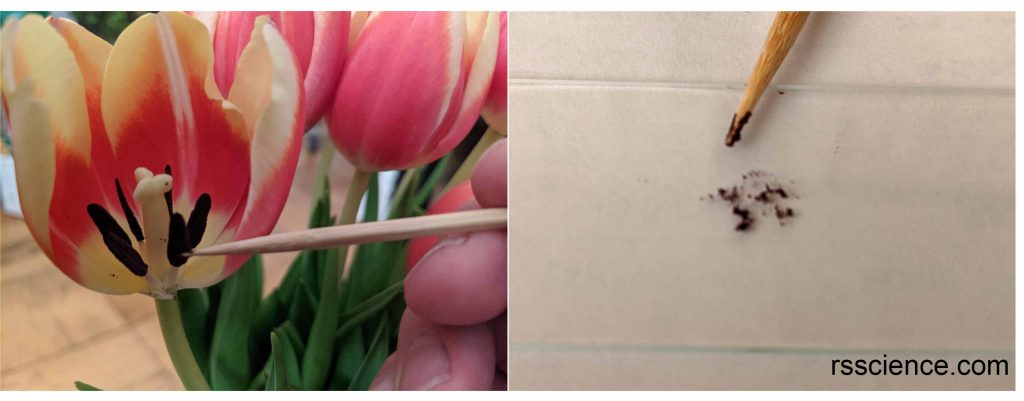
[In this figure] Collect tulip pollen grains using a toothpick to a microscopic slide.
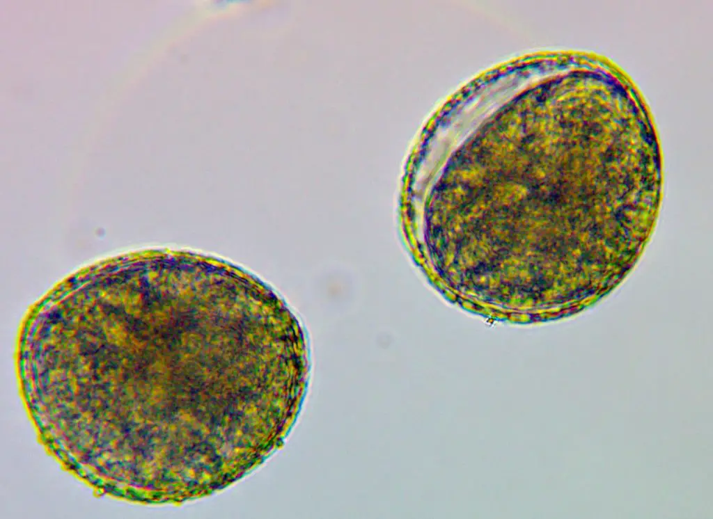
[In this figure] Tulip pollen grains under a microscope.
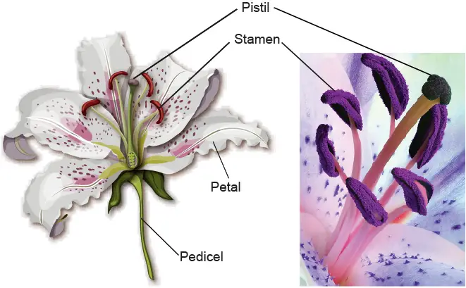
[In this figure] The anatomy of a lily flower.
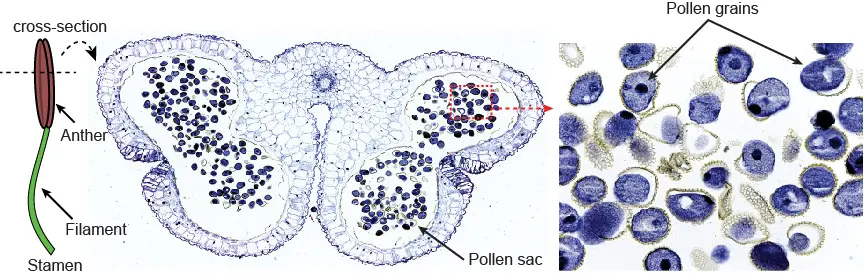
[In this figure] Look at pollen grains inside a lily anther.
You can purchase this slide in Rs’ Science 25 microscope prepared slide set.
9. Salt, sugar, and pepper
When you look for stuff to see under your microscope, do not forget all kinds of everyday things in your kitchen! You will be amazed by how beautiful they are. For example, I have two kinds of salts in my kitchen. They look similar and also taste similar. However, you will find they are quite different under the microscope.
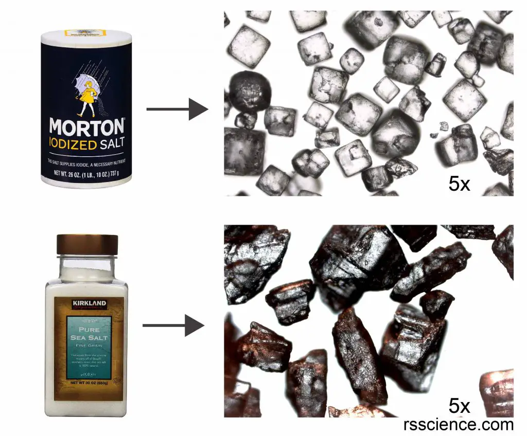
[In this figure] Microscopic view of salt and sea salt.
Notice that their appearances are quite different under the light microscope.
The Morton salt is finer and the salt grains look like white crystals. In contrast, the sea salt grains are bigger with a rock-like texture. Although the sea salts are white in color, some of them shine orange color by reflecting the light. I guess that the color comes from the minerals in the sea salts. When I use higher magnification to see the sea salts, I feel like I am looking at a meteorite or an asteroid.
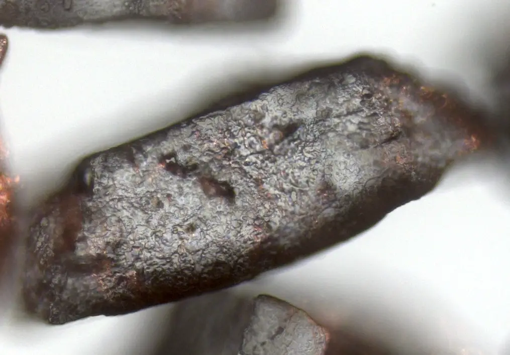
[In this figure] High magnification of Microscopic view of sea salt.
10. Soap foam
Do you wonder why I say you should look at soap foam under the microscope? Am I CRAZY???
Don’t worry; I am totally normal. Trust me; soap foam will be one of the most fun and amazing things to see under your microscope.
First, collect different kinds of soap, dishwasher detergent, laundry detergent, etc. Mix them in a jar with water and put them on a lid. Shake the jar to form finer foam. Then use a dropper or your finger to transfer some foam onto a slide and cover it with a coverslip.
Now, under the microscope, you will see the foam is made of many bubbles. These bubbles are very dynamics (or unstable). Some of them will move toward a direction. Some of them will collide and fuse into a bigger bubble. Some of them will suddenly burst. Try a different proportion of soap and water and the bubbles you get will be totally different.
[In this video] Bubble dance under the microscope.
Summary
This is just a short list of everyday things you should take a look at under the microscope. In fact, we just scratch the surface of the amazing microscopic wonder world. If you don’t want to miss any of our future updates, please sign up on our email list!
Related posts
How to Find Tardigrades (Water Bears)
Lesson 2: Mount a Slide & “Look at Your Cheek Cells“