
This article covers
What are rotifers? A quick overview
Rotifers are microscopic aquatic animals of the phylum Rotifera. Rotifers got their name from the corona: a rotating, wheel-like structure covered with cilia at their heads. Rotifers also have a jawed mouth and complete digestive, sensory, and reproductive organ systems. They are “small,” but not “simple!”
[In this video] A video capturing the “rotating wheels” of a rotifer and won the 2012 Nikon Small World in Motion Competition.
Rotifers are filter feeders that eat dead material, algae, bacteria, and other microscopic living organisms and are essential components of aquatic food webs.
Quick facts of rotifers:
- Rotifers are multi-cellular animals with a microscopic-sized body.
- Rotifers are aquatic invertebrates that constitute the phylum Rotifera.
- There are three classes of Rotifers: Bdelloidea, Monogononta, and Seisonidea.
- Rotifers have a complete digestive tract.
- Most rotifers are free-living. They can swim or walk by anchoring their feet.
- Rotifers can reproduce both asexually and sexually, depending on environmental conditions.
- Cryptobiosis allows rotifers to survive through extremely harsh conditions.
If you want to know the Rotifers or “Wheel animals” more in detail, keep reading this article.
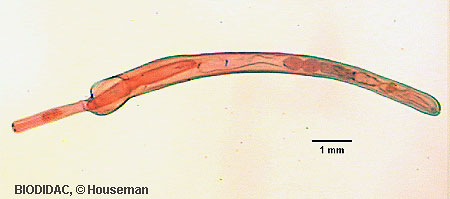
[In this figure] Electron microscope image (fake color) of a cute Bdelloid rotifer.
Part 1. The Basic of Rotifers – Name, Size, Classification, and Evolution
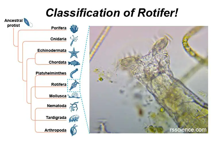
[In this figure] Rotifer taxonomy – the position in the phylogenetic tree.
Rotifers are multi-cellular, invertebrate animals. Comparing to other microorganisms, rotifers are pretty advanced in the aspect of evolution.
Classification of rotifers
Although their taxonomy is currently in flux, scientists now place the Rotifers in the Kingdom of Animalia as listed below:
Scientific classification
Kingdom: Animalia
Subkingdom: Eumetazoa
Clade: ParaHoxozoa
Clade: Bilateria
Clade: Nephrozoa
(unranked): Protostomia
(unranked): Spiralia
Clade: Gnathifera
Phylum: Rotifera (Cuvier, 1798)
Class: Bdelloidea, Monogononta, Seisonidae
(Reference: https://en.wikipedia.org/wiki/Rotifer)
The Phylum of Rotifera and examples
Rotifers occupy their own Phylum, Rotifera, and there are currently over 2,200 recognized species and 120 genera. Under the Phylum of Rotifera, the members can be further classified into three classes: Bdelloidea, Monogononta, and Seisonidea. The largest group is the Monogononta, with about 1,500 species, followed by the Bdelloidea, with about 350 species. There are only two known genera with three species of Seisonidea.
Acanthocephala (Spiny-headed worm), previously considered to be a separate phylum, has been demonstrated to be modified rotifers and may be combined into the Phylum of Rotifera in the future.
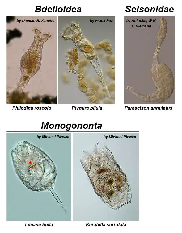
[In this figure] Examples from three classes of rotifers are shown.
Species from the class Bdelloidea are characterized by a large corona and telescopic body. Rotifers from the class Monogononta have a smaller corona than Bdelloid rotifers and have a single gonad (body segment), which gives the class its name. Seisonidae rotifers are saltwater rotifers found on the gills of Nebalia, a marine crustacean. They have a large body and elongate neck with reduced corona.
Photo source: Monogononta.
[In this figure] Acanthocephala (spiny-headed worms or thorny-headed worms) is a group of parasitic worms. Recent genome analysis has shown that they are descended from highly modified rotifers.
Photo source: Tree of life web projects.
Are rotifers animals? Are rotifers single-celled or multicellular organisms?
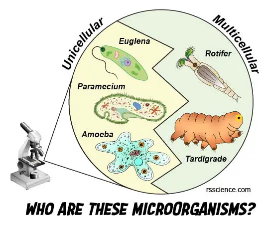
Rotifers are multicellular animals; instead, amoeba, euglena, and paramecium are single-celled organisms. Most rotifers are composed of an average of about 1,000 cells. Rotifers are small but fully functional organisms with several specialized organ systems. For example, rotifers have a complete digestive tract that includes both a mouth and anus. Since these characteristics are all uniquely animal features, rotifers are recognized as animals, even though they are as small as unicellular protists.
[In this figure] Many microorganisms fall in a size that ranges 100-500 μm. However, they can be totally different. Some microorganisms (like Euglena, Paramecium, and Amoeba) contain only one gigantic cell (called unicellular or single-celled) and are classified as protists. Others (like Rotifer and Tardigrade), however, contain thousands of cells and belong to multicellular animals or micro-animals.
A list of commonly found microscopic creatures:
| Unicellular | Multicellular |
| Euglena | Rotifer |
| Amoeba | Tardigrade |
| Paramecium | Hydra |
| Heliozoan | Copepod |
| Vorticella | Daphnia |
| Stentor | Flatworm |
| Coleps | Nematode |
| Dinoflagellates | Gastrotricha |
How big are rotifers?
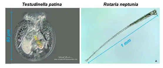
Although rotifers are multicellular creatures, they make their living at the scale of unicellular protists. Most species of rotifers are about 200 to 500 μm long. Some large stentors (unicellular ciliates) can easily grow several times bigger than that.
[In this figure] Size ranges of rotifers.
Different species of rotifers vary in their sizes. Here are two examples. Left: Testudinella patina – nicknamed “Turtle Rotifer” has a spherical body around 50-80 µm in length. (Modified from Dr. Robert Berdan). Right: Rotaria neptunia has an unusually long foot. The total length of some individuals may exceed 1 millimeter. (Photo source: The Quekett Microscopical Club).
Where did the name “Rotifer” come from?
The name “Rotifer” came from the Latin word meaning “wheel animalcules. ” This name represents the most interesting feature of a rotifer – the crown of cilia around the mouth very well. The rapid movement of the cilia of rotifers makes them appear to whirl like a wheel.
[In this video] A video of rotifer showing its cilia movement like two rotating wheels.
Who discovered rotifers? – the history
Rotifers were one of the first organisms observed by Antony Van Leeuwenhoek in the late 1600s. He noticed the swirling, rotating cilia on the crowns and named these organisms “wheel animalcules.” “Animalcules” means little animals (from Latin animal + the diminutive suffix -culum).

[In this figure] Left: The simple microscope used by Antony Van Leeuwenhoek to discover the microscopic organisms. Right: A 1795 illustration of van Leeuwenhoek’s animalcules by an unknown artist.
Photo source: wiki.
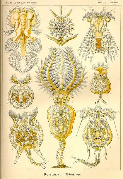
[In this figure] The beauty of rotifers from the book “Art Forms in Nature” by Ernst Haeckel, originally published in 1904. This is an example of how the 19th century biologist’s illustrations of microlifes bring art and science together.
Part 2. The Anatomy Structure and Organ Systems of Rotifers
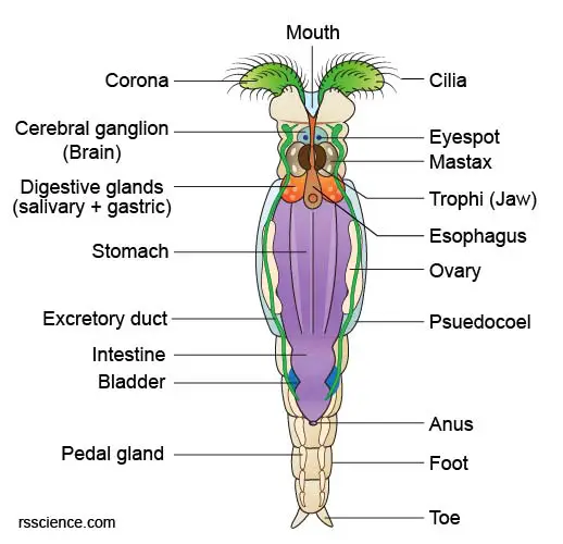
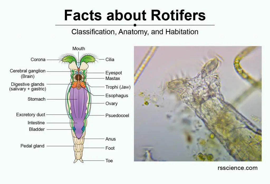
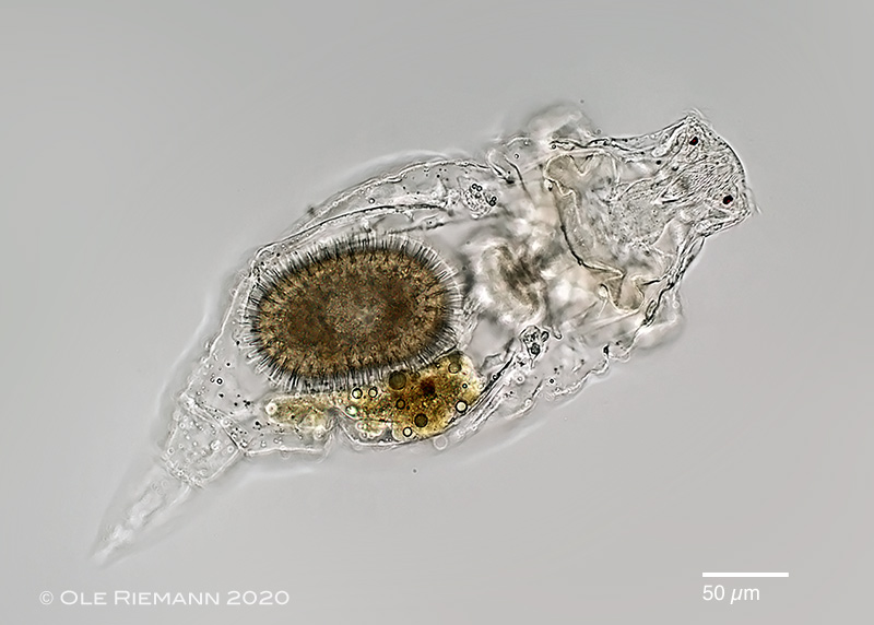
[In this figure] The anatomy of a female Bdelloid rotifer with the internal organs labeled.
The body of rotifers consists of a head (which contains two coronae), a trunk (which contains the organs), and the foot.
Rotifers’ digestive system
Rotifers have a pretty complete digestive system (very similar to humans) in their microscopic bodies. The body cavity (like our digestive tract) opens from the mouth to the anus, which is partially lined by a mesoderm.
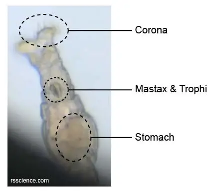
Rotifer’s crown
The head carries the coronae (singular: corona) with the mouth opening between the coronae. The cilia on the coronae draw a vortex of water into the mouth, where the rotifer sifts for food.
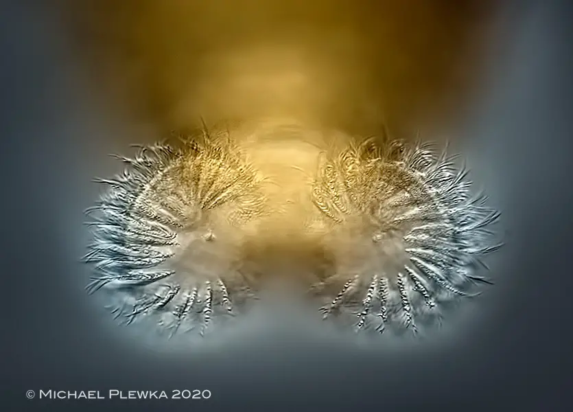
[In this image] A front view of Bdelloid Rotifer’s wheels and cilia.
Photo source: Plingfactory.
Actually, the cilia do not rotate but wave back and forth in a sequential pattern, giving rise to the illusion of rotation. The cilia are arranged around the coronae in double rows contained an inner ring and an outer ring (called trochal discs).
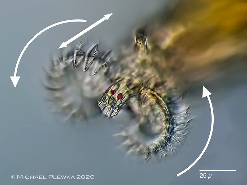
[In this image] The axis of rotation of the cilia is parallel to the longitudinal axis of the body. Two small red dots are eyespots.
Photo source: Plingfactory.
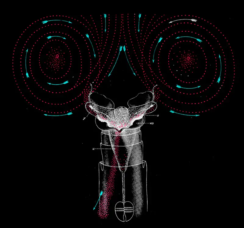
[In this image] A diagram showing the water flow caused by the cilia movement.
The turquoise arrows mark the direction of flow. The small red dots represent the food particles collected into the rotifer’s month by the water flow.
Photo source: Plingfactory.
Rotifer’s chewing jaw
After passing the mouth, food is transported to a muscular pharynx, called the mastax. This mastax contains jaw-like hard structures, called the trophi, and associated chewing muscles. The food particles will be ground by the trophi into smaller pieces.
Pay attention to three labeled parts of a feeding rotifer in the video below. You can see the muscles of mastax driving the consistent chewing of jaw-like trophi.
[In this video] Rotifer moves its cilia like a rotating wheel to create a vortex and suck in food.
The trophi are found in almost all rotifers and are characteristic organs of the phylum Rotifera. The trophi are also the only hard part of rotifers that might be preserved in the fossil record.
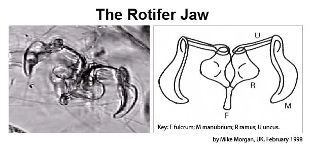
[In this figure] The jaw of rotifers, called trophi, consists of several parts to cut and chew the food particles.
Photo source: Microscopy UK.
[In this figure] Scanning electron microscopy (SEM) showing a morphological variation of Bdelloid rotifers and their jaws.
Photo source: Diego Fontaneto.
Rotifer has a complete digestive tract just like human
Ground food then passes through the esophagus into the stomach. There are two kinds of digestive glands, salivary and gastric glands, that produce digestive enzymes to facilitate the process in the stomach. Digested food then moves into the intestine for nutrient absorption. Excretory wastes are collected in a bladder before being released out the anus.
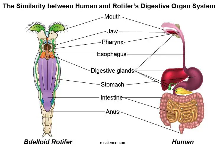
[In this figure] The similarity between human and rotifer’s digestive organ system.
This labeled diagram helps us understand how the rotifer’s digestive tract work. Rotifers have almost the same digestive organs like us. The major difference is that we have more organs (including the salivary gland, liver, pancreas, stomach, and gallbladder) to secrete different digestive enzymes (because our food sources are more complicated).
The body of rotifers
The body of the rotifers is a telescopic shape (for Bdelloid rotifers). The external of the body is covered with a layer of transparent, semi-flexible, extendible cuticle. The composition of the rotifer’s cuticle suggests rotifers are close relatives of roundworms and arthropods.
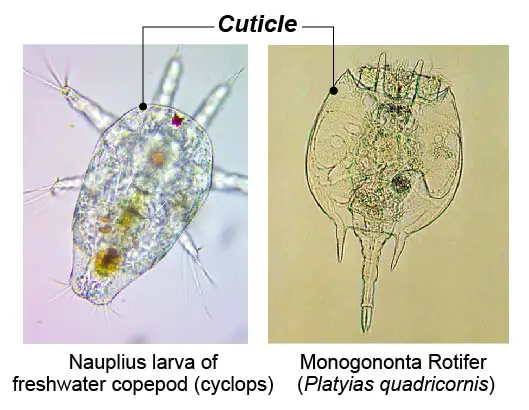
[In this figure] The body of rotifers is protected by a shell of a cuticle, which could also be found on arthropods.
The body of Bdelloid rotifers looks segmented externally. However, they don’t have segments inside the body cavities.
The feet and toes of rotifers
The last part of the rotifer body is its foot with toes. For Bdelloid rotifers, the feet and toes work just like their name. These rotifers anchor on the surface like standing on their feet. The feet also allow them to locomote like a leech.
There are pedal glands that can produce a cement-like sticky substance which allows the rotifer to attach itself to objects in the water.
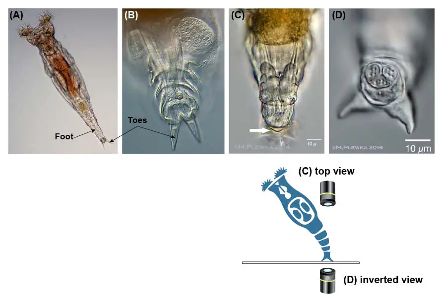
[In this figure] (A, B) Typical images of Bdelloid rotifers’ feet and toes. (C) Top view of an anchored foot: The white arrow indicates the anchorage of Rotifer’s foot on the surface. (D) Bottom view of an anchored rotifer’s toes by an inverted microscope.
Photo source: Cloudynights, Plingfactory.
For Monogononat rotifers, the feet and toes may not use for anchoring. Instead, these parts function as a tail for free-swimming.
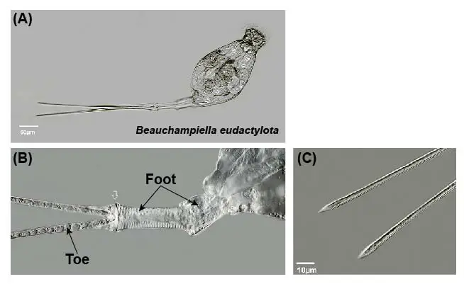
[In this figure] Beauchampiella eudactylota (a Monogononat rotifer) swims using its long foot and toes.
Photo source: Plingfactory.
Do rotifers have brains and eyes?
Yes, the microscopic body of rotifers even contains a primitive central nervous system. Rotifer’s head contains neuronal organs in the form of a bi-lobed brain (Cerebral ganglion) and two small eyespots near the coronae.
Rotifers have also been shown to possess a few sensory organs to detect changes in pressure, light, or chemical compounds. These sensors allow rotifers to move towards favorable areas (with food, etc.) or away from predators.
Part 3. The Habitation of Rotifers – How do they move, eat, reproduce, and perform other interesting behaviors
Can rotifers sense danger?
Yes, rotifers can sense their surrounding environment by sensors on their head. When they sense danger, the head retrieves and hides inside the body. They can even contract into like a ball. Only when the rotifers feel safe, the coronae will fully extend to start feeding.
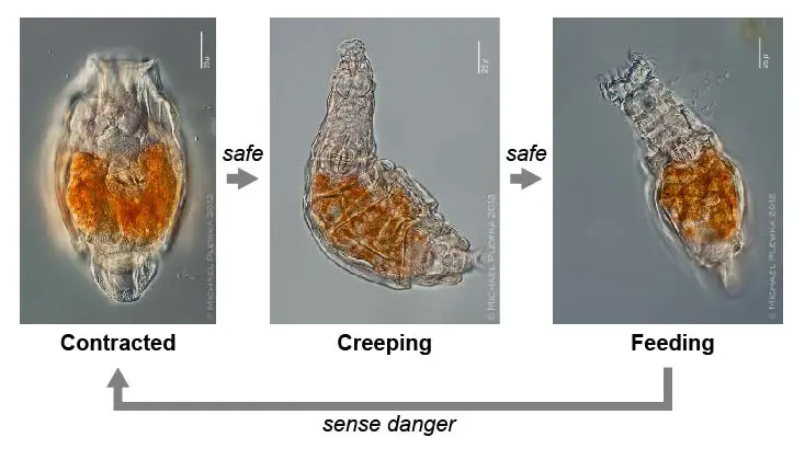
[In this figure] Rotifers consistently sense their environment. If a danger sign is found, a rotifer retrieves its head and contracts like a ball. Once the environment is back to safe, it slowly exposes its head and resumes its feeding.
Where do rotifers live?
Rotifers can be found in many freshwater environments, such as pond bottoms and flowing water, such as rivers or streams. Rotifers are also commonly found on tiny plants like mosses and lichens growing on tree trunks and rocks or on mushrooms growing near dead trees. They also like to inhabit moist soil or leaf litter in rain gutters and puddles. What they need is just a thin film of water surrounding soil particles. They can even use their feet to anchor on the surface of freshwater crustaceans and aquatic insect larvae. Some species of rotifers live in saltwater environments. Rotifers have even been found in the waters of Antarctica!
Most rotifers are free-living plankton. Some species of rotifers like to stay together (a colony) with their feet attached to each other.
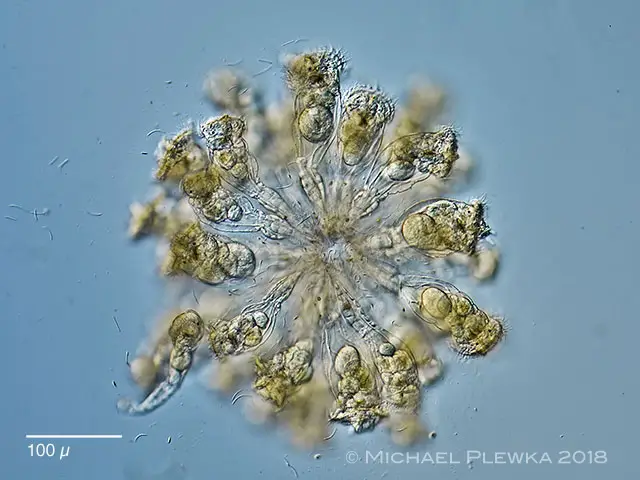
[In this image] A colony of sea rotifers, Conochilus hippocrepis.
Photo source: Plingfactory.
Many rotifer species live in close association with plants or other animals (for example, growing by attaching to the surface). However, few rotifers are true parasites, actually harming their hosts. One example is the parasite rotifers living inside of a beautiful alga called Volvox. The Volvox is a colony of algae cells that live together in a sphere. It looks like a green globe spinning slowly through the water.

[In this image] Volvox is a multicellular colony of green algae growing like a sphere.
Photo source: National Geographic.
The parasitic rotifers live and lay their eggs inside the Volvox. They also eat algae as food, slowly destroy the perfect globe shape of Volvox. When the rotifer has eaten enough of its host, it escapes and swims off to find another victim.
How do rotifers move?
Leech-like locomotion
The term “Bdelloid” means “leech-like”. Bdelloid rotifers got their name from their leech-like appearance and the manner in which they can move. Basically, Bdelloid rotifers move by looping along a surface, alternately attaching the head and foot ends. When they are moving by this inchworming movement, their coronae retrieve and hide inside the body.
[In this video] The video showing how a Bdelloid rotifer moves on a surface by inchworming.
Swimming
Rotifers can also swim with the aid of the ciliated coronae, using the coronae to pull them through the water much like an airplane propeller.
How do rotifers eat?
Most rotifers are filter feeders. Their cilia on the coronae move to create a water flow and bring the food into the mouths.
[In this video] Once a Bdelloid rotifer finds a good place to anchor, it extends its coronae and starts filtering the surrounding food. Rotifer moves its cilia like a rotating wheel to create a vortex and suck in food.
[In this video] Rotifers under the microscope showing their filter-feeding.
You can also see the chewing of its trophi jaw.
The diet of rotifers commonly consists of dead or decomposing organic materials and unicellular algae, bacteria, and other small phytoplankton. As rotifers are microscopic animals, their diet must be small enough to fit through their tiny mouths during filter feeding.
What is the role of rotifers in ecology?
Since rotifers feed primarily on decomposing organic materials and tiny microorganisms, this makes rotifers an important primary consumer. Rotifers are preys for carnivorous secondary consumers. Many animals eat rotifers, including shrimps, crabs, water fleas, tadpoles, aquatic insects, fishes, clams, ducks, great egrets, and other wading birds. A balanced population of rotifers is a key indicator of a healthy aquatic ecosystem. Thus, rotifers play an important ecological role.
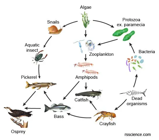
[In this figure] Food web of the aquatic ecosystem.
Every member of the ecosystem is essential. Rotifers and other zooplankton function as bottom consumers and food sources for bigger animals.
How rotifers reproduce?
Rotifers are dioecious organisms (having either male or female types). The females are always larger than the males (sexual dimorphism). In some species, the female can be up to ten times bigger than the male. In many species, males are short-lived and with no digestive system (males are degenerate due to the loss of half part of DNA). These males only have a single testis as the internal organ.
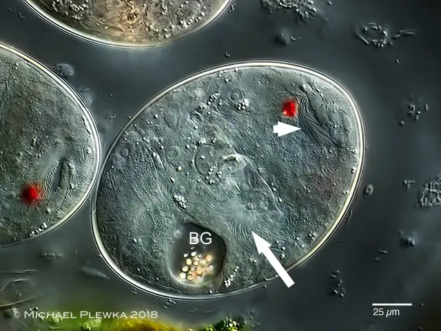
[In this figure] A female rotifer (Rhinoglena frontalis) carrying a resting egg.
Photo source: Plingfactory.
Two types of reproduction have been observed in rotifers. Some species consist only of females that produce their daughters from unfertilized eggs, a type of reproduction called Parthenogenesis. In other words, these parthenogenic species can develop from an unfertilized egg asexually.
[In this figure] Hatching eggs.
The white arrows point to the visible ciliated parts of a developing embryo. Red eyespots are also visible.
Other species reproduce by Sexual reproduction. Gender determination of these rotifers is unusual. A fertilized egg develops into a female and an unfertilized egg develops into a male. In this case, the male rotifers have only a half copy of genome DNA (called haplodiploidy) but still can produce sperm.
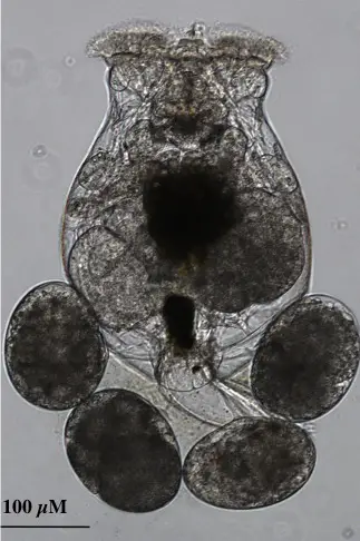
[In this figure] A female rotifer carrying 4 eggs.
Photo source: Developmental biology interactive.
Mating between females and males results in fertilized eggs developing within the female rotifer. The eggs are released and hatch in the water. These resting eggs are pretty resistant in a dormant form that can survive when the local water supply dries up. If the eggs develop in the summer, the eggs may remain attached to the rotifer’s posterior end until hatching.
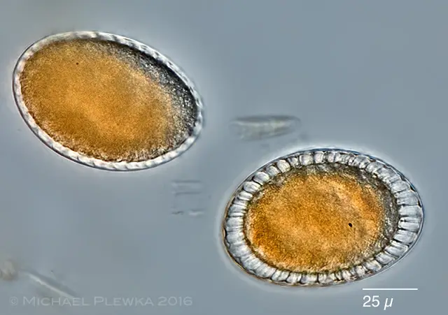
[In this figure] Two resting eggs of rotifers.
The thick shell around the eggs provides great protection.
Photo source: Plingfactory.
Many species can switch between Asexual Parthenogenesis and Sexual Reproduction according to the condition of habitation. This forms a Life Cycle of rotifers. Usually, sexual reproduction happens when the environment becomes harsh (dry, cold, or lack of food). The rotifer species can be preserved as the resting eggs in a resistant dormancy until the condition becomes habitable.
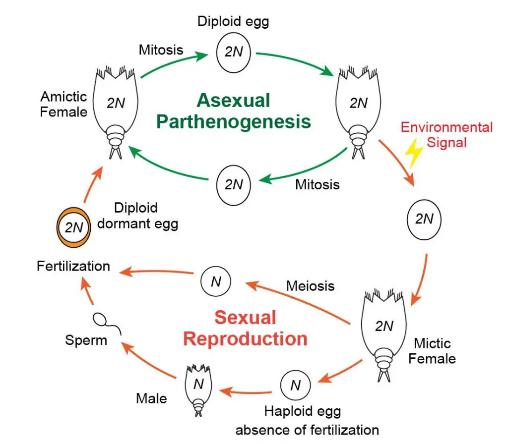
[In this figure] The reproduction life cycle of rotifers.
In a nutrient-rich environment, rotifers reproduce by Asexual Parthenogenesis with only females. Upon encountering an environmental cue (becoming harsh; for example, during the winter), rotifers adapt to Sexual Reproduction. To do so, asexual females produce sexual daughters via meiosis. If unfertilized, these haploid eggs develop into males who fertilize sexual females to produce diploid dormant eggs. These resting eggs can survive until the condition is turning favorable (for example, spring) and then develop into asexual females. 2N means diploid (a pair of each chromosome) and N means haploid.
Rotifers are unique in that they are born with all their cells. Unlike most animals that grow by adding new cells, rotifers grow by increasing their cells’ size.
Rotifers are considered extremophiles like Tardigrades!
Ideally, rotifers thrive in humid conditions and mild to warm temperatures. During the harsh condition like winter or dry seasons, rotifers can survive through a process known as Cryptobiosis (another famous example is Tardigrades, a.k.a, water bears). Rotifers enter cryptobiotic life by stopping all metabolic processes and shrinking into a dormant state. When environmental conditions return to being hospitable, the rotifers can quickly return to their normal state. Rotifer eggs can also withstand drying by staying dormant for many years. Rotifers are also superior to survive radiation damages due to their DNA repair capability. In many aspects, rotifers are truly extremophiles (meaning organisms with the ability to thrive in extreme environments).

[In this figure] Tardigrades or water bears are the superstars in the microscopic world and are known for their ability to survive in extreme environments. However, many people don’t know that rotifers are also pretty tough, too.
Part 4. Observing Rotifers under the Microscope
Observing rotifers is relatively easy. Rotifers are almost everywhere. Finding rotifers is possible in specimens collected from all kinds of humid environments. You can try collecting water samples with bottom sediments from scums and ponds. Rotifers also like to grow on decayed leaves and aquatic plants. Mosses and lichens are great places to find rotifers, too. You can place a piece of plant samples in a petri dish with some water for several days and observe rotifers migrate out. For example, I successfully saw rotifers and water bears recovered from a piece of frozen moss collected during the winter. You may even see rotifers growing from hatching eggs.
[In this video] Seeing rotifers with a foldscope.
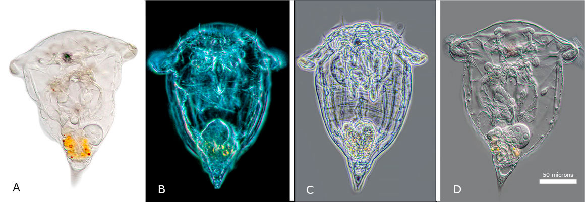
[In this figure] A comparison of Synchaeta sp (a Monogononta rotifer) under (A) bright field, (B) darkfield, (C) phase contrast, and (D) DIC microscopy.
Photo source: The Canadian Nature Photographer.
[In this video] Stephanoceros fimbriatus – the most beautiful rotifer.
Rotifers can be cultured to feed fish larvae or to use as a stock for research/teaching. Here is a great resource to do so.
Rotifers in science
Rotifers are important model organisms to study genetics. Scientists are fascinated by how rotifers developed so many different species and morphologic diversity through asexual reproduction.
Recently, scientists found that bdelloid rotifers can incorporate foreign DNA from fungi, plants, and bacteria into their genome and creating a mosaic of DNA. This kind of horizontal gene transfer is rare in multi-cellular eukaryotes, but it has been found that bdelloid rotifers contain a high proportion of horizontally transferred genes. Scientists believe the contribution of foreign DNA resulted in the diversity of the rotifer family.
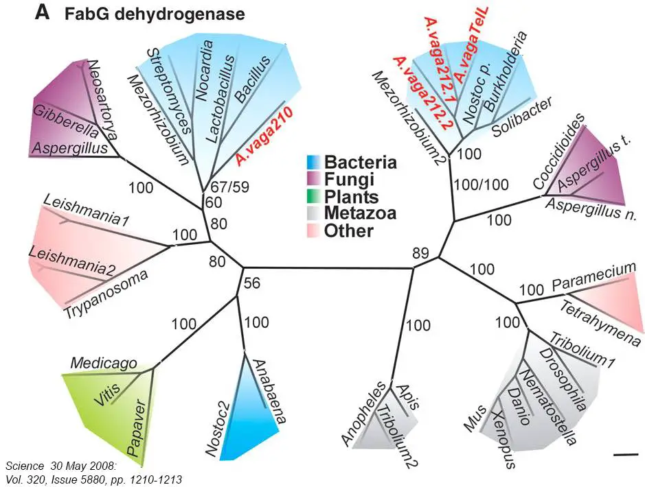
[In this figure] Recent genomic analysis surprisingly found that many genes in bdelloid rotifers appear to have originated in bacteria, fungi, and plants. Some of the foreign genes were defective, whereas others were intact and active. Massive horizontal gene transfer may represent an important force in bdelloid evolution.
Photo source: Science.
Rotifers are also a great model organism to study aging. Rotifers have many advantages including, ease of culture, a short life span of 2 weeks – 2 months, and simplified organ systems analogous to humans. For example, scientists studied calorie restriction (reducing average daily caloric intake) can extend the life span in rotifers.
Interesting facts about rotifers
Scientists recently discovered a substance made by Bdelloid rotifers (Rotaria rotaria) that can paralyze parasitic flatworms that cause schistosomiasis, a dangerous infection that affects 200 million people worldwide. Schistosomiasis is also known as snail fever and bilharzia, a disease caused by parasitic flatworms called schistosomes. If the substance produced by rotifers can be developed as a drug, it will be a huge success in treating parasitic diseases.
Summary
- Rotifers are multi-cellular (around 1000 cells) animals of microscopic sizes (100-500 μm).
- The phylum Rotifera includes three classes of Rotifers: Bdelloidea, Monogononta, and Seisonidea.
- Rotifers live in various aquatic and humid environments.
- Rotifers have several organ systems, including a complete digestive tract.
- Rotifers got their name from the unique structures of coronae of cilia around the mouth. The rapid movement of the cilia makes them appear to whirl like a wheel.
- Rotifers have a chewing jaw (trophi) to grind the food before passing to the stomach.
- Rotifers have a brain, eyespots, and several sensory organs.
- Most rotifers are free-living. They can swim and walk by anchoring their feet.
- Rotifers can reproduce both asexually and sexually, depending on environmental conditions.
- Some species of rotifers are all female due to asexual reproduction, called Parthenogenesis.
- Cryptobiosis allows rotifers to survive through extremely harsh conditions.
References
“Introduction to the Rotifera”
“Nano-Animals, Part I: Rotifers”
“An introduction to bdelloid rotifers and their study” by Aydin Örstan and Michael Plewka
“The Rotifer Jaw” by Mike Morgan
“Phylum Rotifera”
“Frontal aspects of bdellod rotifers” by Michael Plewka
“Photographing Rotifers” by Dr. Robert Berdan
“Bdelloid Rotifers: Female Filter Feeders” by Thomas Webster
“Chapter 36 – Rotifers as a Model for the Biology of Aging” by Kristin E.Gribble and Terry W.Snell
Gallery of rotifer images
“Life in Water” by Michael Plewka