
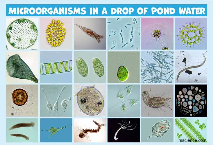
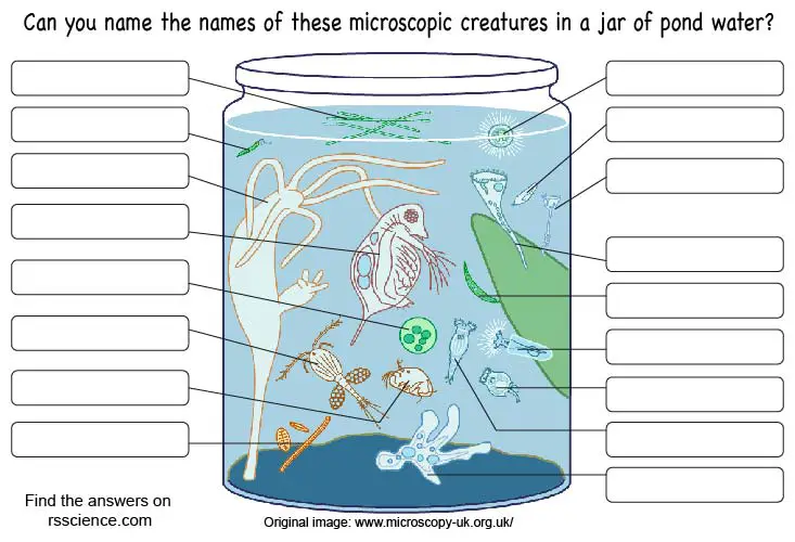
Within every drop of pond water lurks an invisible world, alive with an amazing variety of microscopic creatures. You can find simple life forms such as bacteria, great oxygen-producers like algae, all kinds of alien-like protozoans, and cute microscopic animals like water bears.
With the help of this guide and a microscope, you can bring these tiny creatures into focus and discover the secrets in which they live.
Let’s see what you will find in a drop of pond water!
This article covers
What are microorganisms?
Microorganisms or microbes are microscopic organisms that can be found all around the world. They can be unicellular or cell clusters. As such, they are only visible under a microscope.
They are largely composed of the members of the Archaea and Bacteria kingdoms (both are prokaryotic cells) and unicellular Protists (belong to eukaryotes). Within the umbrella of Protist, the members can be further divided into:
• Protozoa – “animal-like”, like Paramecium and Amoeba;
• Protophyta – “plant-like”, like Diatom, green, red, and brown algae;
• Molds – “fungus-like”, like water mold and slime mold.
Typically, pond water contains a variety of microorganisms. They could be free-living single cells or microorganisms that cluster together in large numbers (colonies). Sometimes, you will find microscopic animals and plants that consist of hundreds or even thousands of cells. We will also include these multicellular organisms in this guide.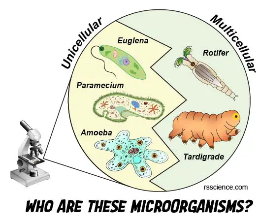
[In this figure] Many pond creatures fall in the size range between 100-500 micrometers (μm). However, they can be totally different. Some microorganisms (such as Euglena, Paramecium, and Amoeba) contain only one gigantic cell (called unicellular or single-cellular) and are classified as protists. Others (such as Rotifer and Tardigrade), however, contain thousands of cells and belong to multicellular animals.
How small are these microorganisms?
Before we start hunting for microorganisms, we should have a good idea of how small they are and the relative biology scale.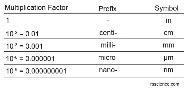
[In this table] The biology scale can span a huge range: from an atom to an elephant. To express numbers that are too large or too small to be conveniently written in decimal form, it is common to use “Scientific notation” in biology and other science branches. Here is the list of units that describe the size (in length) of biological objects. “m” = meter.
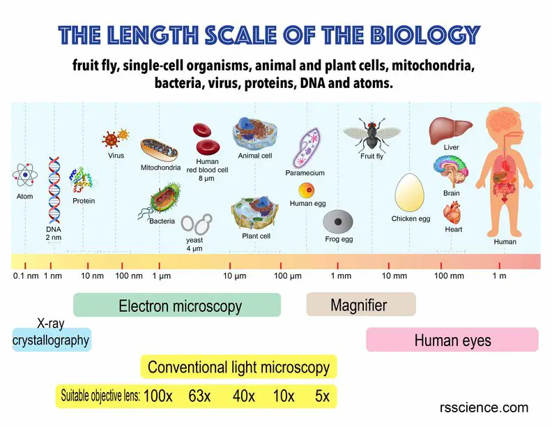
[In this figure] Knowing the exact dimension of the specimen is critical for a biologist. Here, we summarize a biological scale to give you a sense of the sizes of different biological objects. Generally, the microorganisms we can find and see under a light microscope fall in the range between 0.5 – 1000 µm.
Why are microorganisms important?
Every organism plays an essential role in the ecosystem and it is also true for microscopic organisms. Autotrophic microorganisms like algae are the foundation and producers of nutrients for all other living creatures. They are eaten by primary consumers like protozoans, which then become the food of larger predators. This builds up a food web of the aquatic ecosystem.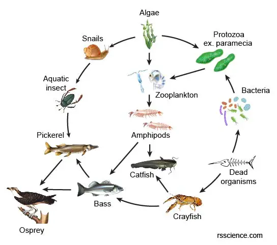
[In this figure] Food web of the aquatic ecosystem.
How do we name and classify these microorganisms?
To classify, categorize, and organize all the microorganisms, we need a naming system that everyone can follow. Taxonomy is the science of biological classification that provides species with names. It can help to distinguish how similar or different living organisms are to each other.
Taxonomy works very similarly to the library. Inside the library, books are divided into individual areas: the children’s books in one section, the adult books in another, and the teen books in another section. Within each of those sections, there will be more categories like fiction or non-fiction. There will be even more divisions within those sections, such as mystery, science fiction, and romance novels in the fiction section. Finally, after layer by layer of narrowing, you will get down to a single book.
Taxonomy works the same way. The most common system has 7 major levels. At the top, there are the Kingdoms (animal, plant, or bacteria). Then, it follows by Phylum, Class, Order, Family, Genus, and Species. In the figure below, you can see how this system classifies the human, orange, and gut bacterium (called E. coli.) in their unique place.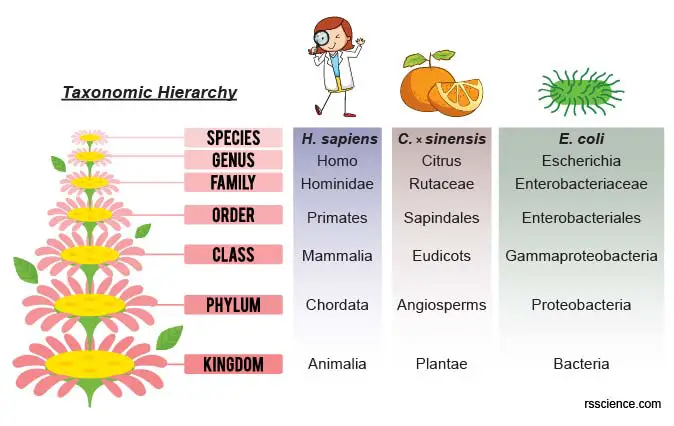
Here, we use the six-kingdom classification system (Archaea, Bacteria, Protista, Plantae, Fungi, and Animalia). The system of taxonomic hierarchy is still under debate and evaluation. Some species, like Euglena, are difficult to classify. You may learn more about this topic on Wikipedia.
Where can you find microorganisms for microscopic projects?
Microorganisms are widespread in all kinds of freshwater environments. Microorganisms stay with their source of food. Ponds or slow-flowing creeks with decayed organic materials in the bottom sediments (like leaves) are ideal habitations to find all kinds of microorganisms.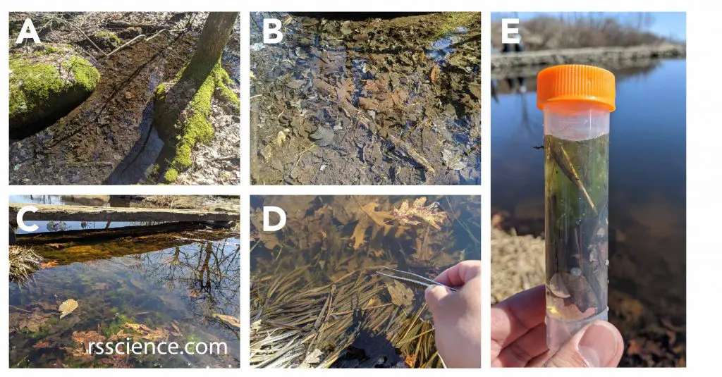
[In this figure] Places to collect microorganisms.
(A-C) Nutrient-rich ponds or slow-flowing creeks are ideal habitations to find all kinds of microorganisms, like paramecia, amoebas, rotifers, water bears, daphnia, and diatoms. (D-E) I used the forceps to collect some decaying leaves and a dropper to collect water with sediments into my sample vial. I brought them home to look for microorganisms under my microscope.
Different microorganisms have their preferred habitations. Therefore, make sure you are looking at the right place. Plankton is the diverse collection of organisms found floating in water that is unable to propel themselves against a current. Phytoplankton is referred to as autotrophic algae near the water surface where there is sufficient light to support photosynthesis. Zooplankton is small protozoans or animals that feed on other plankton. Planktonic microorganisms can be collected by sampling water or using a plankton net.
Some microorganisms like to anchor on the surface of other objects like rocks and leaves of aquatic plants. They can filter-feed or catch passing prey. Also, don’t miss the bottom sediments. There are plenty of microorganisms (called Benthos) that like to stay in this benthic zone where decayed organic materials are rich as food sources.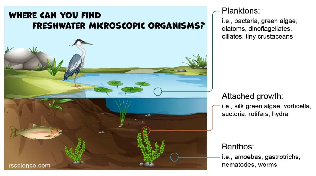
[In this figure] The habitation of microorganisms.
They can be found (1) free-floating in water; (2) staying at the bottom or in sediments; (3) attaching on the surface of rocks or aquatic plants.
What kinds of microorganisms can we find in freshwater?
Within a drop of pond water, we may find species coming from all kingdoms of Earth’s life. The members of Bacteria, Protista, and Animalia are the most common visitors. Archaea or ancient bacteria may be difficult to identify because they are very small and usually live in harsh environments like hot springs.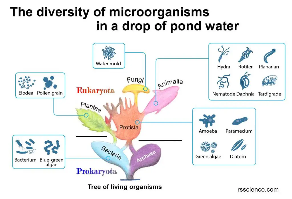
[In this figure] Tree of living organisms showing the origins of eukaryotes and prokaryotes. See where the microorganisms belong when you find them under your microscope.
Modified from wiki.
Let’s see what we can find under the microscope!
I. Prokaryotic microorganisms
These microorganisms are prokaryotes, meaning they do not have a membrane-bound cell nucleus and other organelles.Name Characteristic Taxonomy Size Bacteria 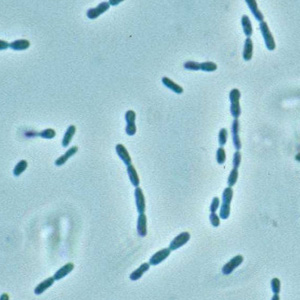
– Prokaryotes
– Single cells
– Rod, sphere, or filament-shapedKingdom: Bacteria 1µm or less Cyanobacteria (blue-green algae) 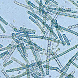
– Prokaryotes
– Free-living photosynthetic bacteria
– Produce the oxygen on EarthKingdom: Bacteria Phylum: Cyanobacteria 0.5 – 60µm
II. Microscopic autotrophic organisms
These microorganisms are single-cellular eukaryotes that can produce food through sunlight by photosynthesis (i.e., algae).Name Characteristic Taxonomy Size Chlamydomonas 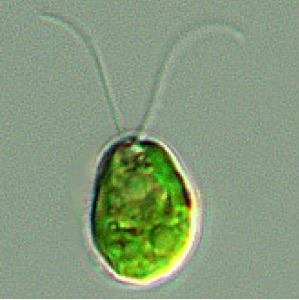
– Green algae with two flagella
– Small single cells or clusters
– Rapid movementClass: Chlorophyceae
i.e., Chlamydomonas sp.< 50 µm Volvox 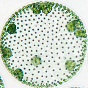
– Green algae with two flagella
– Spherical colony formed by single cellsClass: Chlorophyceae
i.e., Volvox sp.500µm – 2mm Pediastrum 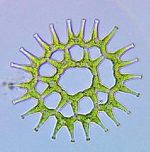
– Green algae (no flagella)
– Single cells or clusters in arrangement
– No movementClass: Chlorophyceae
i.e., Pediastrum sp.< 500 µm Spirogyra
(water silk)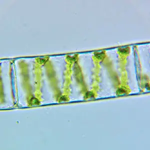
– Filamentous green algae
– Non-branching chains of single cellular units
– No movementClass: Zygnematophyceae i.e., Spirogyra sp. 100µm – several cm Closterium 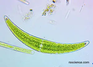
– Green algae (no flagella)
– Single cells, new moon-shaped
– No movementClass: Zygnematophyceae i.e., Closterium sp. 100 -1000 µm Desmidium 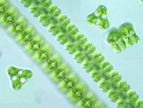
– Green algae (no flagella)
– Single cells or assembled into long filaments
– Various shapesClass: Zygnematophyceae i.e., Desmidium sp. 100 -1000 µm Diatom 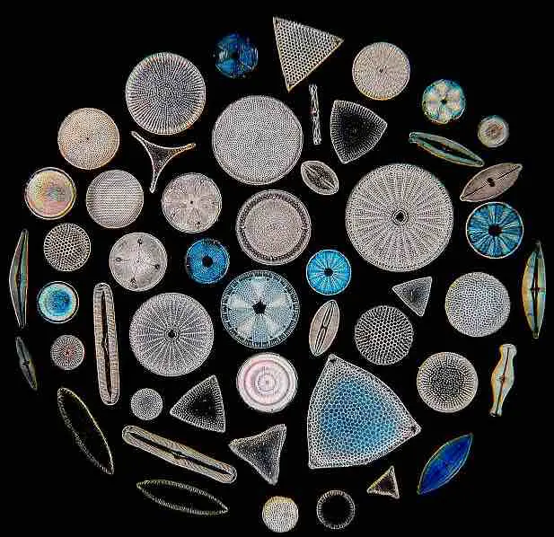
– Brown algae
– Slow gliding motion
– Cell wall (frustules) made of silicaClass: Bacillariophyceae i.e., Pennales < 500 µm Synura 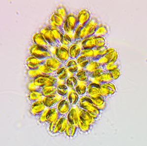
– Brown algae
– Cluster of cells
– Rotating with flagellaClass: Synurophyceae
i.e., Synura sp.< 500 µm Euglena 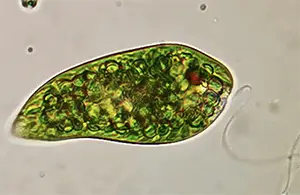
– Contain chloroplasts
– Move by flagella
– Red eye spot
– Have features of animal and plantPhylum: Euglenozoa
i.e., Euglena sp.< 400 µm Dinoflagellate 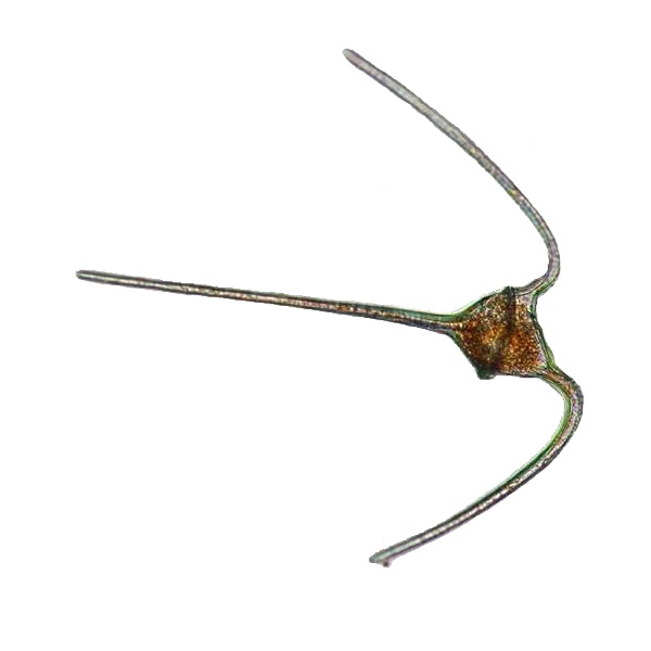
– Free swimming with flagella
– Tough armorClass: Dinoflagellata
i.e., Ceratium sp.< 400 µm
III. Heterotrophic protozoa
These microorganisms are singled cellular eukaryotes that obtain nutrition from outside sources. They are classified in the kingdom of Protista and are commonly referred to as “Protozoa.”Name Characteristic Taxonomy Size Amoeba 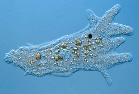
– Slow movement by pseudopodia
– Engulfs food by phagocytosisPhylum: Amoebozoa i.e., Amoeba proteus 250 -750 µm Shelled Amoeba 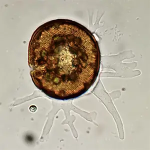
– Testate amoeba with a shell
– Slow movement by pseudopodiaPhylum: Amoebozoa i.e., Arcella sp. 50 – 300 µm Heliozoa 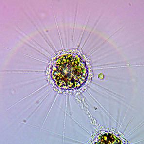
– Spherical
– Radiating hair-like pseudopodsPhylum: Sacrodina Order: Heliozoa 50 – 1000 µm Paramecium 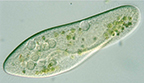
– Free-living ciliates
– Movement by waving ciliaPhylum: Ciliophora Order: Peniculida
i.e., Paramecium sp.50 – 300 µm Didinium 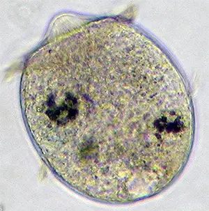
– Free-living ciliates
– Capture paramecia using the mouthPhylum: Ciliophora Order: Haptorida
i.e., Didinium nasutum50-150 µm Stentor 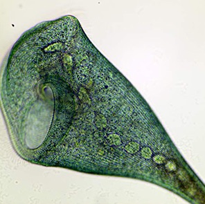
– Large, free-living ciliates
– Contractile
– Collect food by cilia on the mouth endPhylum: Ciliophora Order: Heterotrichea
i.e., Stentor sp.100 µm – 4 mm Suctoria 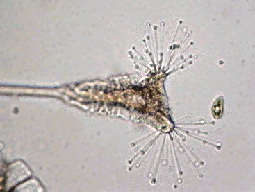
– Ciliates with sticky tentacles
– Attached to a surface of a substratePhylum: Ciliophora Order: Eridogenida
i.e., Tokophyra sp.<500 µm Vorticella 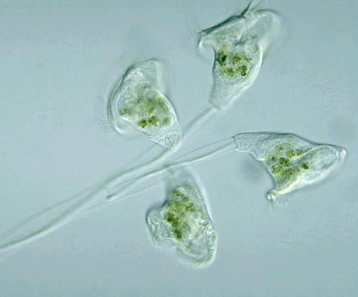
– Bell-shaped ciliates
– Cilia around the mouth
– Often colonial
– Attached to a surface of a substratePhylum: Ciliophora Order: Peritrichida
i.e., Vorticella sp.<500 µm Coleps 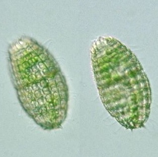
– Barrel-shaped ciliates
– Bodies covered by platesPhylum: Ciliophora Order: Prostomatea
i.e., Coleps sp.<100 µm Lacrymaria 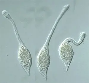
– Ciliates with a long neck Phylum: Ciliophora Class: Litostomatea
i.e., Lacrymaria olor100-150 µm
IV. Microscopic animals
These organisms are tiny animals. They have functional organ systems consisting of many specialized cells. However, many of them may be smaller than some giant single-celled protozoans.Name Characteristic Taxonomy Size Rotifer (Bdelloid) 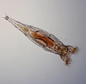
– Corona with cilia
– Attached on a surface
– Move like a leech
– Filter-feedingPhylum: Rotifera
Class: Bdelloided100-500 µm Rotifer (Monogononta) 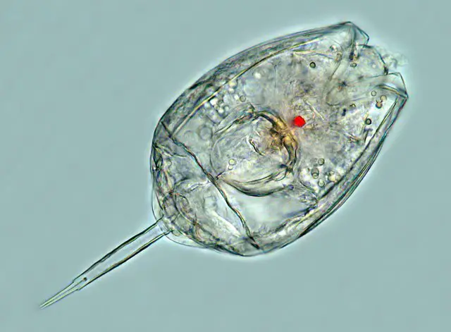
– Free swimming rotifers
– Filter-feedingPhylum: Rotifer
Class: Monogononta50-150 µm Hydra 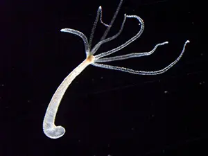
– Green, brown or colorless body
– Use tentacles to catch preys
– Attached on a surfacePhylum: Cnidaria
Class: Hydrozoa
i.e., Hydra sp.200 µm – several cm Planarian (flatworm) 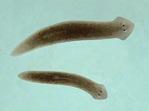
– Flattened body
– Two eyespots
– Move in gliding motionPhylum: Platyhelminthes
Class: Turbellaria
i.e., Planaria sp.500 µm – 2 cm Nematode (roundworm) 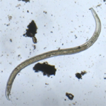
– Round body
– Move in rapid “s” form
– Has anterior and posterior openingsPhylum: Nematodes Class: Chromadorea
i.e., C. elegans1-10 mm Gastrotrich (hairyback) 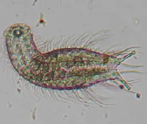
– Mainly benthic
– Hair-like bristles
– Eat algae, bacteria, protozoaPhylum: Gastrotricha
i.e., Lepidodermella sp.100 µm – 3 mm Tardigrade (water bear) 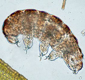
– Head and 4 trunk segments
– 4 pairs of legs
– Two eyespots
– Survive harsh conditions by CryptobiosisPhylum: Tardigrada Class: Eutardigrada, Heterotardigrada 0.5 – 1.5 mm Oligochaeta (earthworm) 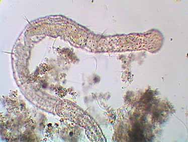
– Segmented
– Worm motion
– Hair bundlesPhylum: Annelida
Class: Oligochaetafew mm to 1 cm
V. Arthropods (crustaceans and insects)
There are a huge variety of microscopic arthropods in freshwater that are worth discussing separately. They are multicellular animals with segmented bodies covered by exoskeletons. They could be tiny crustaceans or larvae of aquatic insects.Name Characteristic Taxonomy Size Daphnia (water flea) 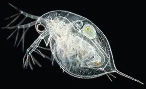
– Antennae
– Large compound eyes
– HoloplanktonClass: Crustacea
Order: Cladocera
i.e. Daphnia sp.0.2 – 3 mm Copepod 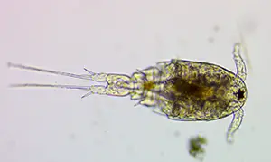
– Long antennae
– Tiny eyespots
– HoloplanktonClass: Crustacea
Order: Copepoda1 – 2 mm Ostracod
(seed shrimp)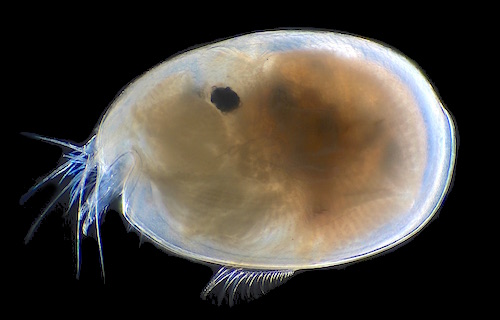
– Bean-like shell
– Filter feeders
– Bivalve carapaceClass: Crustacea
Order: Ostracoda i.e. Cypris sp.1 – 5 mm Amphipod 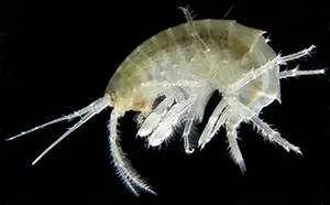
– Curved, compressed body
– Humped back
– ScavengersClass: Crustacea Order: Amphipoda <10 mm Mosquito larva (wiggler) 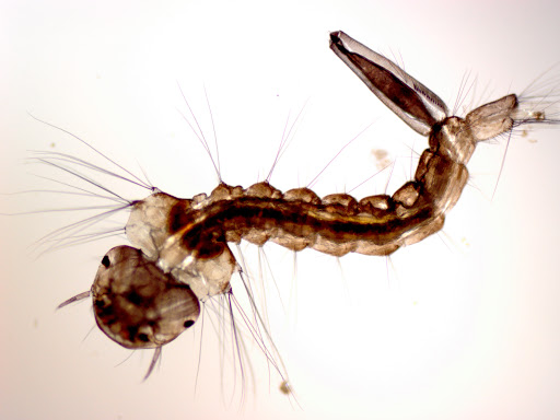
– Long slender body
– Move in undulating “s” curvesClass: Insecta Order: Diptera <10 mm
Introduction of pond microorganisms – Definition, biology, and fun facts
Bacteria
Bacteria (singular: bacterium) are single-celled organisms that thrive in diverse environments, including a freshwater pond, lake, and swamp. Bacteria are prokaryotes (pro-KAR-ee-ot-es) that don’t have the membrane-bound nucleus and other organelles. Bacteria are small, simple cells, measuring around 0.1-5 μm in diameter. They have diverse shapes, including spheres, rods, and filaments. Some of them can swim around by waving their tail-like flagella. Some bacteria can cause diseases in humans. However, they are also essential nutrient sources for many protozoa.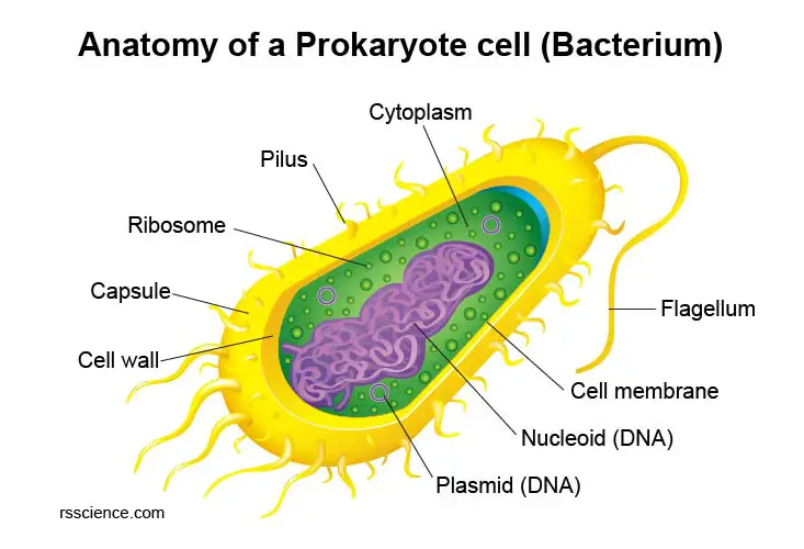
[In this figure] Anatomy of a bacterium.
The key structures in a prokaryote cell are nucleoid, plasmid, cytoplasm, flagellum, pilus, ribosome, capsule, cell membrane, and cell wall.
Extended read:
Cyanobacteria
Cyanobacteria, also known as “blue-green algae,” are a phylum of prokaryotes consisting of free-living photosynthetic bacteria. Cyanobacteria are autotrophic and can obtain their energy through photosynthesis. Scientists believe that cyanobacteria played a significant role in Earth’s history by producing the largest source of O2 in the atmosphere today. However, an overgrowth of cyanobacteria called cyanobacteria bloom is harmful. Cyanobacteria produce a range of toxins known as cyanotoxins that can pose a danger to humans and animals. Cyanobacteria range in size from 0.5 to 60 μm, which represents the largest prokaryotic organism.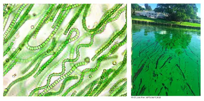
[In this figure] Left: Microscopic images of Cyanobacteria, showing many single cells assembled into long chains. Right: A picture of the cyanobacteria bloom.
Photo source: cyanobacteria, cyanobacterial bloom
Protophyta
Protophyta is an informal term to describe a group of “plant-like” protists. Common examples include green, red, brown algae, and diatoms. Most of them are autotrophic and can produce their energy through photosynthesis in chloroplasts.
Green algae
Algae (singular alga) is an informal term for a large, diverse group of organisms that can obtain the energy to grow through photosynthesis (similar to plants). Algae include members ranging from unicellular microalgae, such as Chlorella and Diatoms, to multicellular forms, such as the giant kelp (a large brown seaweed that may grow up to 50 m in length)! Most of them are aquatic (living underwater) and contain chloroplasts. They also lack the various structures that characterize land plants, such as the roots, leaves, stomata, and vascular bundles. Most algae belong to the Protist kingdom, which includes any eukaryotic organism that is not an animal, plant, or fungus.
Green algae are excellent examples to learn the diversity of living organisms in nature and they are also easy to collect. You can collect different types of algae from a pool or lake. For instance, Spirogyra and Zygnema are filamentous green algae like a brush of green hairs. Under the microscope, you can easily see how their cells arrange into the long fiber shape.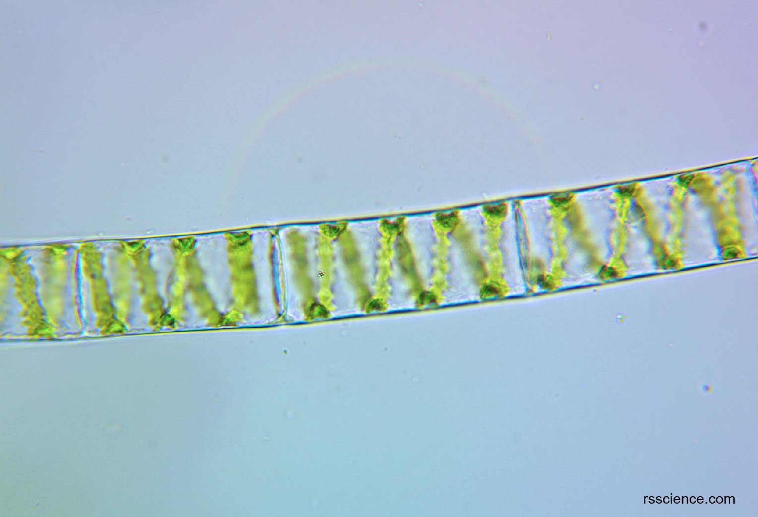
[In this figure] Spirogyra under a microscope.
One of the spirogyra’s characteristics is its helical arrangement of chloroplast strands.
You can also collect free-floating green algae that are in the form of single cells or colonies with a beautiful arrangement. Some of them are consistently twitching or rotating by waving their flagella.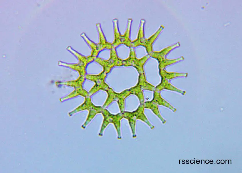
[In this figure] Pediastrum under a light microscope.
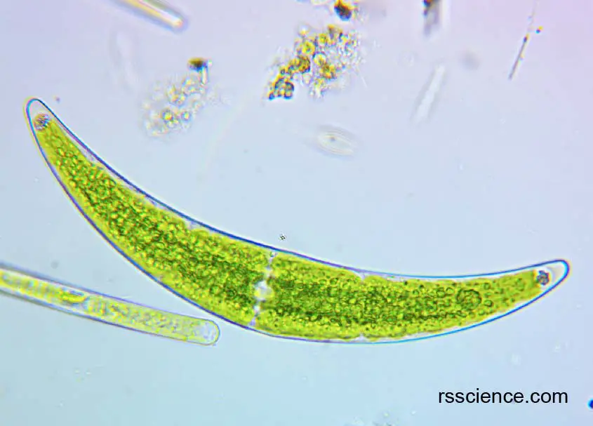
[In this figure] Closterium under a light microscope.
[In this video] Volvox forms spherical colonies of up to 50,000 cells.
[In this video] Chlamydomonas are single-cellular green algae with two flagella.
Extended read
Diatom
Diatom is a big group of microalgae. They are free-floating unicellular algae found in both the oceans and freshwater. A unique feature of diatom cells is that they are enclosed within a cell wall made of silica (like glass) called a frustule. The photonic structures in the frustules such as pores and chambers on the micro to nanoscale interact with the visible light spectrum, creating colorful, shining, and opal-like appearances. Therefore, they are described as “jewels of the sea.” The abundance of diatoms has a huge impact on the earth. One-fifth of the photosynthesis on the earth is done by diatoms.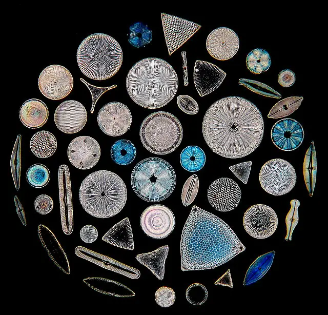
[In this figure] The beauty of diatoms on a microscopic slide.
Photo credit: micromagus.net
Extended read
Synura
Synura is a small group of golden-brown algae containing chloroplasts, found mostly in freshwater. They are covered in silicate scales and of yellowish color. A group of Synura cells tends to aggregate and assemble into a cluster. Each cell has its two flagella facing outward. When they wave their flagella together, the entire cluster starts to rotate. It is super cute and fun to see them rolling this way! I called them microscopic “Roasted Corn on the Cob.”
Synura sometimes forms blooms (usually in the spring) and releases ketones and aldehydes that can give the water an unpleasant fish-like odor or taste.
[In this video] An funny video showing the assembly and rotation of Synura.
Extended read
Euglena
Euglena (Greek: “eu” = true, “glene” = eye-ball) is a genus of single-cell eukaryotes with flagella and chloroplasts. Euglena, indeed, confused the biologists when considering its biological classification. Euglena shares both characteristics of plants and animals. For example, euglena contains chloroplasts; thus, they can make their own food, a characteristic of plants. In contrast, euglena can also move using its flagella and consume food through phagocytosis, which is animals’ characteristic. Euglena also lacks a cell wall.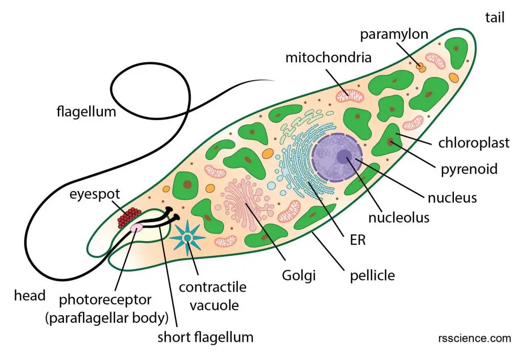
[In this figure] Euglena anatomy and its organelles.
[In this video] Euglena under a microscope.
Scientists suggested that Euglena may become a future food source due to its high nutrient value. I surprisingly found that there is a Japanese company selling Euglena cakes and energy bars. Does anyone want to try?
Extended read
Dinoflagellate
Dinoflagellates are unicellular protists wearing tough armors that exhibit a great diversity of forms. Dinoflagellate is another organism like Euglena that gives the scientists big trouble. Many dinoflagellates are photosynthetic, manufacturing their own food using the energy from sunlight, and providing a food source for other organisms. However, there are some dinoflagellates that are parasites on fishes or on other protists. So, are they plants or animals?
[In this figure] Electron microscopic images showing various types of Dinoflagellates.
Photo source: medium.com
Some species of dinoflagellates are capable of producing their own light through bioluminescence, which also makes fireflies glow. Tiny dinoflagellates can make a huge environmental impact. Some coastal marine species “bloom” during the warm months of summer, producing a “red tide“. When this happens, many surrounding marine lives suffer, from the dinoflagellates producing a neurotoxin that affects muscle function. Even humans could also be affected by eating fish or shellfishes containing the toxins.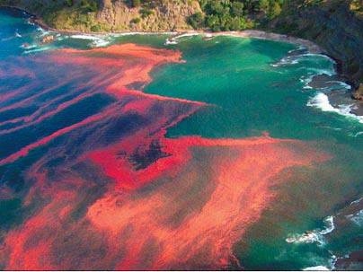
[In this figure] Red tide caused by Dinoflagellate bloom.
Photo source: Dinoflagellate bloom.
[In this video] Dinoflagellate bioluminescence stimulated by hand.
Protozoa
Protozoa is an informal term for “animal-like” unicellular protists. They are heterotrophic and have to eat other microorganisms like bacteria to obtain energy.
Amoeba
Amoeba is a group of primitive protists. Among the big family of Amoebas, Amoeba proteus is probably the best-known member – common in classrooms and research laboratories. Amoeba proteus is known for how they move, a transformative crawling manner – through extension and retraction of “false feet” (or pseudopods) over varied substrates. Amoeba proteus does not have a fixed shape – it constantly changes because it extends its pseudopods.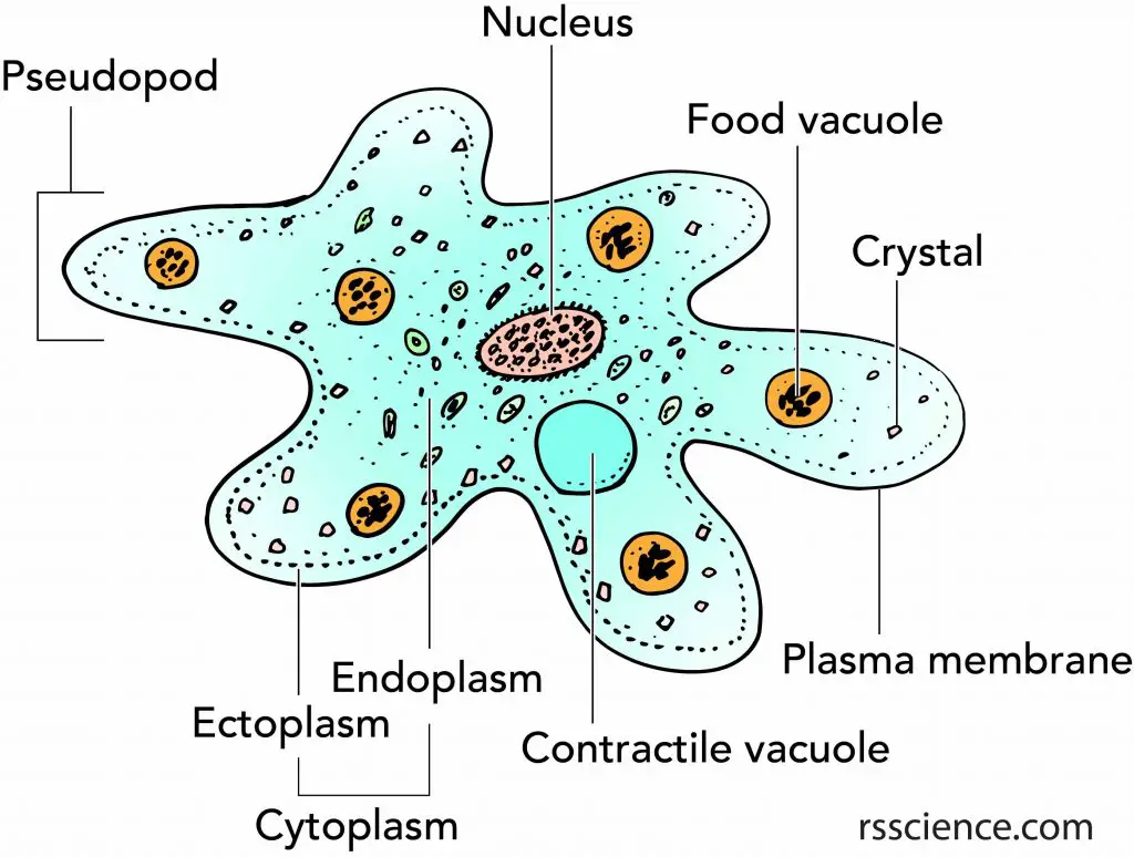
[In this figure] The structure of Amoeba.
An amoeba has a single granular nucleus, containing most of the organism’s DNA.
Amoeba is a giant eukaryotic cell. It has a membrane-bound nucleus and many organelles such as contractile vacuole and food vacuole. Amoeba can eat, reproduce, and even sense its environment (i.e., light, temperature, and electric field). It is tiny but not simple!
[In this video] The Microanatomy of Amoeba.
Extended read:
Shelled Amoeba
Do all amoebas look like Amoeba proteus? The answer is no.
Surprisingly, some species of Amoebas make protective shells, called “tests,” around their cells. Some shelled Amoebas make the tests entirely by themselves, and the materials could be organics, siliceous (containing silica), or calcareous (containing calcium carbonate) components produced by the Amoebas. Some shelled Amoebas prepare their tests by collecting particles of sediment around them and gluing these mineral particles together with slime ingredients secreted from the cells.
[In this video] A shelled amoeba called Arcella.
Heliozoa
Heliozoa is commonly known as sun-animalcules. Its stiff arms radiate from a spherical body, which is responsible for its common name. These arms can be used for capturing food, sensation, movement, and attachment. I personally like to call Heliozoa a microscopic “Uni” (sea urchin in Japanese)! Heliozoa is a cousin of Amoebas.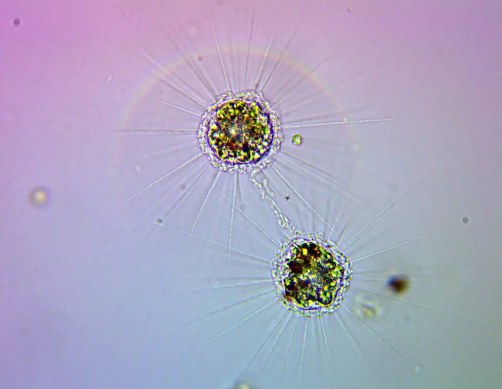
[In this figure] Two Heliozoans under the microscope.
Ciliate
Many microorganisms you can find in pond water belong to “ciliates.” The ciliates are a group of protozoans characterized by the presence of hair-like organelles called cilia. Cilia are structurally similar to flagella but are in general shorter and present in much larger numbers.
Paramecium
Paramecium (pair-ah-me-see-um; plural, Paramecia) is a unicellular ciliate with a shape resembling a slipper. Although paramecium is small and has only one cell, it can do everything that a living creature can do: Paramecium can swim, digest food, and reproduce. Paramecium’s cell contains several complex organelles performing specific functions to make its survival possible.
Paramecium collects foods via its mouth, called the oral groove. The food materials enter the cell body and then are digested in food vacuoles. Paramecia eat other microorganisms such as bacteria, yeasts, and algae.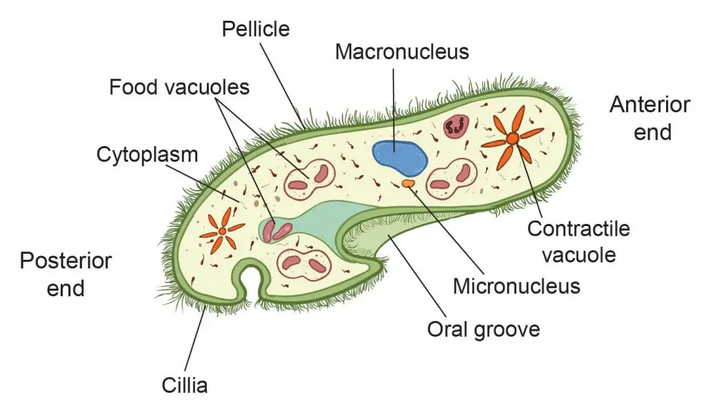
[In this figure] The organelles of a paramecium.
Paramecium constantly moves by beating rows of microscopic hairs, called cilia. The cilia of paramecium move like many tiny oars, propelling the organism through the water at a rate that is “four times its body length per second”. That said, if a paramecium is as tall as a human being, it can swim four times faster than Olympic gold medalist Michael Phelps.
[In this video] The movement of Paramecium caudatum under a microscope.
Extended read:
Didinium
Didinium is a genus of free-living ciliates that are well-known predators of paramecia. Didinium has a barrel-shaped body encircled by two ciliary bands, which can move Didinium through water by rotating the cell around its axis. Didinium has a cone-shaped mouth that can capture and paralyze its paramecium prey. Once captured, the prey is engulfed through Didinium’s mouth.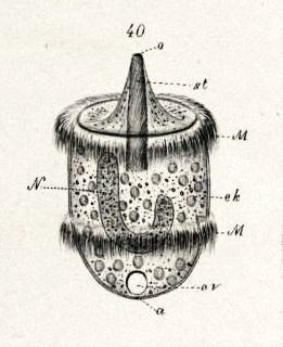
[In this figure] A 19th-century illumination showing a Didinium nasutum with two bands of cilia and a mouth protrusion.
[In this video] Didinium eats paramecium.
Stentor
Stentor, sometimes called trumpet animalcules, are a genus of filter-feeding ciliates. Stentors can reach lengths of two millimeters; as such, they are among the biggest known unicellular organisms. Their bodies are generally horn-shaped. A ring of cilia around the anterior “mouth” sweeps in food and aids in swimming.
[In this video] Stentors under a dark-field microscope.
Extended read:
Vorticella
Vorticella is a genus of bell-shaped ciliates that have a stalk to attach itself to a surface of a substrate. The stalk can contract, allowing it to pull the cell body back when sensing danger. Vorticella and stentor are cousins. They both use cilia to collect food particles.
[In this video] Vorticella looks like tiny tulips under the microscope.
Extended read:
Suctoria
Suctoria looks like Vorticella, which also has a stalk to attach itself to substrates. Instead of filter-feeding by cilia, Suctoria feeds using several specialized tentacles. These tentacles have toxic extrusomes called haptocysts at the tip. Suctoria uses its tentacles to catch prey and then sucks the prey’s cytoplasm directly into a food vacuole inside the cell, where it digests and absorbs the contents. Functionally, Suctoria hunts like a hydra (will discuss later). However, a hydra consists of hundreds of cells, but Suctoria is only a single cell!
[In this video] Suctoria preys and paralyzes a rotifer by its tentacles.
Coleps
Coleps is a genus of ciliates with barrel-shaped bodies and a shell made of mineralized plates. Coleps swim around by cilia and feed on bacteria, algae, flagellates, and other ciliates. Like Suctoria, Coleps can paralyze and capture the prey by toxicysts, which are organelles containing poison from their oral area.
[In this video] Colep uses its ciliates to swim.
Lacrymaria
Lacrymaria is a group of ciliates found in freshwater ponds. Its name means “swan tear” in Latin and refers to its general shape: namely, a teardrop-shaped cell with a small “head” at the end of a long slender “neck.” This protist is notable for its ability to extend the “neck” of the cell up to 7 times its body length and manipulate it in many directions — even around obstacles — to capture its food.
[In this video] Lacrymaria olor – One of Earth’s Strangest Creatures
Micro-animal
Micro-animals are animals so small that they can be visually observed only under a microscope. Unlike most other microorganisms, that are unicellular, micro-animals are unique because they are multicellular, like all other animals.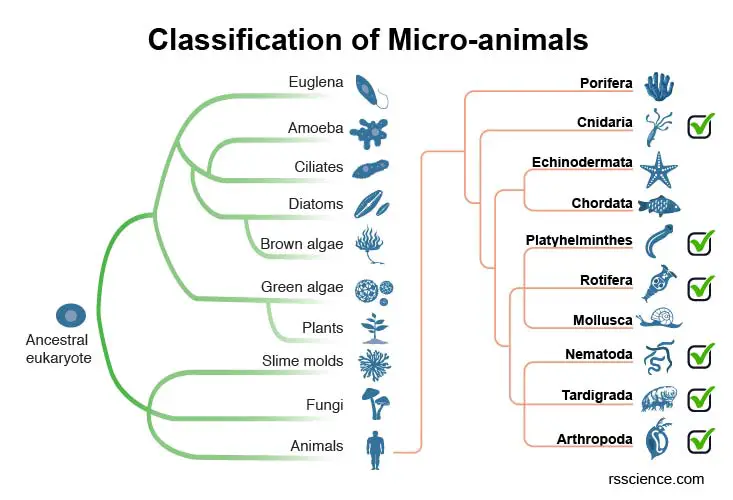
[In this figure] The animal kingdom Animalia contains approximately 35 phyla (singular: phylum). The micro-animals we discussed here belongs to 6 phyla. By the way, we (humans) are in the phylum Chordata (possessing a notochord or a dorsal nerve cord).
Rotifers
Rotifers are microscopic aquatic animals. Rotifers got their name from the corona: a rotating, wheel-like structure that is covered with cilia at their heads. Rotifers also have a jawed mouth and complete digestive, sensory, and reproductive organ systems. Rotifers are filter-feeders that eat dead material, algae, bacteria, and other microscopic living organisms, and are therefore very important components of aquatic food webs.
[In this video] A video capturing the “rotating wheels” of a rotifer and won the 2012 Nikon Small World in Motion Competition.
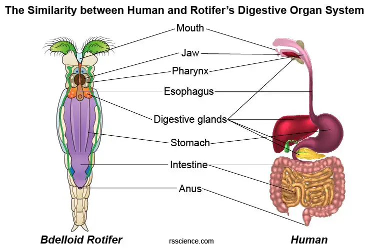
[In this figure] Don’t underestimate a rotifer. It has a complete set of the digestive organ system that is very similar to ours packed in a tiny body.
There are three types of rotifers: Bdelloidea, Monogononta, and Seisonidea. Among them, “Bdelloid” rotifers are the most common microorganisms that you can find in all kinds of humid environments.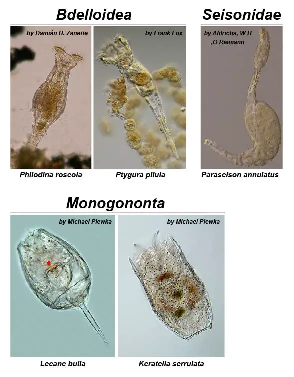
[In this image] Examples from three classes of rotifers.
Species from the class Bdelloidea are characterized by a large corona and telescopic body. Rotifers from the class Monogononta have a smaller corona than Bdelloid rotifers and a single gonad (body segment), which gives the class its name. Seisonidae rotifers are saltwater rotifers found on the gills of Nebalia, a marine crustacean. They have a large body and elongate neck with reduced corona.
Photo source: Monogononta.
Extended read
Hydra
The name “Hydra” came from a snake-like water monster in Greek and Roman mythology, called The Lernaean Hydra. Hydra has a cylindrical, radially symmetric body with a mouth opening surrounded by several (6-10) tentacles. These tentacles can extend and capture the prey by specialized stinging cells called cnidocytes. Upon contact with the prey, the contents of the cnidocytes are explosively discharged, firing a dart-like thread containing neurotoxins. Paralyzed preys will then be engulfed within the body cavity for digestion. Most of the time, a hydra remains attached by the basal disc to some suitable object in the water. However, occasionally hydra can move quite readily, especially when hunting.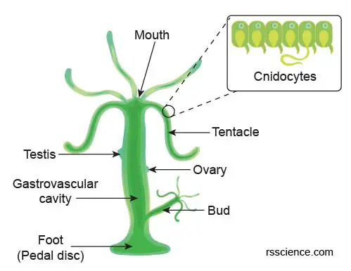
[In this figure] An anatomy structure scheme of hydra.
Extended read
Planarian
Planarian is one of many flatworms that belong to the phylum of Platyhelminthes. Many flatworm species are parasitic (i.e., beef tapeworm, an intestinal parasite in humans causing taeniasis), but planaria are free-living and harmless. Planaria are commonly found in aqua tanks. They have two eyespots that can detect the intensity of light. They have primitive brains and nervous systems. They are also very interesting scientific objects to study because of their “super regeneration capability”. For example, a planarian cut into three pieces (head, body, tail) will regenerate into three separate individuals. You can even create a three-headed planarian if you make two incisions on its head.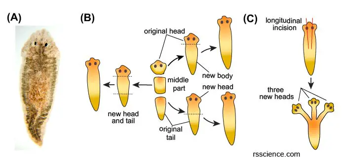
[In this figure] Planarian regeneration.
(A) The most frequently used Planarian in high school and first-year college laboratories is the brownish Girardia tigrina. (B-C) Diagrams of the main planarian regeneration experiments.
Extended read
Neonates
Although many nematode worms are parasitic, you still can find free-living nematodes like Caenorhabditis elegans (or C. elegans) in the pond water. C. elegans is a transparent nematode about 1 mm in length that lives in temperate soil environments. It moves like a snake and feeds on bacteria. C. elegans is a nematode species with special scientific importance. Because of its transparent body, the internal cells of C. elegans are easy to observe under a microscope. Scientists learned many biologies using C. elegans, including gene regulation, programmed cell death, and aging. The identification of the first long-lived mutants in C. elegans led to the discovery of a genetic pathway (insulin pathway) that regulates aging. This research may in the future extend the human’s lifespan!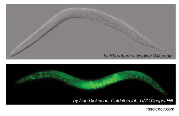
[In this figure] C. elegans is a model organism studied in many laboratories around the world.
Upper image: An adult C. elegans under a stereo microscope. Because of the transparent body, the cells inside the worm can be easily studied. Lower image: A C. elegans was genetically labeled with green fluorescent proteins (GFP) in all the cells. GFP labeling with the fluorescence microscope is a very powerful tool to study the function of specific cell types in biomedical research.
Gastrotrich
Gastrotrichs, commonly referred to as hairybellies or hairybacks, are a group of microscopic, worm-like, animals. Gastrotrich has a head with a brain and sensory organs, a body with a simple gut, and skin covered by many cilia. This gave Gastrotrich a “hairy” looking. They are mostly benthic (living at the bottom) and feed on detritus, sucking up organic particles with their muscular pharynx.
[In this video] Gastrotrich under a microscope.
Tardigrades
Tardigrades, also known as water bears or moss piglets, are fascinating organisms. Tardigrades look like chubby, microscopic bears walking slowly with eight short legs. Of no doubt, Tardigrades are the cutest tiny creatures you can find under a microscope.
Tardigrades represent a very successful group of animals. They have been studied for their fascinating ability to survive in extremely harsh conditions. Tardigrades can perform Cryptobiosis and turn into a dried-up dormant tun to survive for years without water. The tun is resistant to extreme high (150oC) and low (-270oC) temperatures, pressure (higher than the bottom of Mariana Trench), irradiation, and freezing. Once encountering water, the tardigrades rehydrate and come back to life in a few minutes.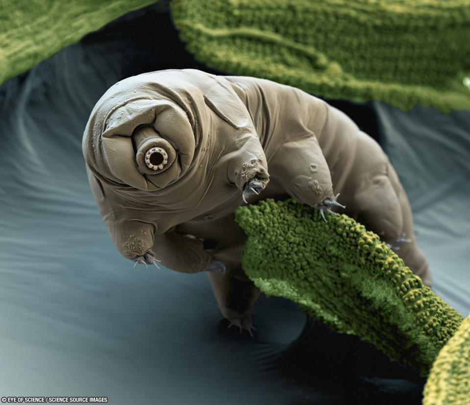
[In this figure] A colored scanning electron microscopic (SEM) image of a Eutardigrada tardigrade (Macrobiotus sapiens) on a piece of moss. Photo by Nicole Ottawa & Oliver Meckes.
Extended read
Oligochaeta
Oligochaeta is a subclass of animals in the phylum Annelida. Earthworms are typically large, terrestrial Oligochaeta or worms. There are some species of smaller aquatic worms that you may encounter under the microscope. These worms are usually semi-transparent and have “bristles” on their outer body surfaces.
[In this video] A worm (Oligochaeta) under a microscope.
Arthropods
An arthropod (from Ancient Greek “arthron” = joint and “pous” = foot) is an invertebrate animal having an exoskeleton, a segmented body, and paired jointed appendages. Arthropods form the phylum Euarthropoda, which includes insects, arachnids (like spiders and scorpions), myriapods (like centipedes), and crustaceans (like crabs and shrimps). You may see various tiny arthropods in freshwater specimens. They could be microscopic crustaceans like Daphnia or juvenile larva of aquatic insects and large crustaceans.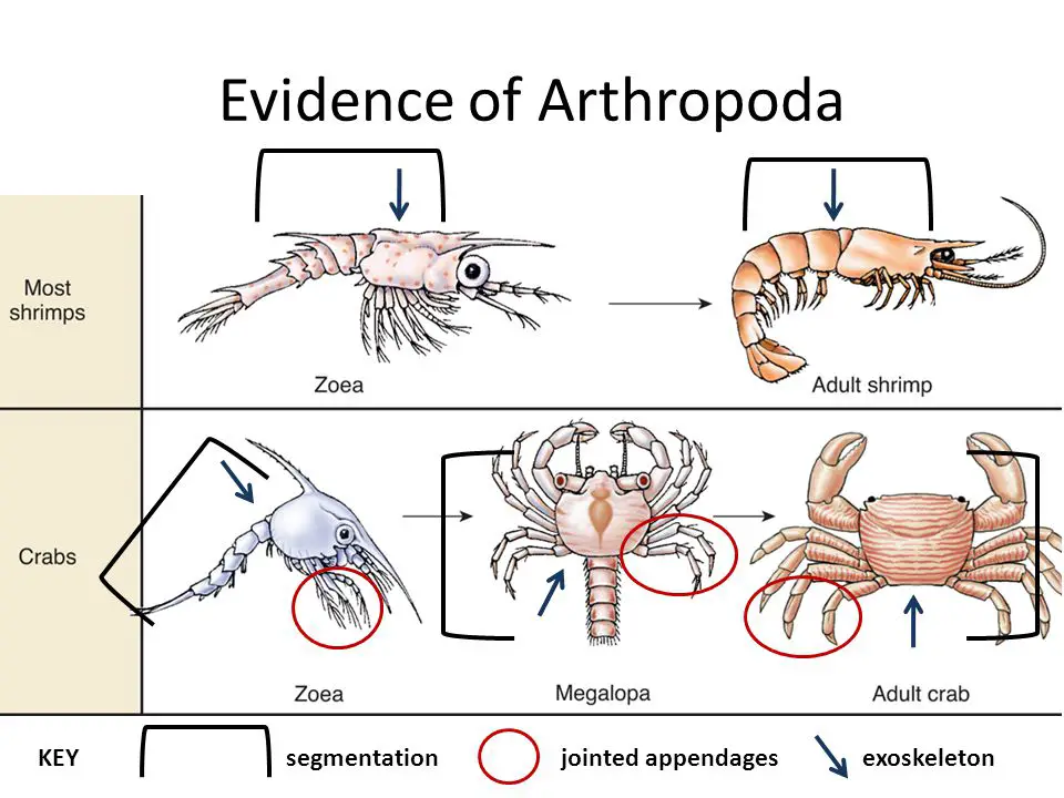
[In this figure] Three major characteristics of arthropods are exoskeleton, a segmented body, and paired jointed appendages.
Photo source: Slideplayer.
Daphnia
Daphnia (also known as water flea) is a genus of small planktonic crustaceans. The daphnia body is usually 0.2-3 millimeters long, which is pretty tiny compared to their cousins in the Class Crustaceans like crabs and lobsters. Daphnia is too small and weak to live in a strong current, which they cannot swim against. However, they are light enough to stay suspended by using their legs and antennae for movement. This is why we classify daphnia as a planktonic animal, meaning they live mainly by drifting in the body of water.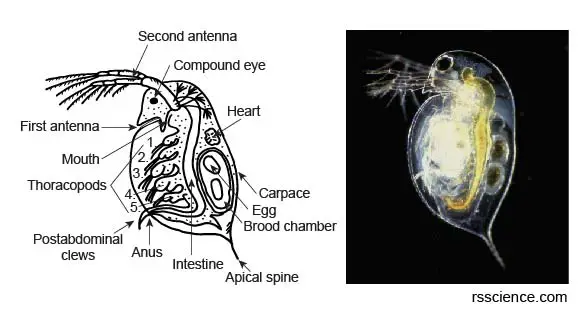
[In this figure] Left: An illustration of body parts of a Daphnia pulex. Right: A dark-field microscopic picture of a Daphnia pulex.
Extended read
Copepod
Copepods (meaning “oar-feet”) are a group of small crustaceans found in nearly every freshwater habitat. Some species are planktonic (drifting in water), some are benthic (living on the floor), and many species have parasitic phases. Copepods are sometimes used as biodiversity indicators.
[In this figure] Copepods of different species imaged by a darkfield microscope with polarized light.
Photo source: wiki.
Ostracod
Ostracods are sometimes known as seed shrimps. They are small crustaceans, typically around 1 mm in size. Their bodies are protected by a bivalve-like “shell.” The hinge of the two valves is on the back of the body.
[In this video] Ostracods under the microscope.
Amphipod
Amphipod is a shrimp-like microscopic crustacean with no carapace (real shrimps have carapaces or shells on the back) and generally with laterally compressed bodies. Amphipods are scavengers and mostly marine animals but are also found in freshwater habitats. Amphipods are popular organisms that can live in a closed Ecosphere.
[In this video] Amazing amphipods.
Mosquito larva
Mosquito larvae commonly called “wigglers,” live in water for 4 -14 days, depending on the water temperature. They must come to the surface at frequent intervals to obtain oxygen through a breathing tube called a siphon. During growth, the larva molts (sheds its skin) four times. When the 4th instar larva molts, it becomes a pupa, which also lives in water.
A mosquito larva eats constantly since it needs a lot of energy to grow. Mosquito larvae eat algae, bacteria, fungi, and other microorganisms in the water. Tiny fan-like brushes filter small food particles toward their mouth.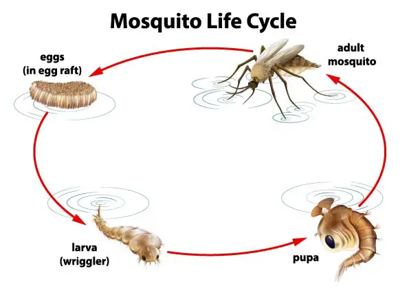
[In this figure] The life cycle of Mosquito.
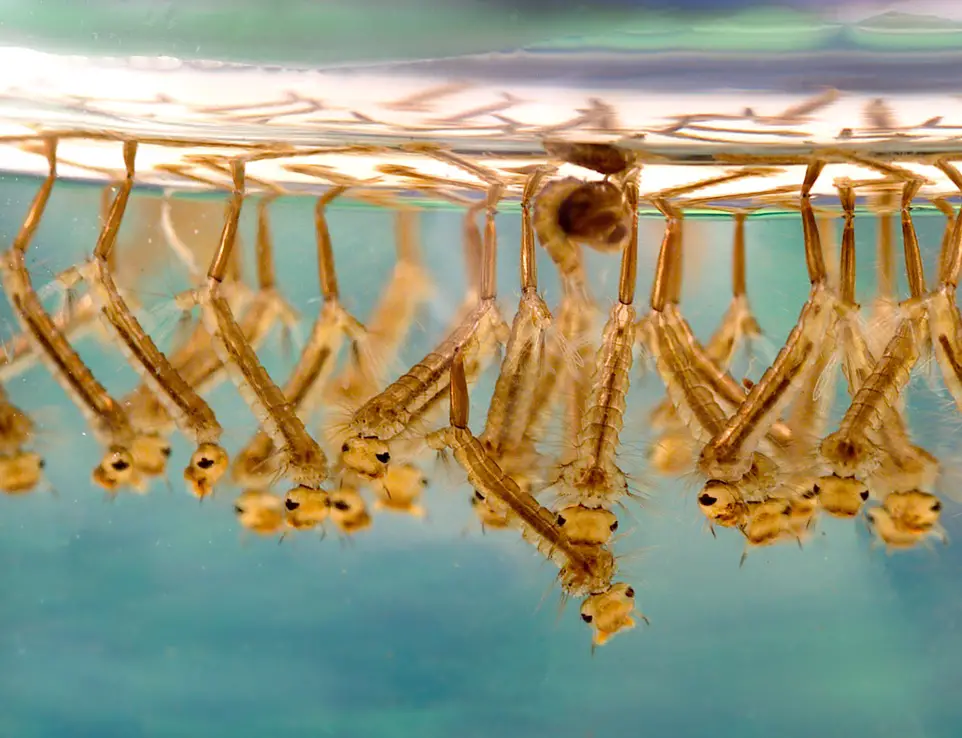
[In this figure] Mosquito larvae breath by their siphons.
Homework
Suggested Books
References
“What are algae blooms and why are they bad?”