
This article covers
What is a Vorticella? A quick overview
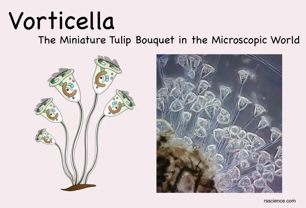
Vorticella (also known as the “Bell Animalcule”) is a genus of the ciliate protozoan. They are tiny, single-cellular, animal-like microorganisms. Vorticella is known for its bell-shaped head with a conspicuous ring of cilia (hair-like processes). Vorticella also has an unbranched stalk that anchors its body on a solid object. The body ranges from 30 to 40 micrometers while the stalk can grow up to 100 micrometers in length. The plural form is Vorticellae.
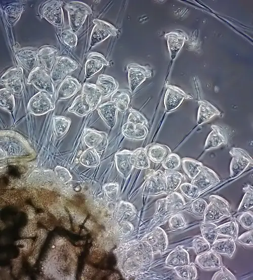
[In this image] A group of Vorticella looks like a bouquet.
This image is taken by Frank Fox from Germany with a Darkfield microscope and 20x objective lens.
Photo credit: Nikon small world
Vorticella can live in a variety of freshwater habitats, including ponds, pools, and ditches. They wave their cilia to bring food into their mouths. Under a microscope, a group of Vorticella looks like a tulip bouquet, making them one of the most adorable and elegant microscopic organisms.
[In this video] The biology of Vorticella under the microscope.
In the video, you can see the vorticella gradually open up and begin whirring their cilia in order to collect food. When disturbed, these creatures close and retract suddenly. You can see its anatomy and its asexual and sexual reproduction in this video.
Classification of Vorticella
Vorticella is a genus of ciliate protozoan under the family Vorticellidae. There are over 150 different species of Vorticella. The common species include Vorticella campanula, Vorticella citrina, Vorticella marina, Vorticella parapulchella, and Vorticella fusca.
Scientific classification
Domain: Eukaryota
(unranked): Sar
(unranked): Alveolata
Phylum: Ciliophora
Class: Oligohymenophorea
Subclass: Peritrichia
Order: Sessilida
Family: Vorticellidae
Genus: Vorticella
(Reference: wiki)
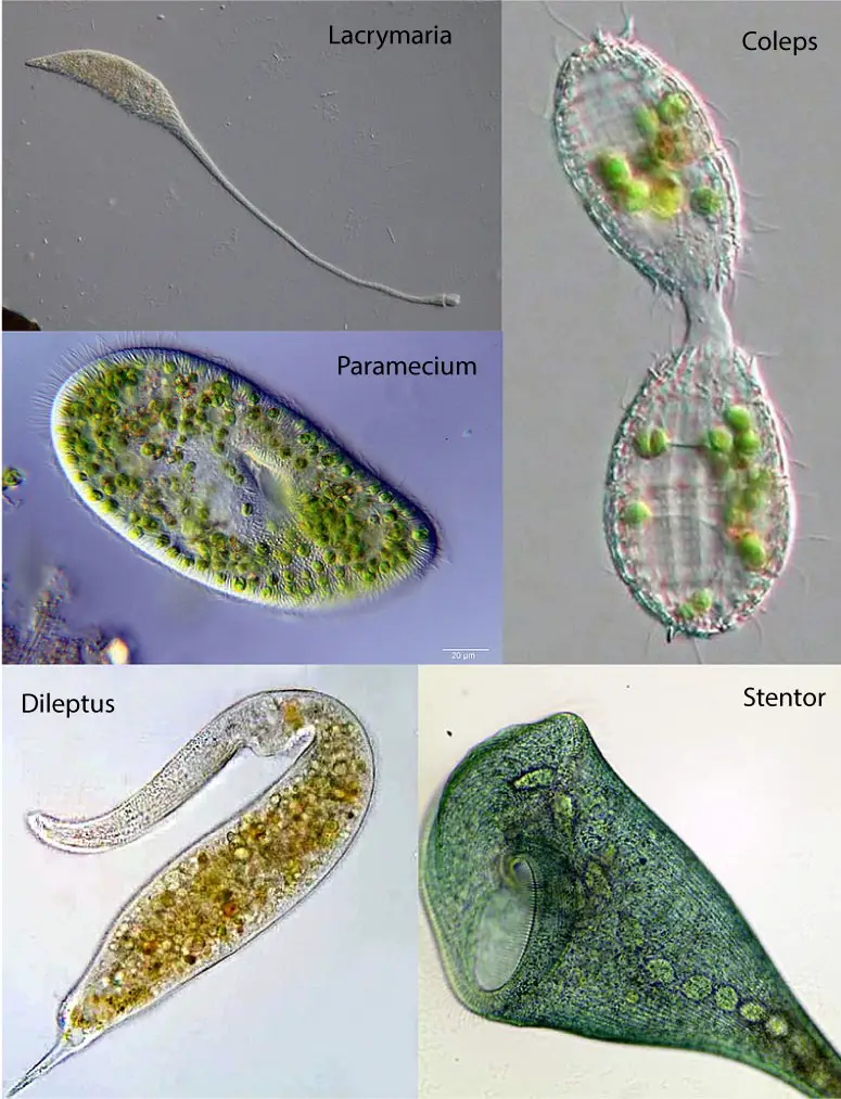
[In this image] A photo collection showing the diversity of ciliates.
Clockwise from top left: Lacrymaria, Coleps, Stentor, Dileptus, Paramecium. The ciliates are a group of protozoans characterized by the presence of hair-like organelles called cilia.
Photo credit: wiki
[In this image] Vorticella convallaria.
Photo credit: Protist Information Server
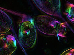
[In this image] Live Vorticella is visualized under a polarized light microscope.
Photo credit: Danielle Cook France, The Marine Biological Laboratory, Woods Hole
The cell structure of Vorticella
An individual Vorticella is a single cell. However, this microorganism has several specialized organelles and cellular structures. Let’s see it one-by-one:
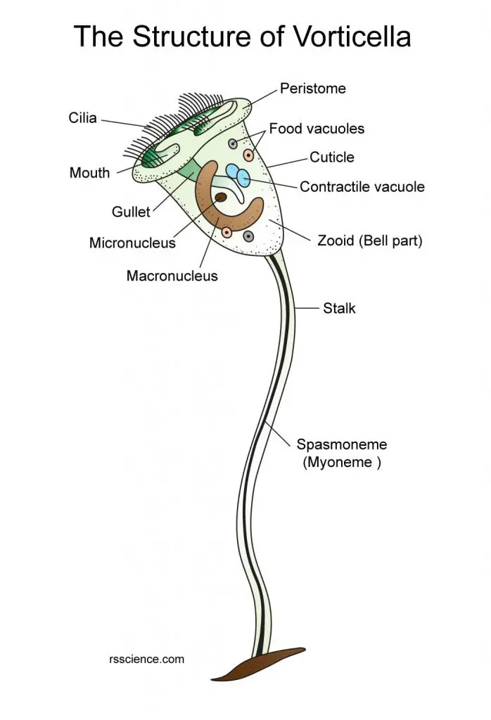
[In this image] The structure of Vorticella.
The inverted-bell-shaped cell body is usually about 30–40 μm in diameter and the stalk is 3–4 μm in diameter and about 100 μm in length.
Mouth and Cilia
The body of Vorticella looks like an inverted bell (named zooid) from the base of which runs a narrow stalk. The margin of the bell is thickened to a rim-like structure, called the peristome. On the top of the bell, there is a disc-like structure. The gap between the peristome and the disc is the opening of the mouth. The mouth connects to a tube-like structure, called the gullet.
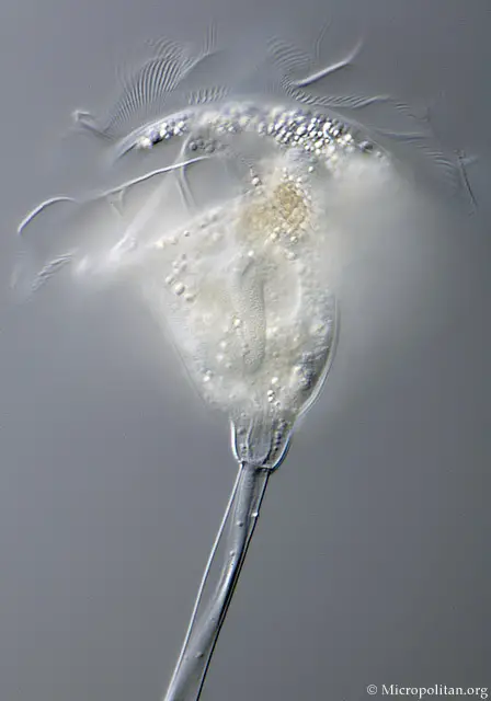
[In this image] The inverted-bell-shaped body of Vorticella.
Photo credit: photomacrography forum
Around the mouth of a Vorticella, there are many cilia arranged in rings. Cilia (singular: cilium) are fine, hair-like structures. A group of cilia can wave back and forth in a coordinated way to create a current or vortex that pulls food towards the mouth. Cilia are the main feature of all ciliates, including Paramecium and Stentor. Click here to learn more about Paramecium’s cilia.
[In this image] A close-up of a Vorticella’s bell showing the rows of cilia.
Photo credit: microscopy-uk
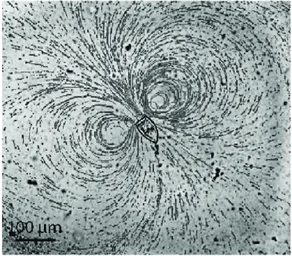
[In this image] The pattern of water flow generated by cilia beating of V. convallaria.
Photo credit: Micromachines 2017, 8(1), 4; https://doi.org/10.3390/mi8010004
[In this video] A Vorticella feeds by the current created by its cilia.
Vorticella are referred to as Peritrichs, meaning that their cilia are only present around the mouth, but nowhere else on the body. However, when Vorticella becomes motile (free-swimming, usually happens in young Vorticella), temporary cilia will form around the body. Once the Vorticella has anchored itself, these cilia will disappear.
Food and Contractile vacuoles
Food vacuoles form by budding from the posterior end of the gullet. Food vacuoles function like our stomach and contain digesting enzymes to break down the food materials into nutrients. The feeding process by engulfing forms food vacuoles, called phagocytosis, is common in single-celled microorganisms such as Amoeba. Click here to see Amoeba’s phagocytosis.
Vorticella usually has a contractile vacuole located next to the gullet. The contractile vacuole is a complex organelle that is unique to free-living organisms. Contractile vacuole acts to regulate the quantity of water inside of a cell. It expels excess water out of the cell by contracting and prevents the cell from absorbing too much water or even burst.
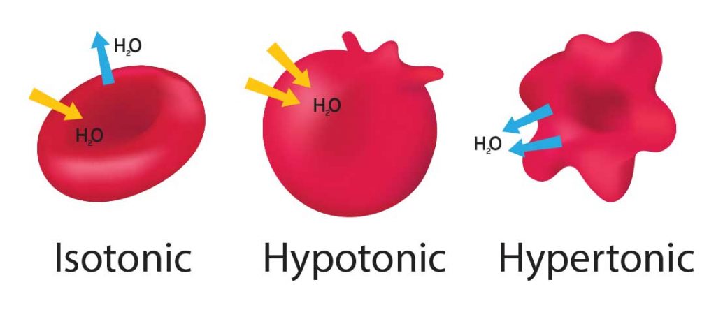
[In this figure] Animal cells (red blood cells as an example in this graph) are sensitive to osmosis pressure.
When our cells are in an “Isotonic” environment (like our blood), the in and out of water molecules are equal, and the cells are safe. If the outside environment becomes “Hypotonic” meaning fewer solutes (minerals) than Isotonic, water will move into the cells to achieve balance. The cells will swell and even burst (lyse) if excess water is not removed from the cell. On the other hand, “Hypertonic” is due to more solutes in the environment and can cause cells to shrink.
Two nuclei
Micronucleus
Vorticella has two nuclei in a cell. The micronucleus is small and rounded. The micronucleus contains all Vorticella’s DNA (called genome). This DNA is passed from one generation to another generation during reproduction. The micronucleus is diploid; that is, it contains two copies of each chromosome (human’s nucleus is also diploid).
Macronucleus
Vorticella also has a horseshoe-shaped macronucleus which is significantly larger than the micronucleus. The macronucleus is responsible for all metabolic activities. The macronucleus contains a subset of DNA from the micronucleus. These DNA fragments are copied from micronucleus to macronucleus because they carry genes that are frequently needed by the Vorticella cell. Genes in the macronucleus are actively transcripted to mRNA and then translated to proteins. The macronucleus is polyploid or contains multiple copies of each chromosome.
Spiral stalk
A long stalk can anchor the bell of Vorticella to a solid surface. The stalk consists of an external sheath that contains fluid and a spirally arranged contractile thread, known as a myoneme or spasmoneme. This unique structure allows the stalk to contract and retract. When the vorticella is contracted; the stalk thread is shortened, and the sheath is coiled like a corkscrew. The full stalk can contract within milliseconds, much quicker than the blink of an eye. It can then extend itself within a couple of seconds. The contract of the stalk may help Vorticella escaping from its predators or create an inward current/vortex to attract suspended food.
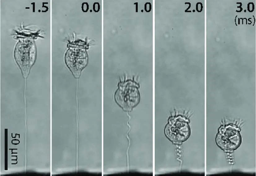
[In this image] Sequential images of stalk contraction of V. convallaria.
The stalk begins to coil at 0.0 and finishes in only 3 milliseconds (ms). A millisecond is a thousandth (0.001 or 1/1000) of a second.
Photo credit: Micromachines 2017, 8(1), 4; https://doi.org/10.3390/mi8010004
How does Vorticella live? – its characteristics and behavior
A Vorticella Bouquet is in fact a group of individual Vorticella
Vorticella lives in fresh or saltwater. Vorticellae (plural) usually anchor themselves to rocks, aquatic plants, surface scum, or aquatic animals. However, they can also swim freely to move to a new place. Although Vorticellae are often found in clusters, each stalk is fastened independently.
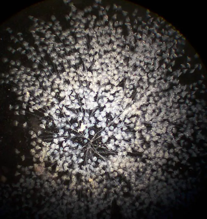
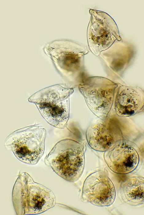
[In this image] A huge colony of Vorticella.
Vorticella is often found in colonies. However, these colonies are not true colonies, because each individual retains its own stalk. Vorticellae, therefore, are free to separate from the colony at any time.
Photo credit: microscopy-uk
[In this image] Vorticellae anchor themselves to a surface by their stalks.
Vorticella feed by their cilia
Vorticella eat bacteria and small protozoans through their oral end. The ring of cilia around their oral opening can create water flow to sweep food down into the gullet.
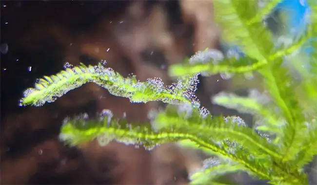
[In this image] Vorticellae grow on aquatic plants.
Photo credit: aquariumbreeder
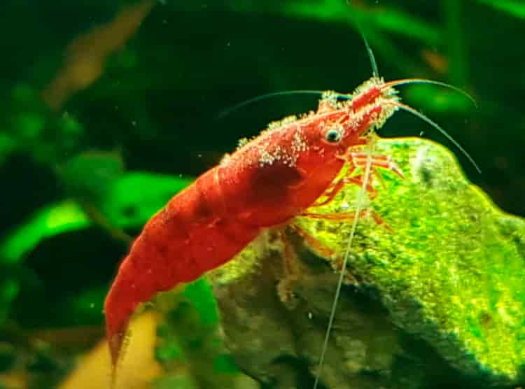
[In this image] Vorticella grows on the head of a shrimp.
Vorticella looks like “fuzzy” light white fungus (although, Vorticella is not a fungus). Normally, Vorticella is harmless to shrimps unless a large number of Vorticella fall into the gills, leading to the suffocation of the shrimp.
Photo credit: aquariumbreeder
Symbiotic Vorticella and green algae benefit each other
Some green algae can live within the bell of Vorticella. They form a symbiotic relationship that can benefit each other. Vorticella provides a safe place for green algae. In return, green algae provide foods for Vorticella.
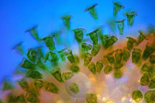
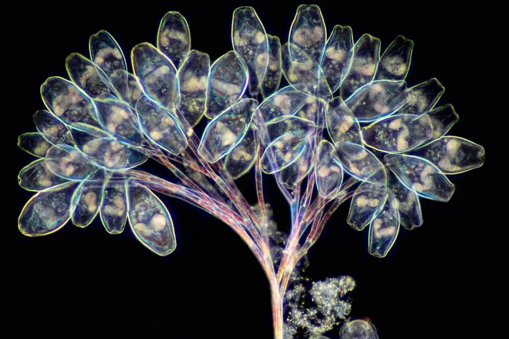
[In this image] Polarized light micrograph of a large group of green Vorticella.
The green color comes from the green algae living inside the Vorticella as their symbiotic partners.
Photo credit: Mauritius images
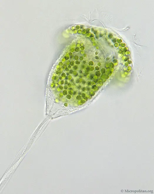
[In this image] A small Vorticella with the symbiotic algae Chlorella inside.
Photo credit: photomacrography forum
Can Vorticella move?
Vorticella are sessile organisms, meaning they like to stay in one fixed place. However, young Vorticella is free-swimming. They will swim away from their parents after reproduction by cell division. Adult Vorticella attaches themselves to substrates with contractile stalks. Adults can also be free-swimming if these stalks are cut. They can also detach themselves if food supplies are scarce and they need to find a new location.
[In this video] Vorticella movement under a microscope.
How does Vorticella reproduce?
Vorticella can reproduce asexually or sexually. The choice of reproduction depends on the conditions where they live. Asexual reproduction takes place under favorable conditions to allow a rapid expansion of the Vorticella population. On the contrary, sexual reproduction occurs at the end of the growing season.
Asexual reproduction of Vorticella
Vorticella can reproduce asexually by longitudinal fission (the split of one cell into two cells). When they undergo fission, they split along the longitudinal axis in a process called budding. When they finally split apart, one of the two daughter cells retains the original stalk. The other grows a temporary wreath of cilia and free swims away (propelling by these cilia). Once the migrant one finds a good place to live, it eventually grows a stalk, attaches to a substrate, and loses its temporary cilia. Asexual reproduction is more efficient, and this is the normal mode of reproduction in Vorticella.
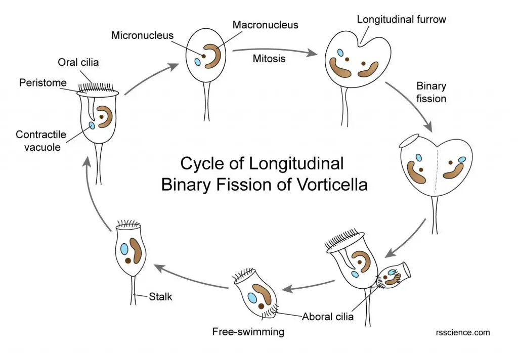
[In this image] Diagram of longitudinal binary fission of Vorticella.
To finish the fission, the Vorticella will go through these steps:
1. Its oral cilia and peristome will temperately disappear. The body becomes round-up.
2. The constriction of the cytoplasm runs inward, each nucleus divides into two; the micronucleus divides by mitosis and the macronucleus divides by vegetative division.
3. Each copy of micronucleus, macronucleus and contractile vacuole move toward the opposite direction, and the cell divides into two daughter cells.
4. One of the two daughter cells gets detached from the stalk, develops a second circlet of aboral cilia at the basal end, resembles the shape of a barrel, and becomes free-swimming (a stage called telotroch).
5. After a brief period of free life, the second circlet of cilia is lost. The migrant one develops a stalk from that end of the circlet cilia, the new Vorticella becomes permanently attached to some substratum.
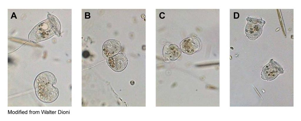
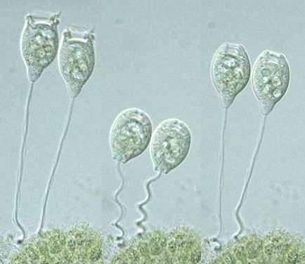
[In this image] The observation of longitudinal binary fission of Vorticella under the microscope.
You can see (A) The lower Vorticella starts its fission by forming a longitudinal furrow at the middle of the cells; (B) One cell divides into two daughter cells; (C) The cilia appear and two daughter cells still attach on one stalk; (D) One daughter cell swim away by its aboral cilia.
Modified from microscopy-uk
Sexual reproduction of Vorticella
The sexual reproduction of Vorticella takes place by conjugation. After several generations produced by binary fission, the Vorticella individuals will appear weakened and unhealthy. To revitalize, the Vorticella will enter a special stage of its life cycle, called conjugation.
The sexual reproduction of Vorticella is anisogamy because their two types of gametes (or sexual cells) are of unequal sizes. Small, migrant microgamete can swim and find an attached vorticella (macrogamete) to initiate the conjugation.
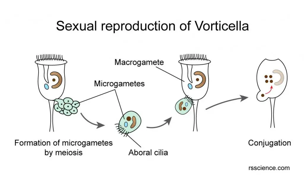
[In this image] The steps of sexual reproduction of Vorticella.
Sexual reproduction initiates when the Vorticella individuals become weakened after several asexual fission. One Vorticella will generate free-swimming microgametes by meiosis. These microgametes will find a macrogamete and attach to its junction between the bell body and stalk. Then, the conjugation will start by transferring genetic materials from the microgamete into macrogamete. The combination of both genetic materials will give rise to a new Vorticella animal.
[In this video] Two Vorticella in the process of conjugation under the microscope.
Microgamete and Macrogamete
The micronucleus of the old, weakened Vorticella will undergo the meiotic division. These daughter nuclei have only half the original number of chromosomes and become microgametes or microzooids. The microgametes swim by their cilia. The microgametes survive only for 24 hours and must conjugate with macrogamete within the period. The stationary macrogametes resemble the normal forms anchored by their stalks. They are able to attract microgametes.
Conjugation
The microgametes swim in the water. Once they find a macrogamete, the microgamete will rest on the macrogamete’s junction of the bell and the stalk. Then, a protoplasmic bridge (a tubular structure extending from the cells and containing the cytoplasm) is established between the two. The nucleus of the microgamete migrates through the protoplasmic bridge to fuse with the nucleus of the microgamete. The cytoplasm of the microgamete is lost and the macrogamete now with the fusion nucleus acts as a healthy ordinary Vorticella.
[In this video] Sexual reproduction of Vorticella.
At 3:07, you can see the microgamete attaching to the base of the bell part of a macrogamete for conjugation.
Why conjugation is important?
The process of conjugation allows the reception of genetic material from another individual. This results in a renewal of vitality, which is essential for rapid reproduction by binary fission.
Who discovered Vorticella?
Dutch microscopist, Antonie van Leeuwenhoek, first reported the Vorticella in his letter dated October, 9 ,1676. Vorticella drew the attention of Leeuwenhoek by its unique motility. He wrote about a small creature with a pair of horns near its mouth which moved like a horse’s ears. It wasn’t until later that this feature was actually the vorticella’s oral cilia working to create a flow of water. Leeuwenhoek also discovered many other microscopic organisms like rotifers and paramecia by using his simple microscopes. Click here to learn who invented the microscope?
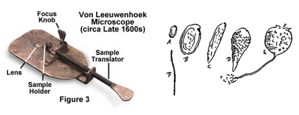
[In this figure] Left: The simple microscope used by Antony Van Leeuwenhoek to discover microscopic organisms. Right: Illustration of Vorticella by Leeuwenhoek.
Photo credit: minst.org
The name “Vorticella” was given by Christian Gottfried Ehrenberg in 1838. Since then, about 80 species of Vorticella have been identified. Today, there are over 150 species that are recognized as Vorticella by the biology community.
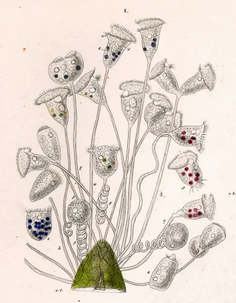
[In this figure] Vorticella nebulifera was painted by Christian Gottfried Ehrenberg, a German naturalist, zoologist, geologist, and microscopist in 1838.
A scientist by the name of Otto Friedrich Müller claimed to have discovered 127 different species of vorticella in 1786. Many of his claims were later reversed as they were actually rotifers. This is not surprising as vorticella physically resembles the phylum Suctoria. Click here to learn what is Rotifer.
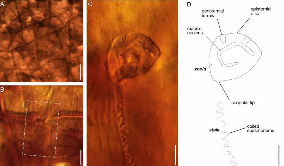
[In this figure] In a recent study, Vorticella was discovered in a fossil that is more than a 200-million-year-old (Triassic period), bolstering the fossil record of soft-bodied organisms.
Photo credit: PNAS
Vorticella: A model organism for bio-inspired engineering
A model organism is a species that has been widely studied in science. Usually, a model organism is easy to maintain and breed in a laboratory setting and has particular experimental advantages. Escherichia coli (a bacterium), yeast, C. elegans (a roundworm), fruit fly, zebrafish, and mice are all famous model organisms.
In fact, Vorticella is also a model organism that helps scientists to study “biological micro-machine” for microscale engineering systems. For example, scientists were fascinated by the Vorticella’s spasmoneme, which can contract in a few milliseconds, coiling the stalk and moving the bell part at 15–90 mm/s. They found the spasmoneme generates a force in the order of 10–100 nN, powered by calcium ion binding. We may learn from the spasmoneme as a model system for biomimetic actuators in microscale engineering systems. This may one day help us to build a tiny robot mimicking this amazing creature.
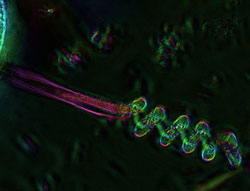
[In this figure] Scientists study coiled Vorticella stalk under a polarized light microscope.
They found the contraction of Vorticella’s spasmoneme to be more powerful than previously thought – weight for weight even more powerful than a car engine.
Photo credit: Danielle Cook France, The Marine Biological Laboratory, Woods Hole
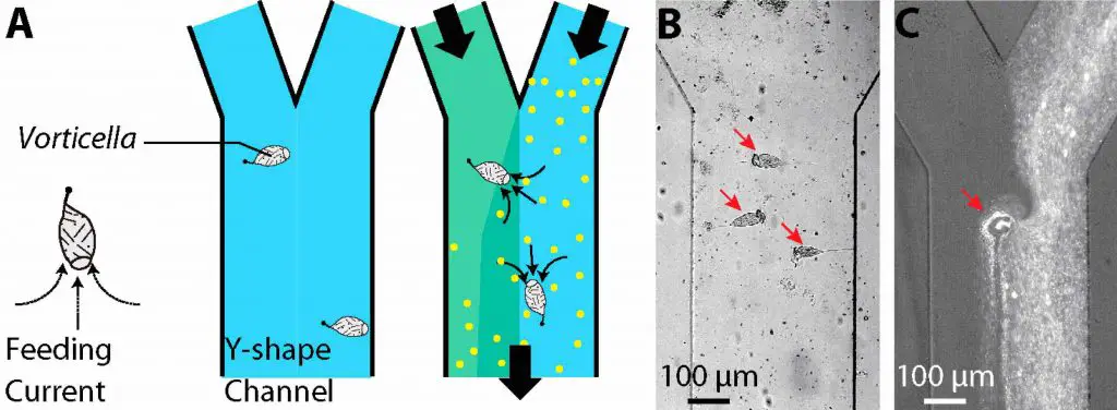
[In this figure] Scientists grow Vorticella in a bio-chip as a microfluidic mixer. Vorticella can mix two streams in the Y-channel.
Photo credit: Sciencedirect
How to find Vorticella for your microscopic project? – habitat
Vorticellas are one of the prettiest microorganisms to observe under the microscope. Compared to other ciliates that may move pretty fast, Vorticella does not move around, making it very easy to observe miniature details.
If you want to observe Vorticella, any pond or lake sediment sample will most likely yield a few good samples. Make sure when you take your samples, you are picking up leaves and sediments from the floor of the body of water as these are typical structures that vorticella will adhere themselves to.
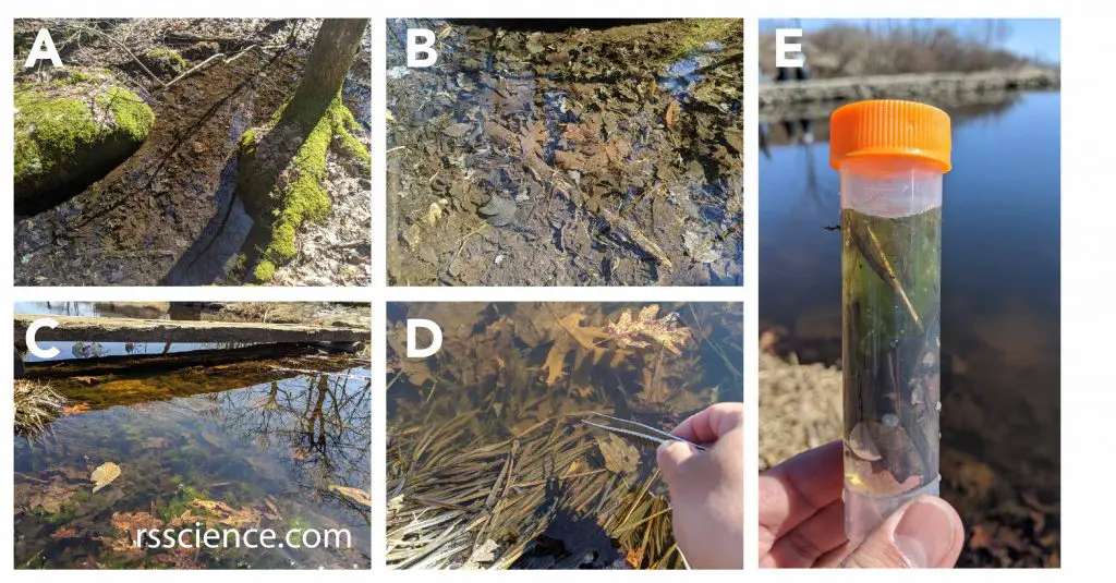
[In this figure] The places to collect Vorticella and other microorganisms.
(A-C) Microorganisms stay with their source of food. Ponds or slow-flowing creeks with decayed organic materials in the bottom sediments (like leaves) are ideal habitations to find all kinds of microorganisms, like paramecia, amoebas, rotifers, water bears, daphnia, and diatoms. (D-E) Vorticella like to anchor themselves on a surface, like leaves. I used the forceps to collect some decaying leaves and use a dropper to collect water with sediments into my sample vial. I will bring it home to look for microorganisms under my microscope.
Summary
1. Vorticella looks like an inverted bell attached to a long stalk. Vorticella likes to anchor itself on a surface by its stalk.
2. Vorticella can grow as a colony like a bouquet. However, individual Vorticella in the colony is an independent organism. Each one has its own stalk.
3. The contraction of the stalk allows a quick retreat of Vorticella’s body away from dangers.
4. Vorticella feed by waving its cilia around the mouth to collect foods. Vorticella can eat bacteria, algae, and other small microorganisms.
5. Vorticella can reproduce both asexually and sexually. The decision depends on the environmental conditions.
6. By asexual reproduction, one Vorticella split into two new daughter cells. One retains the original stalk. The other one swim away by its cilia to find a new place.
7. By sexual reproduction, Vorticella generates microgamete and macrogamete via meiosis. Small microgametes can swim to find a macrogamete. Once they meet, their genetic materials will combine (conjugation) to produce a new Vorticella.
References
“Shrimp Vorticella Parasite. Treatment”
“Triassic leech cocoon from Antarctica contains fossil bell animal”
“Vorticella: A Protozoan for Bio-Inspired Engineering”