
This article covers
What is a Stentor? A quick overview
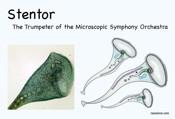
Stentor (also known as the “Trumpet Animalcule”) is a genus of the ciliate. They have a horn-shaped body with cilia uniformly cover the most surface area. Some species of Stentors are among the biggest known unicellular organisms on earth. The species Stentor coeruleus can grow up to 2 mm (0.08 inch) long and has a blue-green-ish color.
[In this image] Stentor is a huge ciliate that you can easily spot under the microscope.
Stentors can live in a variety of freshwater habitats, including ponds, pools, and ditches. They are found either free-swimming or attached to submerged vegetation. Stentors use these cilia to sweep food particles into their mouths. They also swim by waving the cilia that cover their body. They reproduce asexually through binary fission.
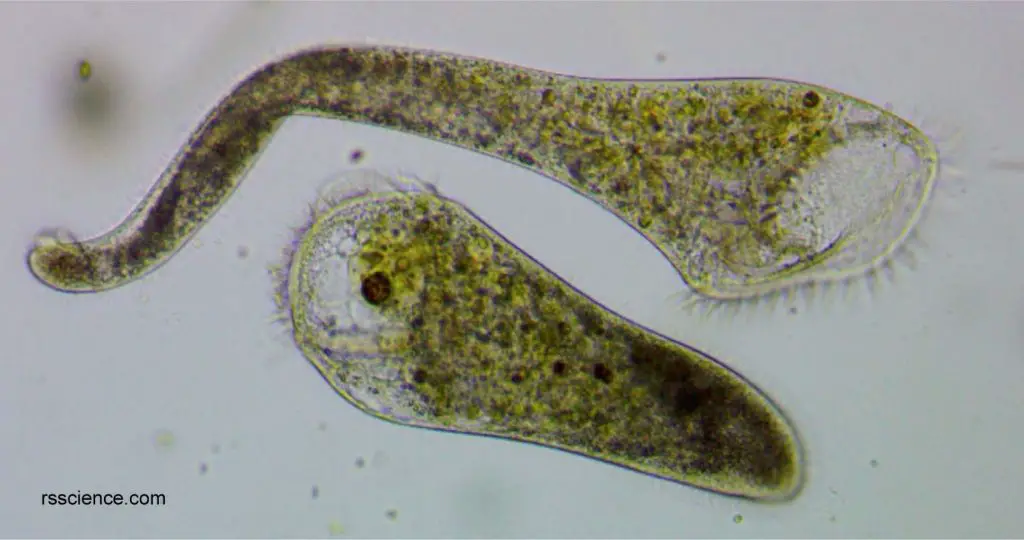
[In this image] Two Stentors. The top one is fully extended with its mouth opening for feeding. The bottom one contracts into a pear shape while traveling.
Stentors have a remarkable “regenerative capability”. Even a small fragment of an adult Stentor can grow back into a complete animal. This unique feature makes Stentor a favorite subject to study the process of regeneration in protozoans.
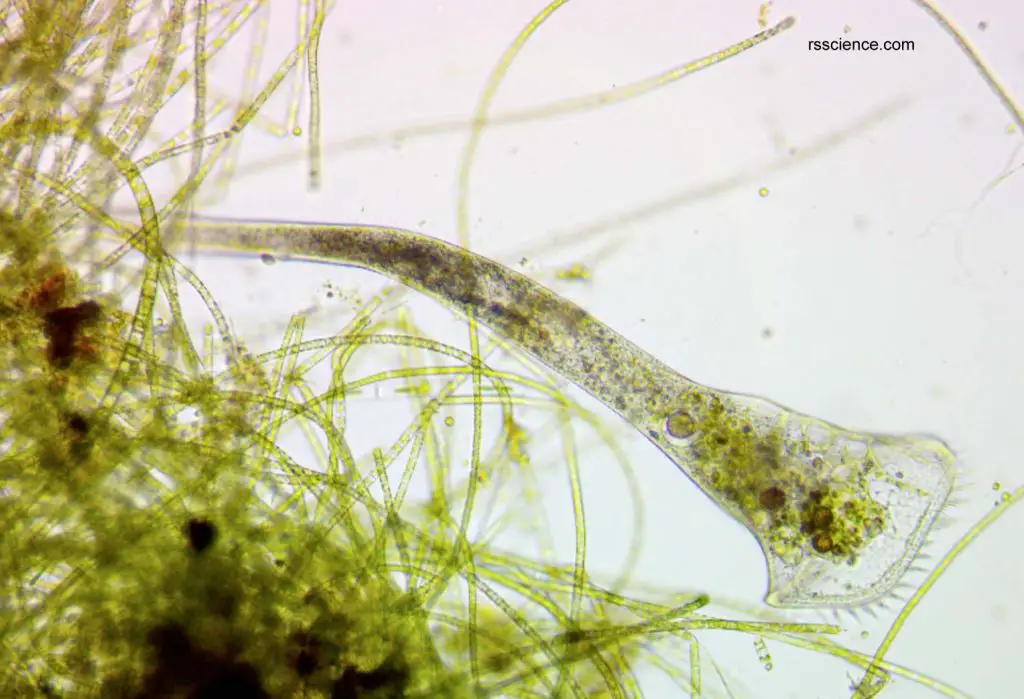
[Video] Stentors under a microscope.
Stentors are huge and you can see their bodies are covered by cilia. The Stentor is a great subject to observe the motion of cilia.
Classification of Stentor
Stentor is a genus of ciliate protozoan under the family Stentoridae. There are over twenty described species of Stentor. The common species include Stentor polymorphus, Stentor coeruleus, Stentor roeselii, Stentor amethystinus, and Stentor muelleri. The genus Stentor was named in 1815 by the German biologist Lorenz Oken (1779–1851).
Scientific classification
Domain: Eukaryota
(unranked): Sar
(unranked): Alveolata
Phylum: Ciliophora
Class: Heterotrichea
Order: Heterotrichida
Family: Stentoridae
Genus: Stentor
(Reference: wiki)
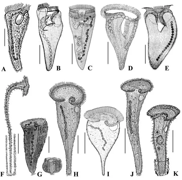
[In this image] A collection of different Stentor species in the literatures:
(A-D) Condylostentor auriculatus; (E) Stentor auriculatus; (F) Stentor muelleri; (G) Stentor introversus; (H) Stentor coeruleus; (I) Stentor caudatus; (J) Stentor polymorphus; (K) Stentor multiformis.
Photo credit: Chen X. et al., Acta Protozoologica, 2007
The ciliates are a group of protozoans characterized by the presence of hair-like organelles called cilia. Other examples of ciliates are Lacrymaria, Coleps, Vorticella, Dileptus, and Paramecium.
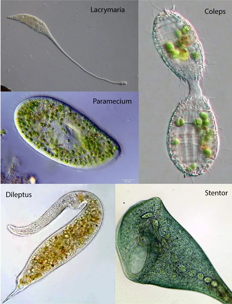
[In this image] A photo collection showing the diversity of ciliates.
Clockwise from top left: Lacrymaria, Coleps, Stentor, Dileptus, Paramecium.
Photo credit: wiki
Colorful Stentors
Stentor can come in different colors: for example, S. coeruleus can appear blue due to the presence of stentorin, a natural pigment. Stentorin can help S. coeruleus respond to visible light stimuli.
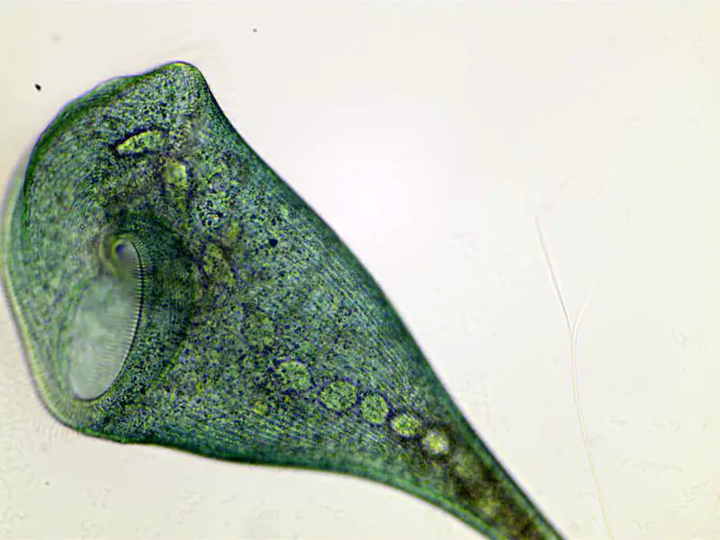
[In this image] Stentor can come in different colors:
For example, Stentor coeruleus (this picture) can appear blue-green-ish due to the presence of stentorin, a natural pigment.
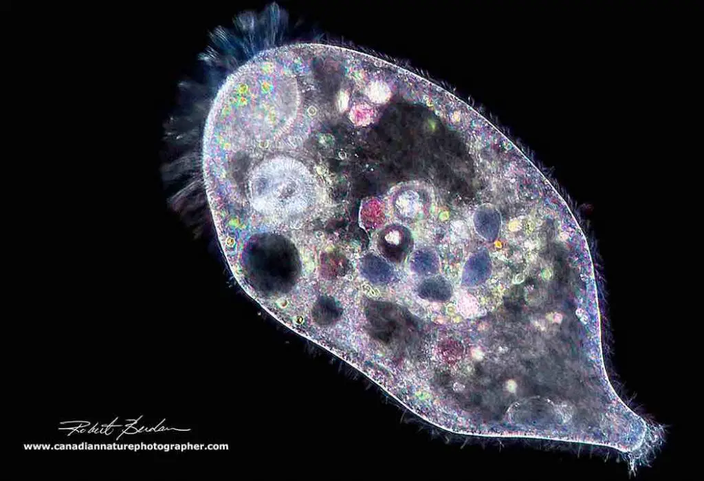
[In this image] A Stentor is viewed under a Darkfield microscopy.
The contents ingested in its food vacuoles show different colors – like a Picasso’s painting.
Photo credit: The Canadian Nature Photographer
The cell structure of Stentors
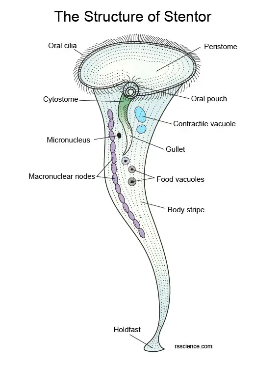
[In this image] The anatomy of Stentor.
This is a fully extended Stentor. The rim of the trumpet is surrounded by a crown of cilia (vibrating hairs) which beat to produce a current that draws particles of food down into the mouth (cytostome). The mouth is connected to the gut (gullet). Let’s look at individual organelles one by one in the next section.
The Crown of Cilia
The most notable part of a Stentor is the “crown” of cilia surrounding the trumpet “bell”. Cilia (singular: cilium) are fine, hair-like structures. A group of cilia can wave back and forth in a coordinated way to create a current or vortex that pulls food closer. Adjacent to the oral opening, the fusion of several cilia to form “super cilia” or membranelles. These giant cilia form a spiral that leads into a small buccal cavity which in turn leads to a cytostome (like its mouth) where food is ingested. Every little while, the stentor will close up the cilia crown and contract, bringing the food down into the tubular cytostome.
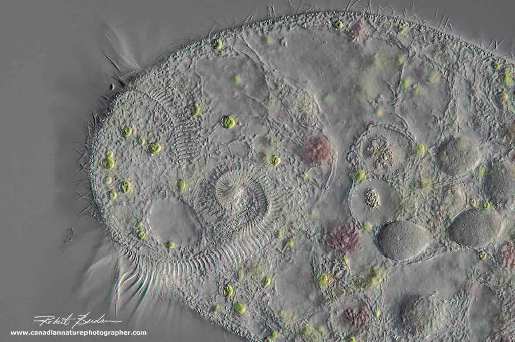
[In this image] On the left are the membranelles that form a circle leading to the cytostome. This image is taken by DIC microscopy at 400x.
Photo credit: The Canadian Nature Photographer
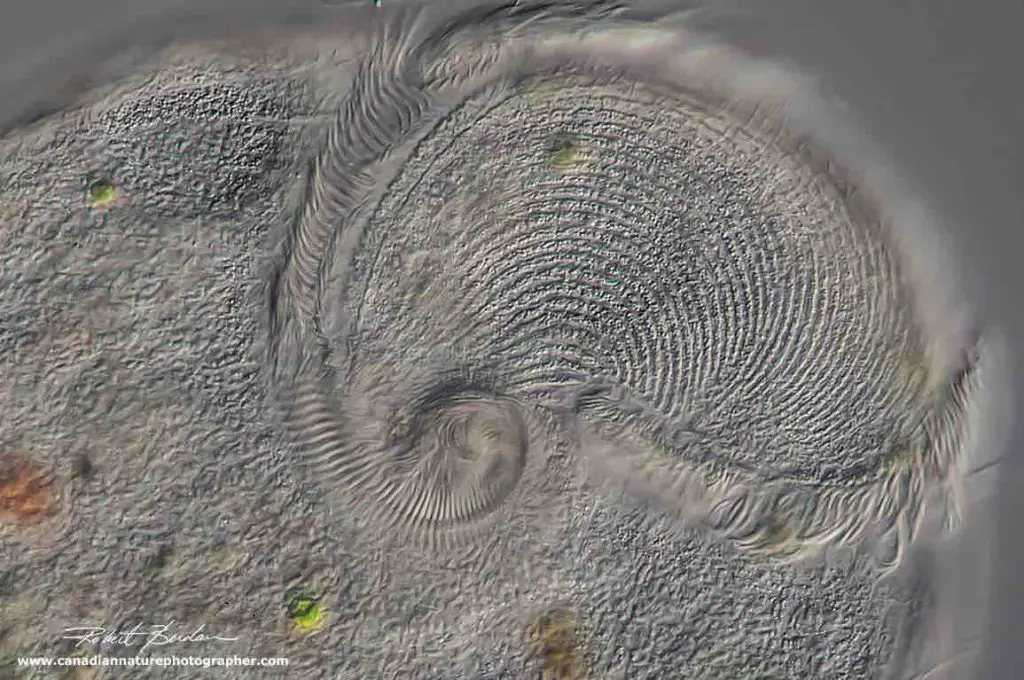
[In this image] A closer view of the oral zone of a Stentor showing how it spirals into the cytostome. DIC microscopy 400x.
Photo credit: The Canadian Nature Photographer
The crown is not the only place having cilia on the Stentor. Its whole body is covered with shorter cilia, which are used for free swimming. When moving, the Stentor will contract into an oval or pear shape instead of the trumpet shape.
[In this video] Under the microscope, you can see these longer cilia forming the “crown” of Stentor’s trumpet. There are also many shorter cilia covering the entire Stentor’s body for free swimming.
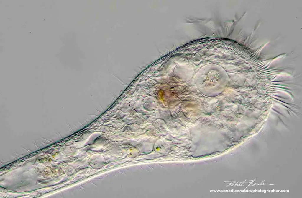
[In this image] This Stentor has large membranelles on the oral zone and smaller cilia covering the rest of its body.
Photo credit: The Canadian Nature Photographer
Cortex and body stripes
The cortex (pellicle) functions like Stentor’s skin. There are parallel stripes running through its body. When you go very close, you can see these stripes are in fact formed by many aligned pigment granules. Pigment granules (0.5 – 2 microns) can be red, blue, green, or brown in color.
[In this image] Stentor viewed by bright field microscopy showing its trumpet form shape and stripes.
Photo credit: The Canadian Nature Photographer
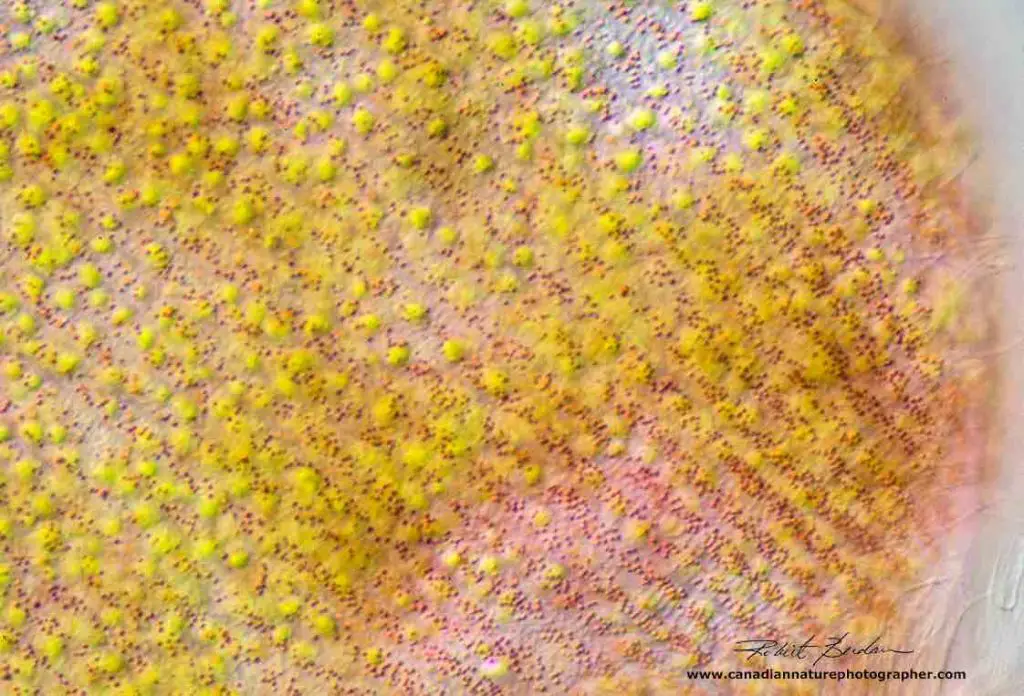
[In this image] A closer view of Stentor’s cortex showing the stripes of granules containing red pigments.
Photo credit: The Canadian Nature Photographer
Food vacuoles
When a Stentor collects its food by cilia (like its hands) into its cytostome (mouth), the food particles will travel down a tube-like structure, called the gullet (esophagus). At the end of the gullet, the cell membrane will envelop the food, and separates (budding) to form round bubbles, called food vacuoles. Food vacuoles function like our stomach and contain digesting enzymes to break down the food materials into nutrients. After the nutrition from the food is extracted, this vacuole moves to the outer cell wall and “pops”, evacuating the remaining debris.
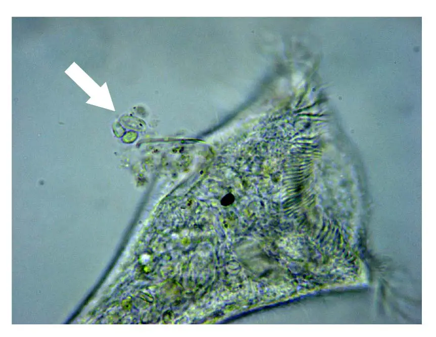
[In this image] The white arrow indicates a food vacuole being evacuated.
Contractile vacuole
Stentors must maintain the quantity of water inside of the cell very carefully. If a Stentor absorbs too much water, it can burst. In order to regulate the “osmotic pressure”, the Stentor cell actively collects the excess water into its contractile vacuole. Contractile vacuole can expel excess water out of the cell by contracting and prevents the cell from bursting.
Two kinds of nuclei
Micronucleus
Like other cousins in the Ciliate family, Stentors also have two types of nuclei in their cells. The micronucleus is small and rounded. The micronucleus stores all of the DNA (called genome) that is present in the organism. These DNA are not active during the normal activity; however, they will be passed from one generation to another generation during reproduction. Each gene/chromosome has two copies in the micronucleus, called diploid.
Macronuclei
Stentors have one or many macronuclei which are significantly larger than the micronucleus. The macronucleus is responsible for all metabolic activities. The macronucleus contains a subset of DNA from the micronucleus. These DNA fragments are copied from micronucleus to macronucleus because they carry genes that are frequently used by the Stentor cells. Genes in the macronucleus are actively transcripted to mRNA and then translated to proteins. The macronucleus is polyploid meaning it contains many copies of each gene. The macronucleus of the Stentor can take on many shapes. It can be a single ball, vermiform, nodular, or moniliform (like a string of pearls).
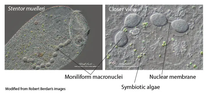
[In this image] Left: The moniliform macronuclei in a Stentor. Right: A closer view to see the beads of the macronuclei linked by nuclear membranes into a string.
Photo credit: The Canadian Nature Photographer
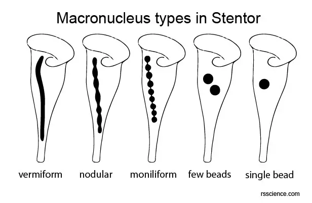
[In this image] The types of Stentor macronuclei.
Note that the moniliform is a regularly spaced chain of beads connected by thread-like elongation of the nuclear membrane.
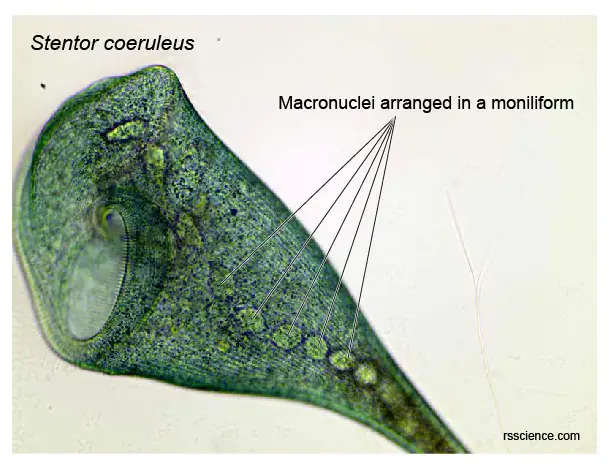
[In this image] Several macronuclei in a Stentor coeruleus form a string of pearls (called moniliform).
Photo credit: Modified from Wikimedia
Holdfast
Stentor is attached to the substrate by a slightly expanded region known as the holdfast. Some Stentors can secret a mucilaginous material (called lorica) surrounding their holdfasts. The lorica can hold a group of Stentors together into a cluster.
How do Stentors live?
Stentors are common worldwide in freshwater lakes and streams; only S. multiformis has been recorded from marine, freshwater, and even terrestrial biotopes.
Blue whale of microscopic world
You can say that Stentors are the blue whale of microorganisms. This is true not only due to the sizes but also the behavior of feeding.
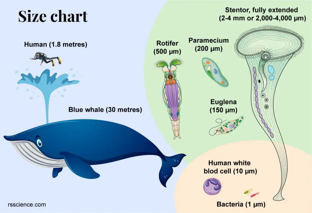
[In this image] Size chart comparison between protozoans.
A Stentor is 2,000 – 4,000 µm in length, about 1-200 times bigger than a paramecium.
Stentors are filter-feeding, heterotrophic ciliates. They eat bacteria, algae, and small organic particles through their oral end. The ring of cilia around their oral opening can create water flow to sweep food down into the mouth. Large stentors are reported to opportunistically eat rotifers or anything else that they can catch.
[In this video] A euglena is trapped by the vortex created by a Stentor’s cilia. Fortunately, the euglena escapes.
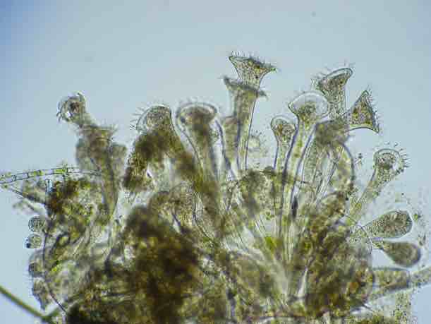
Stentors usually attach to aquatic plants or surface scum when they are feeding. Sometimes Stentors will form social groups of several hundred or more. They can also swim freely to move to a new place for more nutrients.
[In this image] A colony of Stentors is a group of individual organisms that just happen to be located together.
Photo credit: microscopy-uk
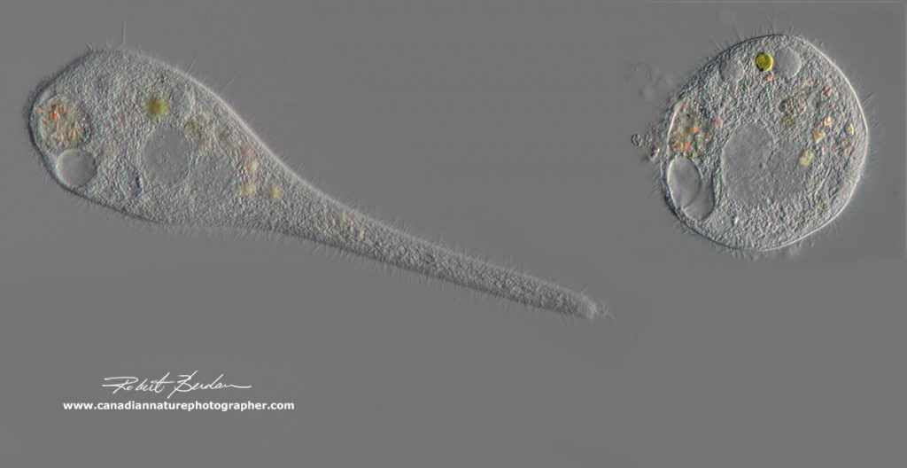
[In this image] When stentors are traveling, they are not in the typical trumpet shape (left). They contract into a more oval shape (right). The cilia around the trumpet bell close up, and the cilia on the body are used for swimming.
Photo credit: The Canadian Nature Photographer
Symbiotic Stentors and green algae benefit each other
Some Stentor species, such as S. polymorphus, can live symbiotically with certain species of green algae (Chlorella). They form a symbiotic relationship that can benefit each other. Stentor provides a safe place for green algae. In return, green algae provide foods for Stentor. Green algae can also absorb and feed on the Stentor’s metabolic wastes.
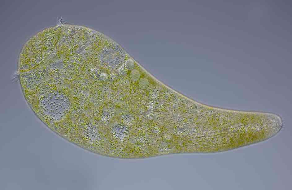
[In this image] Stentor polymorphus with algal symbionts living inside its body.
Photo credit: Mikro-Foto
How do Stentors reproduce?
Stentors can reproduce asexually by binary fission or sexually by conjugation. The choice of reproduction depends on the conditions where they live. Binary fission takes place under favorable conditions allowing a rapid expansion of the Stentor population. In contrast, sexual reproduction occurs at the end of the growing season to revitalize the population.
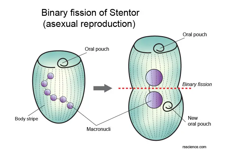
[In this image] Diagram of binary fission of Stentor.
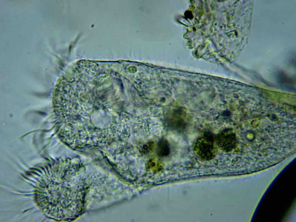
[In this image] A Stentor undergos division.
Photo credit: microscopy-uk
Who discovered Stentors?
German biologist Lorenz Oken (1779–1851) formally recognized these protists and named them Stentor in 1815. The name “Stentor” came from Greek mythology. Stentor was a herald of the Greek forces during the Trojan War. He is mentioned briefly in Homer’s Iliad in which he had a voice as powerful as fifty voices of other men. His voice encouraged the Greeks to fight.
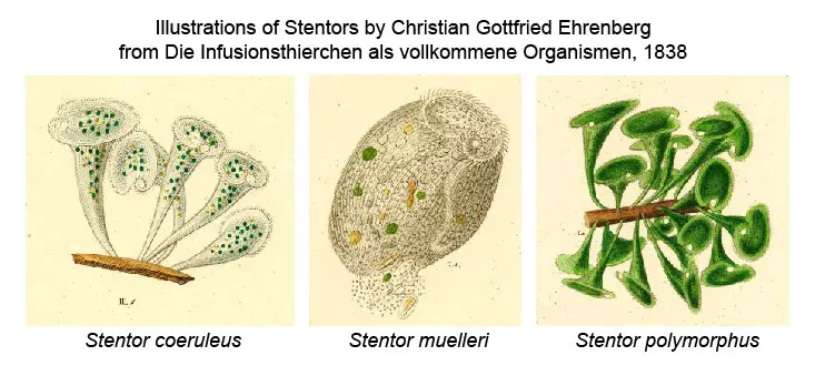
[In this figure] Three species of Stentors were painted by Christian Gottfried Ehrenberg, a German naturalist, zoologist, geologist, and microscopist in 1838. Ehrenberg placed Stentors along with the Peritrichs in his Family Vorticellina.
Fun facts of Stentors
Other than the basic biology of Stentors we mentioned here, there are a few fun facts about Stentors.
- Stentors “surprisingly” use the standard genetic codes
- Stentors have an amazing ability to regenerate
- Can a single cell “change its mind”? Stentor can
How to find Stentors for your microscopic project?
Stentor is one of the largest protozoans found in water and is, therefore, one of the easiest organisms for the microscopist to study. Stentors are usually found in the calm water of ponds and lakes, usually near the surface of leaves or twigs. Collect the specimens with some water in a shallow dish and allow the water to settle for a few minutes. Some Stentors are big enough to be seen with the naked eye. However, a magnifying glass or a microlens attached to your cell phone will provide a better view. A microscope is required in order to see details of the Stentor’s structure and behavior. If a microscope is available, watching a living Stentor can be a very absorbing activity.
Examine the dish with a low magnification lens – any Stentor present will be visible as long trumpet-shaped organisms. Stentors may also be swimming freely when they become more oval in shape. If you are lucky, you may sometimes find a group of Stentors forming a cluster, in which the foot parts of the animals are covered with a mucilaginous substance that they secrete.
Autumn is a good time to find Stentor because falling leaves become the nutrients for the bacteria population thriving. This leads to an increase in protozoa population, such as Stentors, which feed on the bacteria.
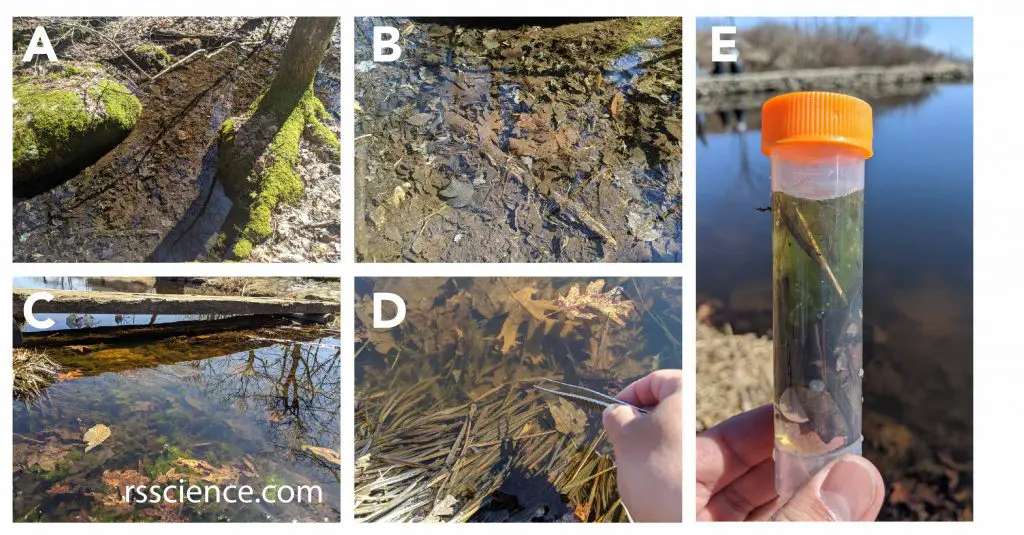
[In this figure] The places to collect Stentors and other microorganisms.
(A-C) Microorganisms stay with their source of food. Ponds or slow-flowing creeks with decayed organic materials in the bottom sediments (like leaves) are ideal habitations to find all kinds of microorganisms, such as paramecia, amoebas, rotifers, water bears, daphnia, and diatoms. (D-E) Stentors like to anchor themselves on a surface, like leaves. I used the forceps to collect some decaying leaves and use a dropper to collect water with sediments into my sample vial. I will bring it home to look for microorganisms under my microscope.
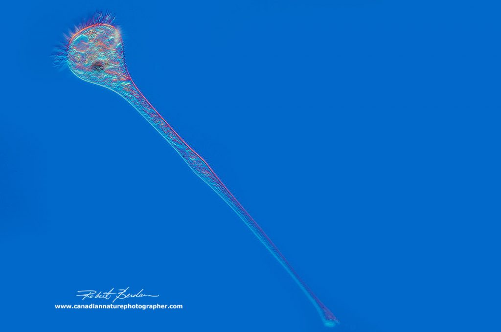
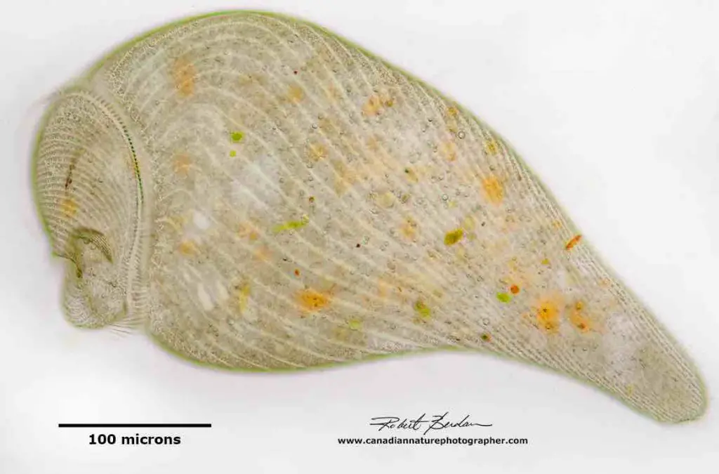
[In this image] Stretched out Stentor resembling a trumpet – some Stentors can reach 2-4 mm in length. DIC microscopy 100x
Photo credit: The Canadian Nature Photographer
Summary
1. Stentors are the largest unicellular microorganisms. Some Stentors can reach a size of up to 4 mm.
2. The Stentor can be very colorful. Stentor coeruleus is famous for its blue color, due to its blue-green-ish pigments, called stentorian.
3. A fully extended Stentor looks like a trumpet. At this stage, the Stentor likes to anchor itself by its holdfast and open its mouth for feeding.
4. The Stentor can contract into a more oval shape while traveling.
5. Stentors feed by waving their cilia around the mouth to collect foods. Stentors eat bacteria, algae, and other small microorganisms.
6. Stentors reproduce both asexually and sexually. The decision depends on the environmental conditions.
7. Stentors have two kinds of nuclei (micronucleus and macronucleus). Some Stentors have very unique shapes of macronucleus – like a string of pearls.
8. Stentors are known for their amazing ability to regenerate. They can regenerate from small fragments into full organisms.
References
“A Beginners Guide to the Collection, Isolation, Cultivation and Identification of Freshwater Protozoa” by A. J. Cowling, A. Rogerson, and B. J. Finlay. Originally published: 1988
“Stentors” by Howard Webb
“The Biology of Stentor” by Vance Tartar, Oxford, New York, Pergammon Press, 1961
“Illustrations of Ciliates”
“Photographing Stentors – A Large Unicellular Ciliate living in Freshwater”