
This article covers
What are bacteria? A quick overview
Bacteria (singular: Bacterium) are single-celled microorganisms. The cell structure of bacteria is much simpler than that of eukaryotic cells. There are no nucleus or membrane-bound organelles in a bacterial cell. Thus, bacteria are classified as “Prokaryotic cells”.
Learn what are the differences between Eukaryotes and Prokaryotes below:
Bacteria are found almost everywhere on Earth – soil, rock, oceans, and even arctic ice. Some species can live in extreme conditions of temperature and pressure. Some live in or on other organisms, including plants and animals. The human body is full of bacteria, and in fact, is estimated to contain 10 times more bacterial cells than human cells in our body. A lot of these bacteria reside in our gut, called the microbiome.
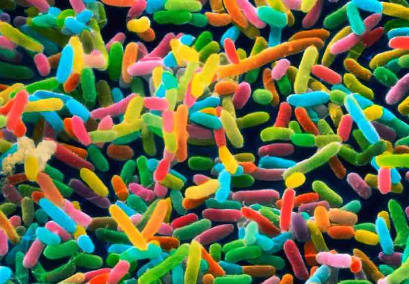
[In this image] Colored electron micrograph of Rod-shaped bacteria.
Photo source: nature
Most bacteria in our body are harmless, and some are even helpful. Some bacteria live in the soil or on dead plant matter, where they play an essential role in cycling nutrients. Some types cause food spoilage and crop damage, but others are incredibly useful in producing fermented foods such as yogurt and soy sauce. A relatively small number of bacteria are parasites or pathogens that cause disease in animals and plants.
Let’s learn more about bacteria below!
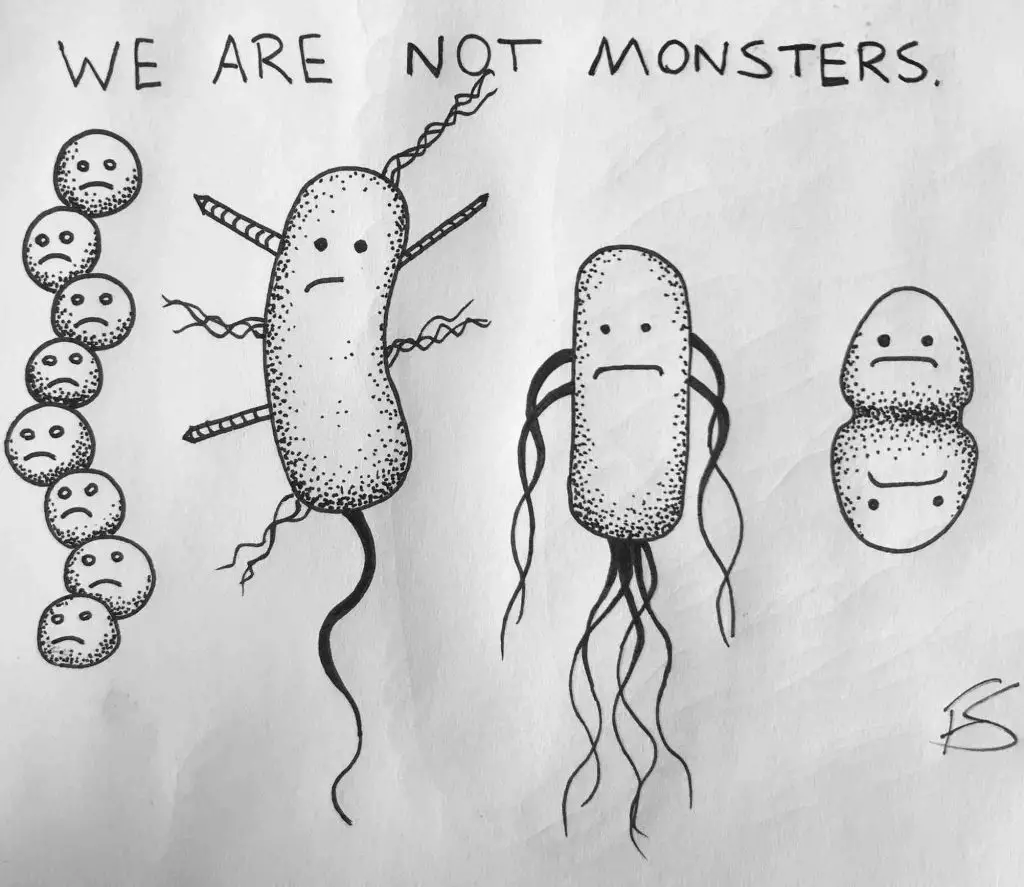
Image source: Frank Santoriello
Fun facts and numbers about bacteria
- Bacteria are thought to be the first living organisms on earth, about 4 billion years ago. The oldest known fossils (found in western Australia) are of cyanobacteria, dated 3.5 billion years old.
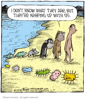
Image source: The comics strips
- Bacteria usually measure a few micrometers (usually 1-1.5 µm) in length and exist together in communities of millions.
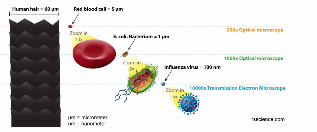
[In this figure] The size comparison between our hair (~ 60 µm) and E. coli (~1 µm).
Notice how small the bacteria are. It requires 1000x magnification to see them well. Learn more about the scale of biology.
- A gram of soil typically contains about 40 million bacterial cells. A milliliter of fresh water usually holds about one million bacterial cells.
- The earth is estimated to hold at least 5 nonillion (5 followed by 30 zeros) bacteria, and much of the earth’s biomass is thought to be made up of bacteria.
Part 1. Cell structure of bacteria
Let’s do physical exams on bacteria to know more about them!
Bacteria are prokaryotes (pro-KAR-ee-ot-es), which means they have no nucleus or membrane-bound organelles (i.e., mitochondria or chloroplasts). Prokaryotic cells tend to be small, simple cells, measuring around 0.1-5 μm in diameter. Bacteria and archaea are two major branches of prokaryotes.
The name, Prokaryote, came from Old Greek: pro- = before; karyon = nut or kernel, referring to the cell nucleus; suffix -otos, pl. -otes).
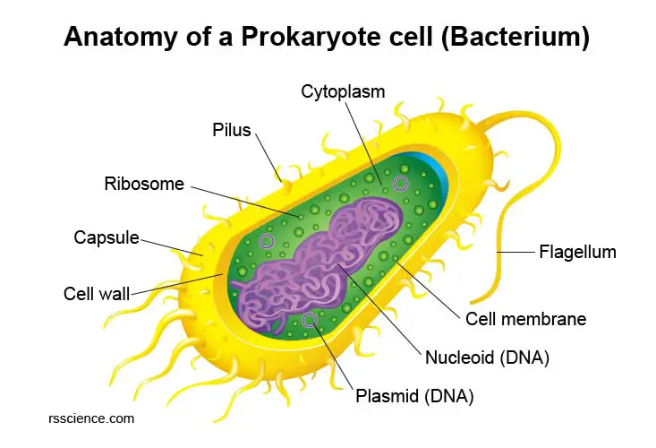
[In this figure] Diagram of a bacterium showing its cellular structures.
While bacteria do not have membrane-bound organelles, they do have distinct cellular structures to survive. Below is a breakdown of what you might find in a bacterial cell.
| Cell Structure | Functions |
| Nucleoid | A central region of the cell contains its DNA. |
| Plasmid | A small circular piece of DNA that is physically separated from chromosomal DNA. |
| Cytoplasm | Cellular fluid hosting all other cellular structures. |
| Cell membrane | Also known as the plasma membrane that separates the cell from the outside environment. |
| Cell wall | Provides structure and protection from the outside environment. Most bacteria have a rigid cell wall made from carbohydrates and proteins called peptidoglycans. |
| Capsule | Some bacteria have a layer of carbohydrates that surrounds the cell wall called the capsule or glycocalyx. The capsule helps the bacterium attach to surfaces. |
| Ribosome | The site for protein synthesis. |
| Pilus | Hair-like structures help with cellular attachment and DNA transfer. |
| Flagellum | Tail-like structure assists in the movement |
Cell wall
The bacterial cell wall is a rigid barrier made of carbohydrate polymers and peptidoglycan proteins (different from the plant and fungal cell walls). It covers outside of the plasma membrane. The cell wall maintains bacteria shape and provides protection.
Plasma membrane
The bacterial plasma membrane is a lipid bi-layer membrane found within the cell wall. The membrane is permeable, which means that substances can pass through it. The bacterial plasma membrane also generates energy and transports chemicals.
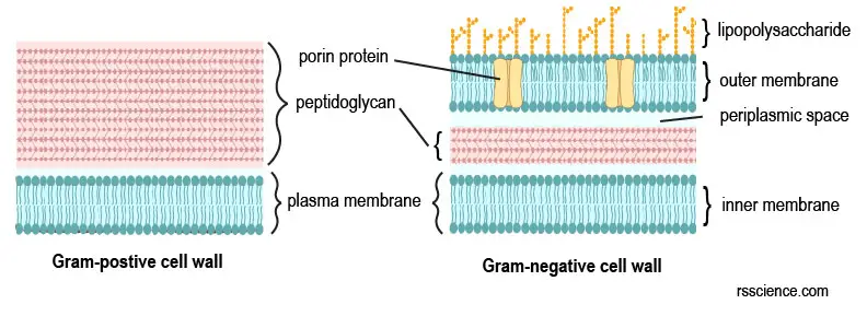
[In this image] Bacteria are divided into two major groups: Gram-positive and Gram-negative based on the difference in their cell walls and membranes.
Both groups have a cell wall composed of peptidoglycan. The cell wall in Gram-positive bacteria is thicker, whereas, in Gram-negative bacteria, it is thinner. In addition, the Gram-negative bacteria have two layers of membranes, sandwiching the thin cell wall and creating a periplasmic space. In Gram-positive bacteria, there is only one layer of plasma membrane beneath the cell wall.
Capsule
Capsule (or glycocalyx) is a layer of complex sugar (carbohydrates) found on the outside of the cell wall in some bacteria. Capsules can help bacteria attach to surfaces. Capsules can also protect a bacterial cell from ingestion and destruction by white blood cells (phagocytosis). The presence of capsules is critical for the capability of infection in several pathogenic bacteria.
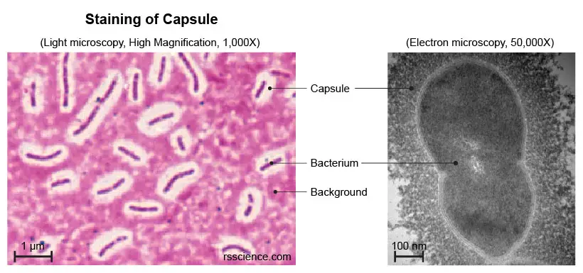
[In this image] Left: Viewing capsules with stains under a light microscope. The thick capsules exclude the stains and appear as clear halos surrounding the bacterial cells. Right: A bacterium with its capsule under a transmission electron microscope.
Photo source: Wen Z. et. al., Molecular Medical Microbiology, 2015
Cytoplasm
The cytoplasm is a gel-like substance inside the plasma membrane. Since bacteria don’t have membrane-bound nuclei and organelles, everything is mixed in the cytoplasm, including genetic materials (bacterial DNA) and ribosomes. The cytoplasm is also the place where many biochemical reactions happen.
Bacterial DNA
Bacterial DNA contains all the genetic information (called genome) used to operate and produce a new bacterium. Bacterial DNA is a long, circular form. It locates inside the cytoplasm. Bacterial DNA is replicated during cell division and transferred to daughter cells (each cell has one copy).
Bacterial DNA occupies an irregularly shaped area in the center of a bacterium. We refer to this nucleus-like (but no nuclear membrane) region as the nucleoid.
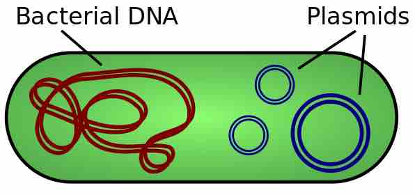
[In this image] There are two types of DNA molecules in a bacterium – Bacterial (chromosomal) DNA and Plasmids.
Photo source: wiki
Plasmids
Some bacteria have extra circles of genetic materials called plasmids. Plasmids are much smaller than chromosomal DNA, and one cell could have many copies of plasmids. Plasmids often contain genes that give the bacterium some advantage over other bacteria. For example, a plasmid may contain a gene that makes the bacterium resistant to a certain antibiotic. Plasmids are also widely used in recombinant DNA technology for genetic engineering.
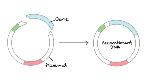
[In this image] “Copy and paste” genes into a bacterial plasmid (called DNA cloning) is a routine in Biotechnology.
Photo source: Khan Academy
Ribosomes
Ribosomes are where proteins are made. Both prokaryotic and eukaryotic cells have ribosomes; however, prokaryotic ribosomes are smaller than eukaryotic ribosomes. Since bacteria lack a nucleus, their mRNAs are transcribed in the cytoplasm and can be translated by ribosomes immediately. As a result, in a bacterium cell, you can even see several ribosomes attached to a single RNA molecule. See “polysomes” to learn more.
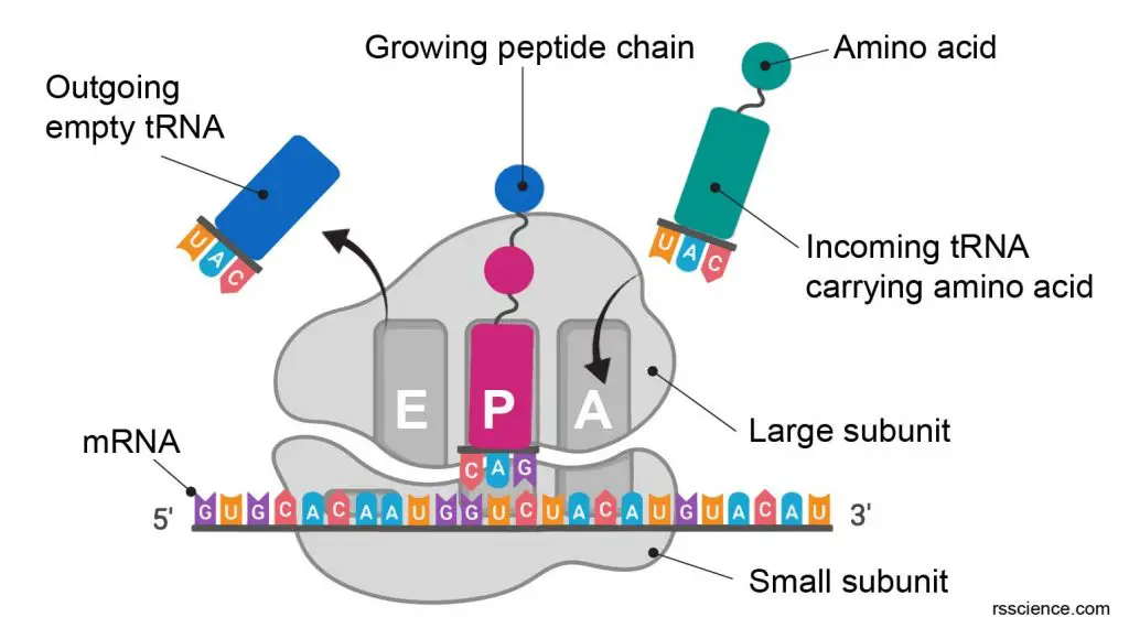
[In this figure] Ribosome.
Ribosomes work like decoding machines to translate the code sequence of mRNA into a protein. Scientists like to call ribosomes, the molecular micro-machines, to admire how exquisite the ribosomes’ design is!
Flagellum
Flagellum (plural: flagella) is a whip-like structure that can propel some types of bacteria to move. Some bacteria can have more than one flagellum.
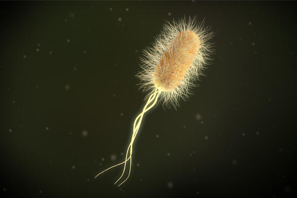
[In this image] A bacterium with three long flagella and many pili covered its cell. This is a 3D illustration of Escherichia coli, not a real picture.
Photo source: Microbiology Society
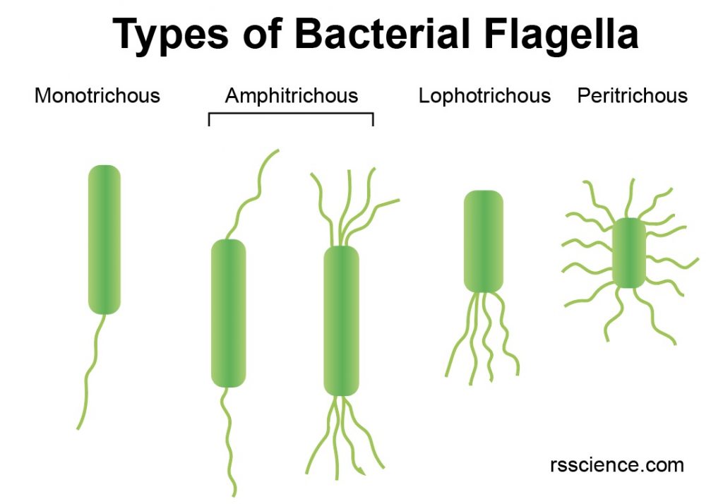
[In this image] Depending on the number and location of bacterial flagella, they can be classified as shown in this image.
Pili
Pili (singular: pilus) are hair-like structures on the outside of bacteria cells. Pili allow bacteria to stick to surfaces, which is critical for infection. Sometimes, special pili can also transfer genetic material from one cell to another (called conjugation). Transferring antibiotic resistance genes can contribute to the spread of illness in humans.
Part 2. Classification and types of bacteria
In the old taxonomic system, Bacteria belong to the Kingdom of Monera. Now, the consensus of the scientific community is creating a level called the Domain above the Kingdom. There are three Domains and six Kingdoms under this system.
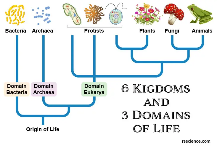
[In this image] The three-domain system is a biological classification introduced by Carl Woese in 1990 that divides life forms into Archaea, Bacteria, and Eukarya domains.
The members of bacteria can be classified by several methods. Here are the examples:
Classification by shapes
Bacteria can be classified into five groups according to their basic shapes:
Spherical (cocci)
Spherical (cocci) – Bacteria shaped like a ball are called cocci (singular: coccus). They can exist as single cells, in pairs, chains, or clusters. The name of the cocci is based on how they reproduce and what they look like after dividing.
For example, cocci that remain in pairs after dividing are called diplococci, and those that divide and remain attached together in a chain are called streptococci (responsible for “strep throat.”). Those that divide into planes and remain in groups of four are called tetrads. Those that divide into three planes and remain in groups of eight are called sarcinae. Those that divide into multiple planes and remain in a cluster like a grape are called staphylococci. These shape characteristics are very useful for the identification of cocci.
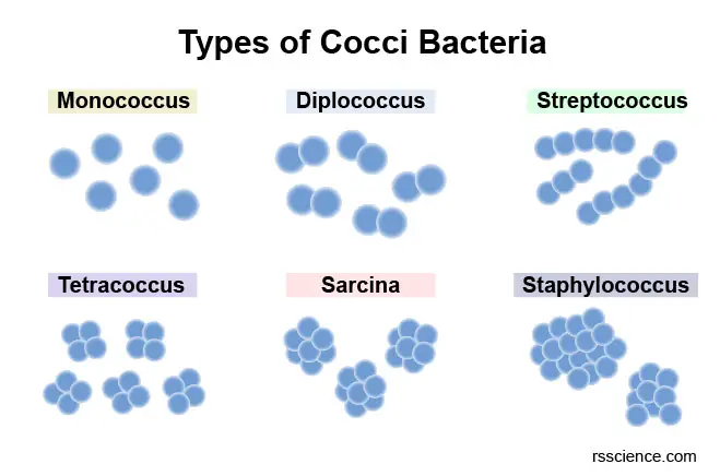
[In this image] Depending on how cells are arranged, cocci can be further divided into these six groups.
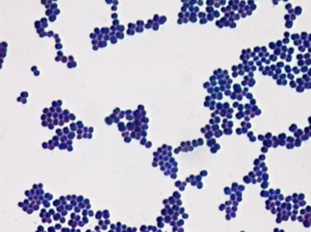
[In this image] Cocci under a light microscope.
Photo source: Neal R. Chamberlain
Rod (bacilli)
Rod (bacilli) – Bacilli (singular bacillus) are rod-shaped bacteria. Examples include Bacillus anthracis, which causes anthrax. Most bacilli are single rods. Those that remain in pairs after dividing are called dipobacilli. Similarly, those that remain together in a chainlike shape after dividing are called streptobacilli. Some bacilli are oval and look like cocci, so they are called coccobacilli.
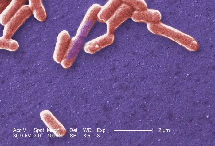
[In this image] Electron micrograph of Escherichia coli (or E. coli).
Photo source: wiki
Comma (vibrios)
Comma (vibrios) – Some rod-shaped bacteria are curved. These are known as vibrios. Several species of which (i.e., Vibrio cholera) can cause foodborne infection, usually associated with eating undercooked seafood.
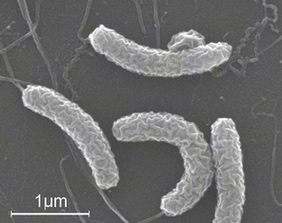
[In this image] Electron micrograph of Vibrio cholera.
Photo source: Louisa Howard
Spiral (spirilla)
Spiral (spirilla) – Spirilla (singular: spirillum) are bacteria with a rigid spiral shape. Helicobacter pylori, the cause of peptic ulcers, is an example.
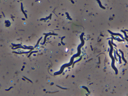
[In this image] Spirilla under a light microscope.
Photo source: wiki
Corkscrew (spirochaetes)
Corkscrew (spirochaetes) – If spiral bacteria have long, helically coiled cells, they are known as spirochaetes. Examples are Leptospira species (causes Leptospirosis), Borrelia species (Lyme disease), and Treponema pallidum (Syphilis).
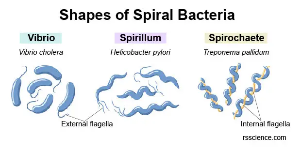
[In this image] Three types of bacteria with curved shapes.
Flagella are another key feature to distinguish spirilla and spirochaetes. Spirilla have several external flagella, while spirochaetes have a single flagellum in the middle of the helically coiled cell.
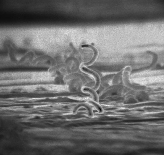
[In this image] Electron micrograph of Treponema pallidum.
Photo source: wiki
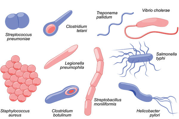
[In this image] Here are several pathogenic bacteria. Could you classify them by their shapes?
Photo source: Microbiology Society
Classification by the usage of oxygen
A bacterium that responds to oxygen can be aerobic, anaerobic, or facultative anaerobes.
- Aerobic bacteria need oxygen to live. They utilize oxygen to metabolize nutrients and generate energy to grow.
- Anaerobic bacteria will die around oxygen. They generate energy by fermentation. In humans, these bacteria are mostly found in the gut. Many of them are beneficial to us by playing critical roles in nutrient absorption. However, some pathogenic ones can cause disease, like appendicitis.
- Facultative anaerobes are bacteria that can grow in both the presence and absence of oxygen. They function best with oxygen; however, without oxygen, they won’t die but just slow down their growth. Many pathogenic bacteria are facultative anaerobes, including E. coli, Pseudomonas aeruginosa, Staphylococcus spp., Listeria spp., and Salmonella.
Classification by the source of nutrients
Autotrophs
Autotrophs are “self-feeders” which produce their own food. Autotrophic bacteria can be two types:
Cyanobacteria (also called blue-green algae) are examples of phototrophic bacteria. They use photosynthesis to convert solar energy and carbon dioxide (CO2) into the nutrients that cells can use.
[In this video] See bacteria, Cyanobacteria, and many other microorganisms under a microscope.
Some bacteria can utilize energy obtained from an oxidative chemical reaction (chemosynthesis). These chemoautotrophs differ from photoautotrophs in that they do not depend on sunlight for energy. Instead, chemoautotrophs use chemicals such as methane or hydrogen sulfide along with oxygen to produce carbon dioxide and energy. As a result, these chemoautotrophs are often found in extreme environments, like deep-sea vents, hot springs, and deep trenches.
Heterotrophs
On the other hand, heterotrophs are “other eaters” which do not produce their own food. Most bacteria that obtain nutrients from their environments or hosts are heterotrophs.
Learn more about Autotrophs vs. Heterotrophs below.
Classification by Gram stain
Other ways of classifying bacteria are learned from pathologists’ experiences, who want to identify the causes of infectious diseases as soon as possible. Gram staining is the best-known example. We can quickly classify the pathogens based on whether or not they are: Gram-positive or Gram-negative.
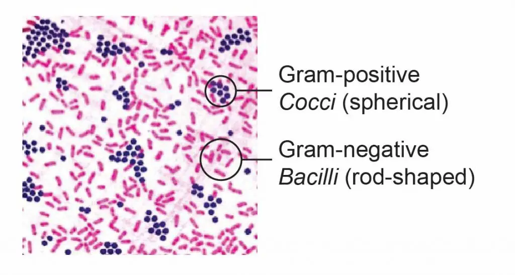
[In this figure] The Gram-positive bacteria stain dark blue or purple while the Gram-negative bacteria stain pink.
Gram stain can distinguish bacteria based on the difference in their cell wall structure. In the hospital, Gram stain is a fast test to identify the type of infection (gram-positive or gram-negative), so the doctor can prescribe which antibiotic to use for treatment.
Learn more about Gram stain steps below.
Classification by ribosomal RNA sequences
Benefiting from the advance of molecular biology and sequencing techniques, scientists now classify bacteria mainly based on their genetic materials, especially ribosomal RNA (rRNA) sequences. This method helps us identify many unculturable species that we had no idea about before.
Part 3. How do bacteria live?
What is life? Is bacterium a living thing?
Bacteria are considered the most basic form of life. How can we say that? Let’s first review what is the definition of life.
Only living things show all of these characteristics:
1. Living things are highly organized in the form of a “cell”.
2. Living things must use energy and consume nutrients to sustain life.
3. Living organisms regulate their internal environment to maintain the conditions needed for living.
4. Living organisms undergo controlled growth.
5. Living organisms can reproduce themselves to create new organisms.
6. Living organisms respond to stimuli or changes in their environment.
7. Populations of living organisms can undergo evolution, meaning that they may change over time.
Bacteria fit all these criteria. They are probably the simplest living organisms.
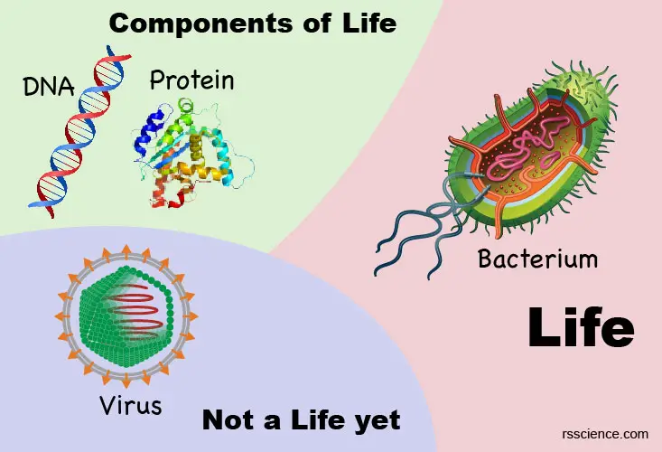
[In this image] The boundary between non-life and life.
DNA and proteins are biological macromolecules that are the components of life. A virus is not a life yet since it requires a host cell to replicate.
How do bacteria obtain energy to grow?
See the part of autotrophic and heterotrophic bacteria.
How do bacteria reproduce?
Bacteria reproduce mainly by binary fission. Binary fission begins when the DNA of the bacterium replicates into two copies. The cell wall then elongates and splits into two daughter cells, each with identical DNA to the parent cell. We can say that each daughter cell is a clone of the parent cell.
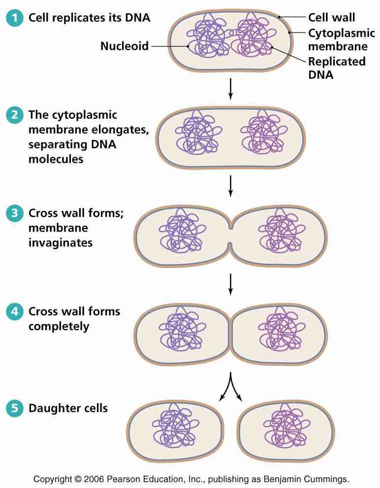
[In this image] Bacteria reproduce by binary fission.
Photo source: Socratic Q&A
When conditions are favorable (right temperature with rich nutrients), bacteria like E. coli can divide every 20 minutes. This means that in 12 hours, one bacterium can divide 36 times and generate 68,719,476,736 bacteria! That’s why we can quickly become ill when pathogenic microbes invade our bodies.
Bacteria survival skills
Some Gram-positive bacteria can form endospores as another way of reproduction during unfavorable conditions. Endospores are resting spores that are extremely resistant to harsh physical and chemical conditions such as heat, UV radiation, and disinfectants. When the condition becomes favorable, these endospores will rapture and give rise to new bacteria.
Many endospore-producing bacteria are nasty pathogens. For example, Bacillus anthracis, which causes anthrax, can form endospores and rest in soil for years.

[In this image] Gruinard Island, which sits just over a kilometer off the coast of mainland Scotland, is still uninhabited due to anthrax spores produced during World War II.
Photo source: Lad Bible
Sexual reproduction of bacteria
Scientists define the sexual reproduction of bacteria when it involves an exchange of genetic information with each other. The exchange and uptake of genetic material allow bacteria to develop new characteristics (for example, the resistance to antibiotics). That way, they can adapt and survive in new environments.
Three are three main methods of genetic exchange (also called genetic recombination):
Transformation – Some bacteria simply take up DNA that is floating around their environment. These DNA molecules could be fragments of chromosomal DNA or plasmids released from a dead bacterium.
Transduction – In this method, genetic material goes from one bacterium to another by the agency of bacteriophages (viruses that infect bacteria).
Conjugation – Using a pilus, two bacteria make contact with each other and exchange genetic material.
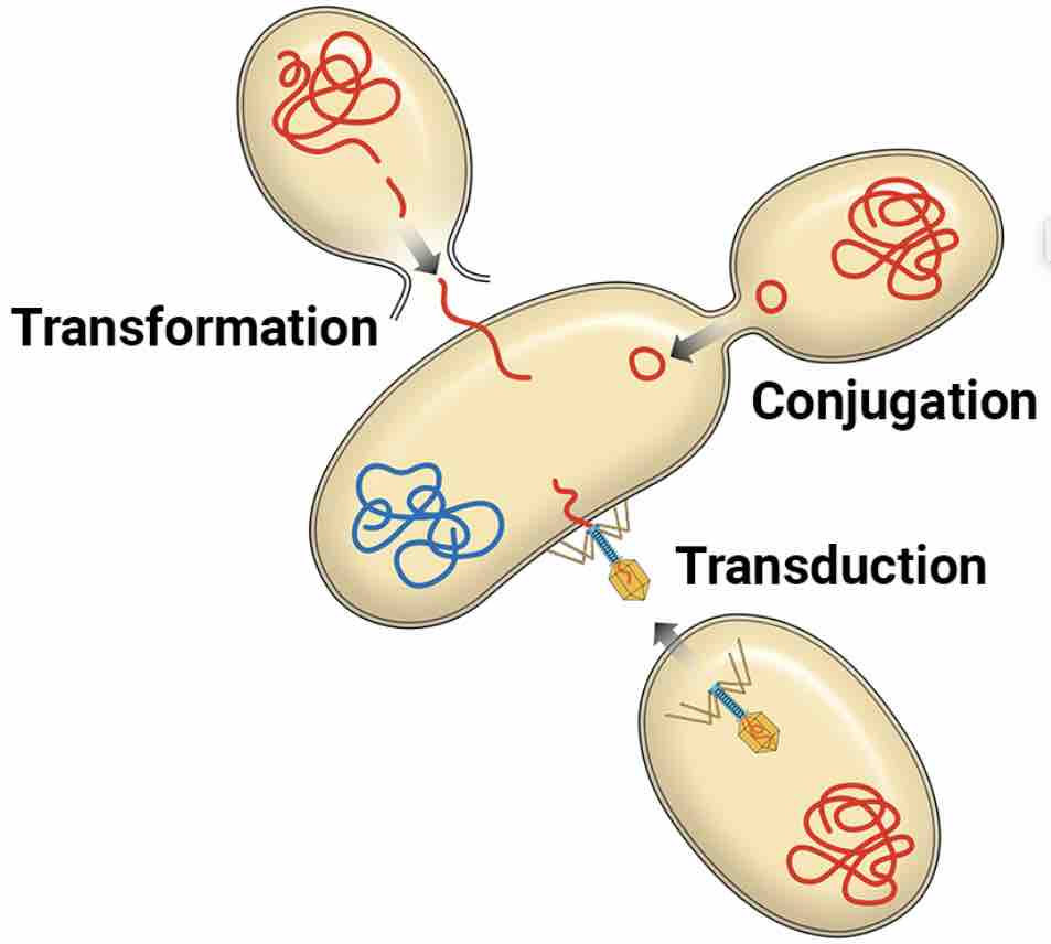
[In this image] Three methods of bacterial genetic recombination: Transformation, Transduction, and Conjugation.
Photo source: Toppr
Part 4. Bacteria and us
What are the benefits of bacteria?
Most bacteria are good for you, including the bacteria (referred to as Microbiota) in your digestive system. These beneficial bacteria help to break down food and keep you healthy. Other good bacteria can produce oxygen (Cyanobacteria) and other chemicals such as ethanol and methane. Some are used to create antibiotics and vitamins. Most interestingly, scientists found some bacteria may one day help us to solve the problem of plastic garbage!
See Plastic Eating Bacteria – how they work.
Bacteria are also used in food production to make fermented foods. For instance, bacteria give the tangy taste in yogurt and the sour in sourdough bread. Fermentation is a type of respiration that doesn’t use oxygen to produce energy.

[In this image] Examples of fermented foods which require bacteria to make.
Photo source: ck12
The ecosystem also relies on bacteria to function properly. For example, bacteria break down dead matter in the environment, like dead leaves, releasing carbon dioxide and nutrients in the process. Without the release of carbon dioxide, plants are unable to grow.
Bad bacteria: pathogens and infectious diseases
Though there are many more good bacteria than bad, some bacteria are harmful. If you consume or come in contact with harmful bacteria, they may grow in your body and release toxins that can damage your health and make you feel ill.
Harmful bacteria are called pathogenic bacteria because they cause infectious diseases and illnesses such as strep throat, staph infections, syphilis, anthrax, leprosy, cholera, tuberculosis, and food poisoning.
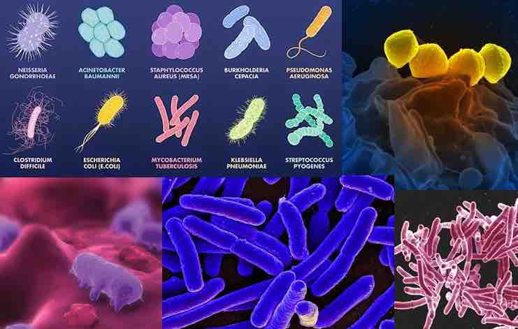
[In this image] Examples of pathogenic bacteria.
Photo source: GloMEc
Fortunately, we have the immune system to defend against bacterial infection. White blood cells, such as neutrophils and macrophages, can engulf entire pathogens by “eating” (or called phagocytosis). At the same time, our immune system also learns and remembers the appearance of these pathogens. If the same bacteria invade our body again, our B cells can produce antibodies to kill these infectious germs more efficiently.
[In this video] See how macrophages eat bacteria under the microscope.
Antibiotic and resistance
Antibiotics are a type of antimicrobial substance against bacteria. They are the game-changer of modern medicine. Penicillin, the first antibiotic, was discovered by Sir Alexander Fleming in 1928. It came from a mold called Penicillium notatum.
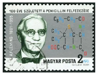
[In this image] A stamp printed in Hungary in 1981 shows Sir Alexander Fleming (1881-1955), Discoverer of Penicillin.
Following the first discovery of penicillin, more and more antibiotics were found and widely used every day in the hospital. However, bacteria also develop the ability to avoid the effect of antibiotics, called antibiotic resistance.
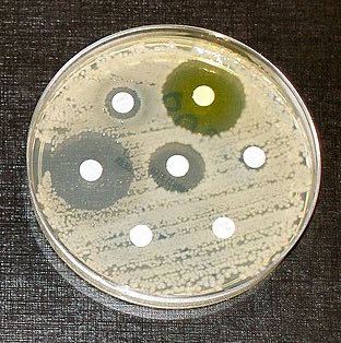
[In this image] Antibiotic resistance tests.
Bacteria are streaked on the plate. Each white paper disk contains a different antibiotic. Clear rings, such as those on the left and middle, show that bacteria can not grow near the antibiotics containing disks. In contrast, the bacteria can grow very closely to the white paper disks on the right corner of the plate, indicating that the bacteria are resistant to those antibiotics tested.
Image source: wiki
Bacteria can become resistant to antibiotics in several ways. Some bacteria can “neutralize” an antibiotic by changing it in a way that makes it harmless. Others have learned how to pump an antibiotic back outside of the bacteria before it can do any harm. Also, they can change their outer structure, so the antibiotic has no way to attach to the bacteria.
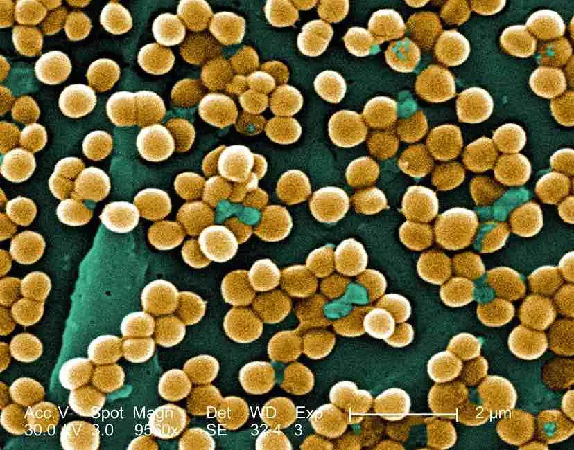
[In this image] Methicillin-resistant Staphylococcus aureus, or MRSA, is a strain of staph bacteria that is resistant to antibiotics.
Image source: Livescience
Bacteria can become resistant through mutation of their genetic material (DNA). After being exposed to antibiotics, some individual bacteria can survive because they found a way to resist the antibiotic. Even if one bacterium becomes resistant to antibiotics, it can then multiply and replace all the bacteria that were killed. In other words, exposure to antibiotics provides selective pressure, making the surviving bacteria more likely to be resistant. These resistance genes may also transfer (through genetic recombination) to other bacteria.
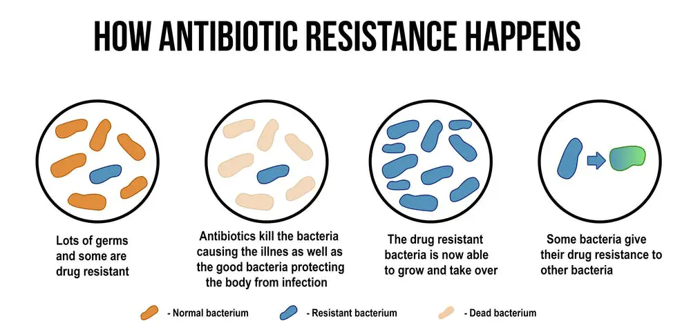
[In this image] Illustration of how antibiotic-resistant bacteria evolve.
Non-resistant bacteria multiply, and upon drug treatment, the bacteria die. Drug-resistant bacteria multiply as well, but upon drug treatment, the bacteria continue to spread and even transfer the antibiotic resistance to others.
Photo source: Public Health Notes
Overuse and misuse of antibiotics can promote the development of antibiotic-resistant bacteria. Every time a person takes antibiotics, sensitive bacteria (bacteria that antibiotics can still attack) are killed, but resistant bacteria are left to grow and multiply. This is how repeated use of antibiotics can increase the number of drug-resistant bacteria.
Biofilms
Although bacteria are single cells, they tend to grow together into a high-density community of cells, called biofilm. Biofilms are structured and organized communities of microorganisms; adhere to the surface. Biofilms could form by a single type of cells or different bacterial colonies together. These cells are embedded in extracellular polymeric substances, a matrix that is generally composed of DNA, proteins, and polysaccharides.
Forming biofilms is an essential characteristic of many pathogens. Living in a biofilm gives individual bacteria many advantages. For example, the matrix protects the cells with high resistance to the penetration of antibiotics.
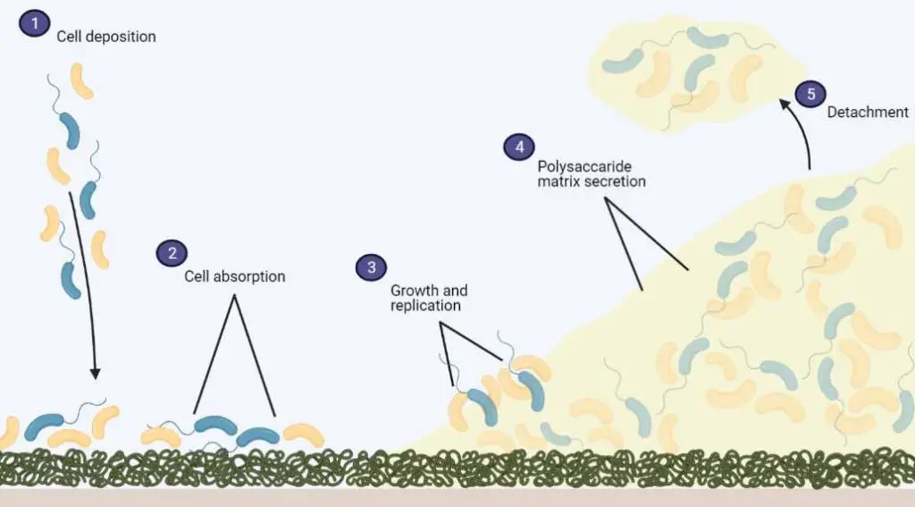
[In this image] Graphic illustration of biofilm formation, the process from bacteria cell attachment to detachment.
Photo source: Particle3D
Part 5. Observing bacteria in a Petri dish or under a microscope
Bacteriology in a Petri dish
A single bacterium is too small to be seen with naked eyes. However, bacteria tend to grow and reproduce as a community, resulting in the formation of bacterial colonies. In the laboratory, we can culture a specimen that potentially contains bacteria in a Petri dish.
This Petri dish is coated with a layer of nutritious substance. We commonly use agar gel mixed with proteins, yeast extracts, and salts (LB agar). In a favorable temperature (37oC), bacteria can rapidly (within a day) grow from one cell into a “colony” that is visible to the naked eye. A colony may contain 107 – 108 cells.
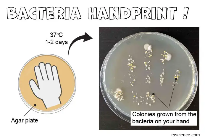
[In this image] A fun scientific project.
See Bacteria on Your Hand (Bacteria Handprint).
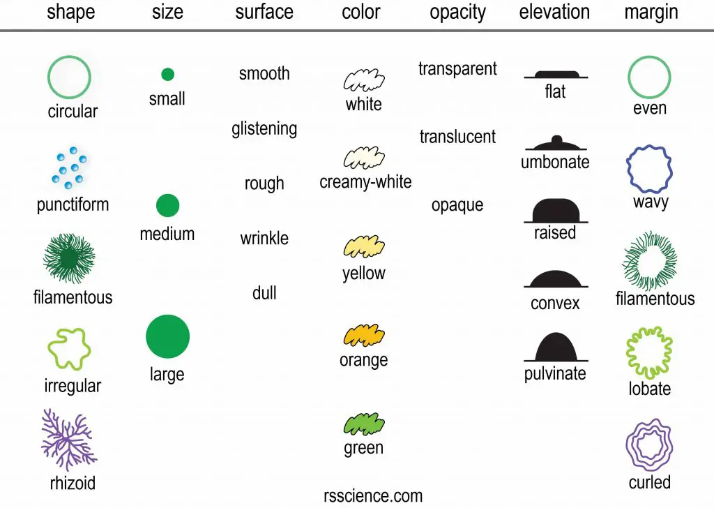
[In this figure] The characteristics to describe the morphology of bacteria colonies. You may roughly classify these bacteria based on the looking of their colonies.
Bacteria under a microscope
Since the average size of a bacterium is only 1-1.5 µm, an optical microscope must have enough power in order to see bacteria. Usually, a high magnification objective lens (63x or 100x) with immersion oil is needed.
Staining could help us see bacteria better. Gram stain is a good example, which can quickly classify bacteria into Gram-positive and Gram-negative groups. Some common stains that you may find at home could also stain bacteria such as methylene blue.
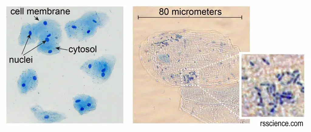
[In this figure] Cheek cells stained with methylene blue.
The left image is a lower magnification. You can see the nuclei stained with a dark blue (because methylene blue stains strongly with DNA). The cell membrane acts like a balloon and holds all the parts of a cell, including a nucleus, cytosol, and organelles inside. The right image is a higher magnification. You can also see some small rod-shaped bacteria. Don’t worry; they are normal oral microbes.
For more detail, see our step-by-step guide to “Look at your cheek cells“.
Detailed structures of bacterial cells, such as flagella and pili, require more advanced microscopy to see. Scanning electron microscopes (SEM) are widely used by microbiologists to study bacteria. SEM images give insight into the surface topography of bacterial cells; therefore, it creates 3-D images.
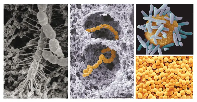
[In this image] Astonishing SEM images of bacteria.
Image source: Royal Microscopical Society
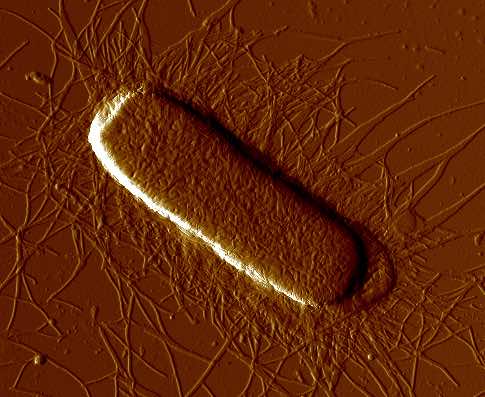
[In this image] Atomic force microscopy (AFM) image of the fimbriated E. coli strain. Fimbriae are pili for attachment.
Photo source: ResearchGate
Frequently asked questions and answers (Q&A)
Are bacteria living things?
Yes, bacteria are living cells. Bacteria are similar to your cells in many ways; yet, bacteria cells also have distinct characteristics. In brief, bacteria fit the criteria of life since they can consume nutrients to grow and divide to produce offspring.
How old are bacteria?
Bacteria lived on Earth for two billion years before the first eukaryotes. During that time, they evolved into millions of different species. You can say they are the most successful organisms on the planet.
What are the differences between bacteria and archaea?
Both bacteria and archaea are single-celled, prokaryotic microbes. However, they branched very early since close to the dawn of life and went through independent evolutionary history. This caused bacteria and archaea to have many differences in the biochemistry of their cells, especially the ways to obtain nutrients and produce energy.
Are bacteria always so small?
Not really. In fact, there are some extremely big bacteria existing in nature as well. Thiomargarita namibiensis, a spherical shape bacterium found in the ocean sediment of Namibia, is called “Sulfur pearl of Namibia” (Thiomargarita means sulfur pearl). It is ~750 µm in diameter, slightly larger than a fruit fly’s eye so it is big enough to be seen with a naked eye. 98% of its cell volume is the vacuole, which is a nitrate reservoir used to fuel sulfide oxidation.
In addition, Epulopiscium spp., intestinal symbionts of certain marine surgeonfish, are the largest known heterotrophic bacteria (meaning they cannot produce their own food). These cigar-shaped cells are up to 600 µm long and 80 µm wide.
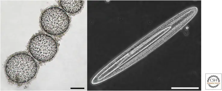
[In this image] Giant bacteria.
On the left is a chain of Thiomargarita namibiensis cells. In this bright-field image, sulfur granules can be seen in the cytoplasm. The panel on the right shows an exceptionally large Epulopiscium cell with two large internal offspring. Scale bars, 100 µm.
Image source: Cold Spring Harb Perspect Biol. 2015 Jul; 7(7): a019216.
What are probiotics?
A healthy digestive system is the foundation of overall health. An imbalance between the good and the bad bacteria could lead to many digestive issues. Probiotic means “for life” as opposed to antibiotics which refers to “against life”. We know that antibiotics can kill bacteria. On the other hand, probiotics help the body bring back the balance in the gut that supports the growth of good bacteria. Therefore, our bodies naturally suppress the bad ones.
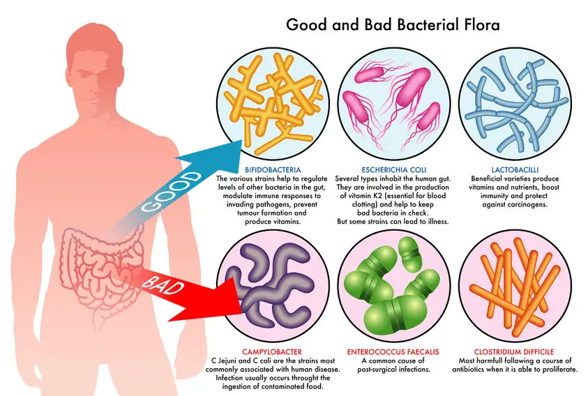
[In this image] Good and bad bacteria flora in the human gut.
Some good bacteria include Bifidobacteria, and Lactobacill. Bad bacteria include Campylobacter, Enterococcus faecalis, Clostridium difficile.
Photo source: MDSupplies
Summary
1. Bacteria are single-celled prokaryotes.
2. Bacteria have no membrane-bound nucleus or organelles in a bacterial cell. Their DNA molecules are floating in the cytoplasm.
3. Bacteria have two types of DNA molecules – chromosomal DNA and plasmids. Both are in circular forms.
4. Bacteria have special cell walls as a protective barrier.
5. Bacteria have filamentous structures – flagella and pili for them to move and attach.
6. Bacteria can be classified into five groups according to their basic shapes – Spherical (cocci), Rod (bacilli), Comma (vibrios), Spiral (spirilla), and Corkscrew (spirochaetes).
7. Some bacteria need oxygen to live (aerobic). But some (anaerobic) can be killed by oxygen.
8. Some bacteria can produce nutrients by themselves (autotrophic) through sunlight or chemical reactions. Others live by decomposing organic substrates to obtain energy.
9. Bacteria can reproduce rapidly by binary fission. They can divide as fast as every 20 mins.
10. During unfavorable conditions, some bacteria can form endospores to survive.
11. Many bacteria are good. They maintain the health in our body (especially in our guts) and the balance of the entire ecosystem.
12. Some bacteria can cause diseases if they successfully invade our bodies.
Related posts
What Living Things You Can See Under a Light Microscope?
Observing Bacteria Under the Microscope – Gram Stain Steps
The Scale of Biology