
This article covers
1. Why the microscope is important for studying biology
Cells are building blocks of life. No only small organisms like protozoa but also large organisms like an elephant, they are all made of cells. However, the size of cells is pretty small, ranging from 1-100 µm (micrometer), which is one-millionth of a meter.
The human naked eye can see an object as small as 0.1 millimeters (100 µm). Therefore, the human naked eye can barely see cells (most of them <100 µm). Thus, we need the amplifying power of the microscope to see cells and even the structure and organelles inside of cells.
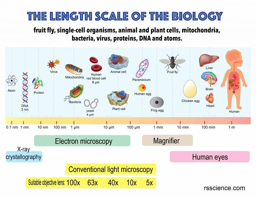
Below is a size and length scale in biology, including eggs, cells, organelles, bacteria, viruses, protein complexes, and atoms. This chart will help you to understand what we can see with a common light microscope (1 -1000 µm). Let’s answer 5 most common questions of what you can see with a light microscope from top-down.
2. Can a light microscope see cells?
Yes! The size of cells ranges from 1-100 µm. Yeast cells are 3-4 µm in diameter. Human red blood cells are 7-8 µm in diameter. Drosophila (fruit fly) intestinal stem cells are around 5-10 µm in diameter and their enterocytes are range from 20-50 µm in diameter. You can see yeast cells, animal cells, and plant cells pretty well with a 400x magnification (assuming 10x eyepiece and 40x objective lens).
See the image below illustrating the human cheek cells about 80 µm wide (scale bar is 50 µm). There are also many blue speckles outside of the cell. These are rod-shaped bacteria. Notice how small the bacteria are. You can barely see them.
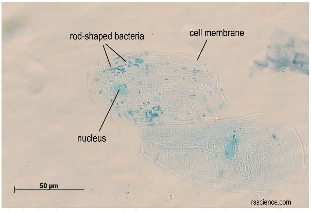
[In this figure] My cheek cells stained with methylene blue.
Surprisingly, the rod-shaped bacteria also got stained with methylene blue and can be visualized.
3. Can a light microscope see mitochondria?
Yes. It is visible. You can see a vague image of mitochondria with very little detail, even with the aid of fluorescence dye. The green color of the image below labeled the mitochondria. Their morphology is tubular, and it is very different from the cartoon illustration, which contains lots of membranes inside.
The transmission electron microscope is required to see the internal structure of the mitochondria, including the inner membrane, outer membrane, intermembrane space, matrix, and cristae. The structure we saw from the transmission electron microscope is more like an illustration image.
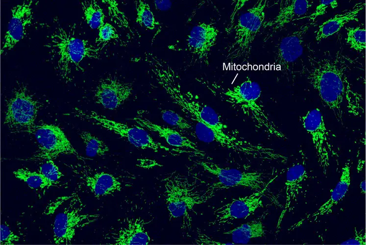
[In this figure] Mitochondria were labeled with green fluorescence.
Mitochondria appear web-like structure. They are surrounded the nucleus (blue) and also in the cytosol.

[In this figure] Electron microscopic image of a mitochondrion.
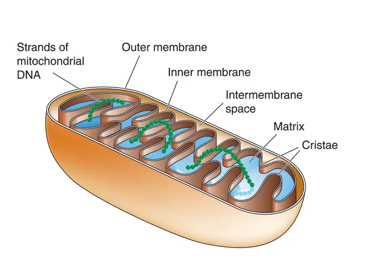
[In this figure] Cartoon illustration of mitochondria.
4. Can a light microscope see bacteria?
Yes, most of the bacteria range from 0.2-2 µm in diameter. The length can range from 1-10 µm for filamentous or rod-shaped bacteria. The most well-known bacteria: E. coli, their average size is ~1.5 µm in diameter and 2-6 µm in length.
As we talked above, you can see some bacteria in my cheek cells sample. They look like a chain of small dots. Most of the bacteria are like that scale with 640x magnification.
The biggest bacteria in the world
In contrast, there are some extremely big bacteria existing in nature as well. Thiomargarita namibiensis, a spherical shape bacterium found in the ocean sediment of Namibia, called “Sulfur pearl of Namibia” (Thiomargarita means sulfur pearl). It is ~750 µm in diameter, slightly larger than a fruit fly’s eye so it is big enough to be seen with a naked eye. 98% of its cell volume is vacuole, which is a nitrate reservoir used to fuel sulfide oxidation.
In addition, Epulopiscium spp., intestinal symbionts of certain marine surgeonfish, are the largest known heterotrophic bacteria (meaning they cannot produce their own food). These cigar-shaped cells are up to 600 µm long and 80 µm wide.
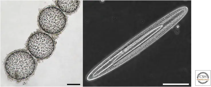
[In this figure] Giant bacteria.
On the left is a chain of Thiomargarita namibiensis cells. In this bright-field image, sulfur granules can be seen in the cytoplasm. The panel on the right shows an exceptionally large Epulopiscium cell with two large internal offspring. Scale bars, 100 µm. This is from Cold Spring Harb Perspect Biol. 2015 Jul; 7(7): a019216. URL: https://www.ncbi.nlm.nih.gov/pmc/articles/PMC4484965/
5. Can a light microscope see viruses?
No, the average sizes of human viruses are around 100 nanometers (0.1 µm), beyond the resolution of the light (optical) microscope (0.5 -1 µm).
You will need an electron microscope to see the viruses. The original electron microscopic image of viruses is black-and-white. The colors are artificially painted post-production for better visualization. We have a detailed post explaining why a light microscope cannot see viruses, what limits the resolution of a light microscope, and how an electron microscope can solve this limit?
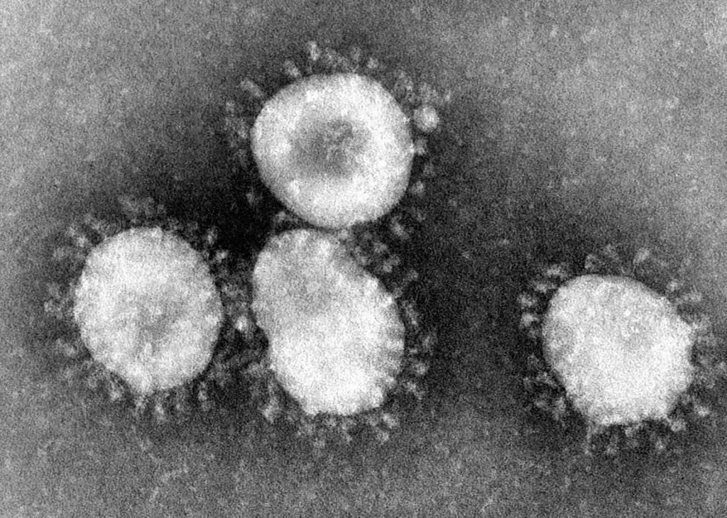
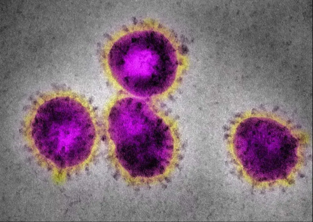
[In this figure] Coronaviruses have a halo or crown-like (corona) appearance.
The new virus, COVID-19, is one kind of coronaviruses. Original electron microscopic images are always black-and-white. The colors are artificially painted for better visualization (right). Photo credit: CDC.

[In this figure] The relative sizes of common human viruses. Source: https://viralzone.expasy.org/5216
6. Can a light microscope see DNA?
No, you can not see the individual double-strand DNA with a light microscope.
The width of the double-strand DNA is about 2 nm, which is beyond the resolution of the light microscope (0.5 µm). However, you might be able to see a condensed chromosome (composed of DNA) inside cells during the cell division with a light microscope like the image below.
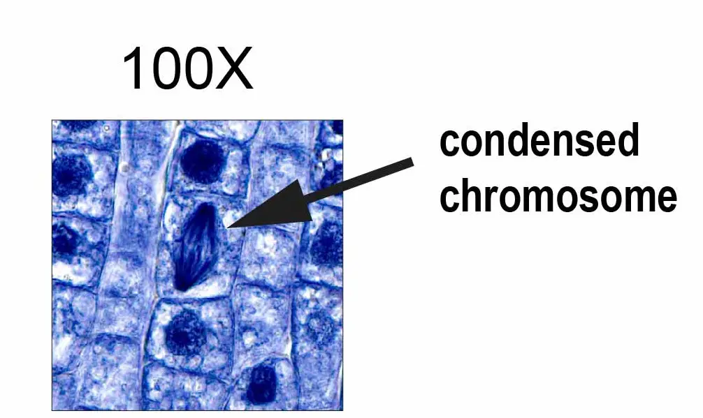
[In this figure] Condensed chromosome with 1000x magnification. The chromosomes (consist of DNA) are condensed when the cells are dividing. Therefore, it can be visualized by blue staining under the light microscope.
Key takeaways
You can see a variety of cells pretty well with the light microscope. To see the cell organelles, you will need to get a higher magnification (usually with a 40x-100x objective lens). In addition, the electron microscope is required to resolve the structure of mitochondria, bacteria, viruses, and large protein complexes.
Do you have any questions you would like to know? please let us know!