
This article covers
Why microscopes are important?
Microscopy makes a big deal out of little things. Thanks to microscopes, scientists have the tools to visualize the microscopic worlds.
The modern microscopy doesn’t only advance our understanding of biology, many other fields, like chemistry, physics, material science, archaeology, and forensic science, all benefit from the incredible capability to see smaller things.
The field of microscopy dates back to the 17th-century. During that time, the optical microscopes were the most advanced scientific tools for the microbiologists (for example, Antonie van Leeuwenhoek) to learn the structure of cells.
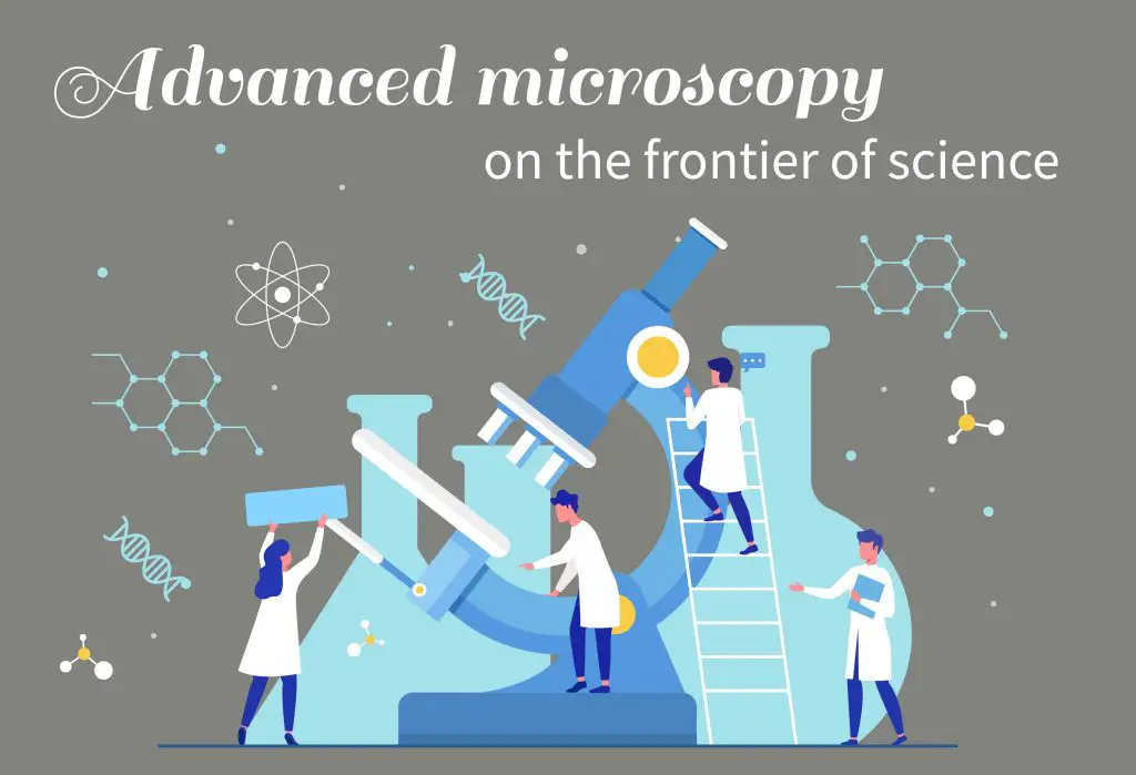
In the 19th-century, following the Industrial Revolution, the glass lens was manufactured, and the optical microscopes became popular in universities around the world. Later, new components (like dark field, phase contrast, and differential interference contrast (DIC), Figure 1) were added to the optical microscope to make better observations of the certain types of specimens. Now, you can purchase a pretty decent optical microscope under $500.
The scientists keep inventing new types of microscopes. Most of these advanced microscopes are extremely expensive and can only be used in research institutes. Let’s see what these microscopes can do: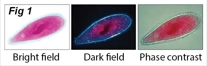
Confocal fluorescent microscope
A fluorescent microscope (Figure 2) is a very powerful tool for biomedical research. Instead of the bright light, a fluorescent microscope uses “fluorescent light” that is emitted by special chemicals or biological molecules to generate an image. Therefore, we can selectively see the specific structures (for example, the cytoskeleton that maintains the shape of cells in green color in Figure 3) inside living cells using specific fluorescent molecules (usually with antibodies).
The additional “confocal” apparatus can further increase the optical resolution and contrast of fluorescent images using a tiny pinhole to block out-of-focus light in image formation. With these high-resolution images, the scientists can learn in detail, for example, how cancer cells grow and how viruses infect healthy cells.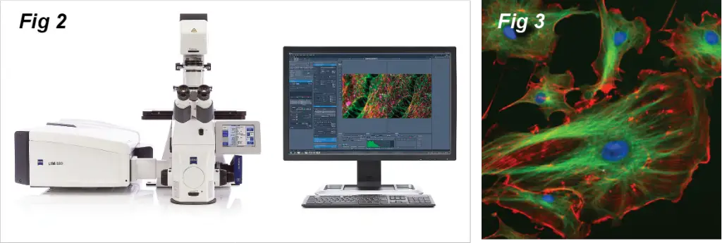
Electron microscope
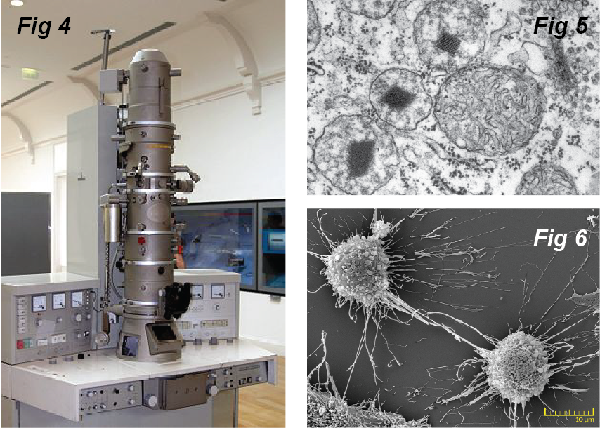
There are two types of electron microscopes (Fig4, EM). The transmission electron microscope (TEM) is used for thin specimens (like tissue sections) because electrons can pass through the specimens to generate a projection image (Fig 5. the organelles inside cells). On the other hand, the scanning electron microscope (SEM) can see the surface of a specimen (Fig 6. the protrusion of human cells).
how does electron microscope work
The resolution of optical microscopes is limited by the wavelength of light. In order to see even smaller features, the scientists have to find an illumination with a wavelength much smaller than the visible light.
An electron has both properties as a particle and a wave. As a result, an electron has a wavelength around ~1 nm depending on its energy states, whereas the wavelength of visible light is between 380 nm to 740 nm. This is why we can use electron microscopes to probe the tiny structure that an optical microscope cannot see.
X-ray crystallography
Strictly speaking, X-ray crystallography (XRC) is not a typical microscope. However, XRC is the most powerful way for us to see the atomic structures of tiny molecules. XRC measures the diffraction patterns that are generated when an X-ray beam passes through a crystal (Fig 7). Then, a special computing software analyzes the patterns to calculate the structures of the molecules.
In this way, scientists have revealed the structure and function of many biological molecules, including vitamins, drugs, proteins, and nucleic acids such as DNA. Figure 8 shows several protein structures that were determined by XRC. The protein structures are usually displayed as colorful strings and ribbons in three-dimensional models. Biomedical researchers can learn how the protein works in our body by looking at their sharp or design a new drug to neutralize its action.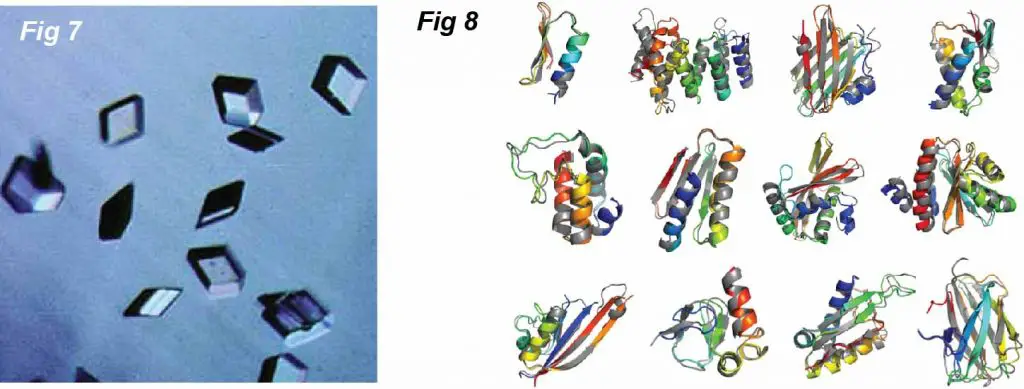
Biological scale
With the knowledge of several advanced microscopies, now you can easily understand the scale of biology and the tools you should use to study them.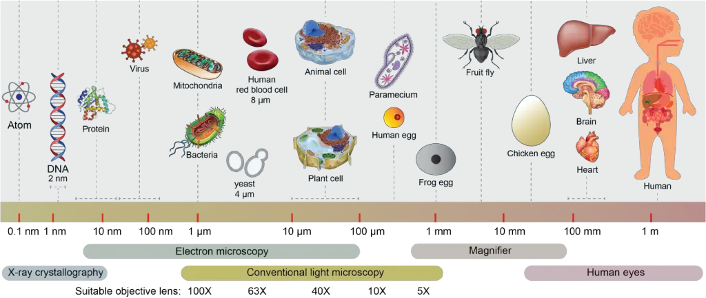
References
BOOKS:
WEBSITES:
- Rs’ Science – www.rsscience.com
- Microscopy-UK – www.microscopy-uk.org.uk
- Microbehunter – http://www.microbehunter.com
- MicroscopyU – www.microscopyu.com
- GeneEd – geneed.nlm.nih.gov