
Have you wondered why the virus infection is so difficult and time-consuming to diagnose?
The quick answer is because THEY ARE TOO TINY TO SEE!
This article covers
The size of the virus
The average sizes of human viruses are around 100 nanometers (1 meter = 1,000 millimeters = 1,000,000 micrometers = 1,000,000,000 nanometer!). No idea how small it is. The illustration below shows the relative size comparison of human hair, red blood cell, bacterium, and virus.
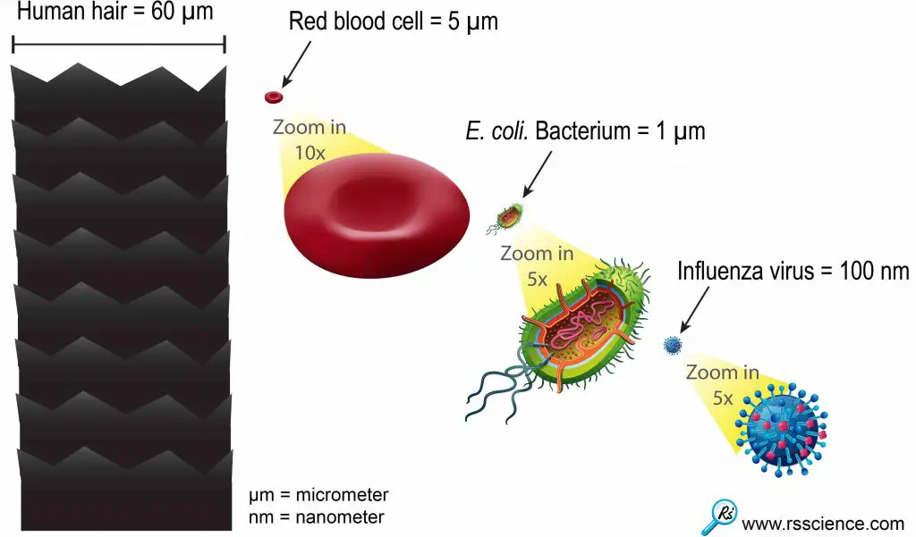
[In this figure] This relative size chart can give you an idea of how small the viruses are.
One influenza or “flu” virus particle is 600 times smaller than the diameter of human hair.
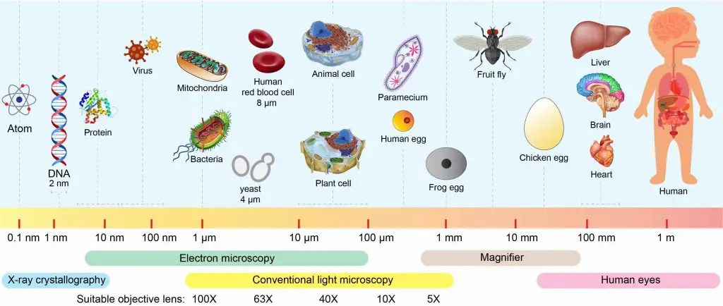
[In this figure] The biological scale.
This biological scale gives you a sense of different sizes of biological objects, such as aminal cells, bacteria, and viruses. It also shows you the microscopic techniques that are required to see the objects. Click here to know more about these advanced techniques.
The resolution limit of the optical microscope
The regular optical microscope (the common ones that we have in the classroom) does not have enough power to resolve things as tiny as a virus (even the highest magnification 1000x). It is because of the limitation of visible light. More specifically, the resolution is limited by the wavelength of the visible light that illuminates the specimen.
The wavelength of the visible light ranges from about 400 (purple light) to 700 (red light) nanometers. Generally speaking, you cannot see the stuff clearly if its size is smaller than half of the wavelength of the visible light, which is about 0.2 – 0.35µm. Therefore, anything smaller than 0.35 µm is not viable using an optical microscope. In summary, 1 µm (the size of a bacterium and mitochondrion) is about the limitation of the “common” optical microscope.

[In this figure] What we can see under an optical microscope with different objective lenses.
The resolution of the electron microscope
In order to see smaller things, scientists have to find an illumination with a wavelength much smaller than visible light. In the early 1930s, they realized that electrons can do so and thus developed the transmission electron microscope (TEM).
According to modern physics, an electron has both properties as a particle and a wave. As a result, an electron has a wavelength (called De Broglie wavelength) depending on its energy (or speed). For example, if an electron is speeded up in a vacuum tube to the speed of 1,000,000 meters per second (equal to 2.2 million miles per hour!), the De Broglie wavelength we get is around one-tenth of a nanometer, which is about the size of an atom. This is why we can use electron microscopes to probe the structure of atoms in a crystal directly. Therefore, seeing viruses with an electron microscope is a piece of cake.
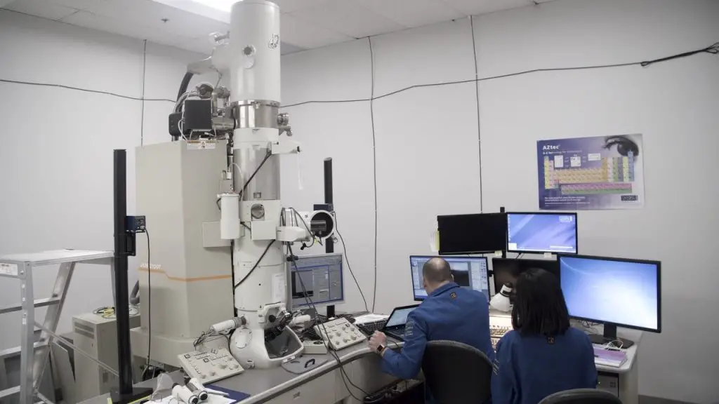
[In this figure] A transmission electron microscope in a dedicated research center. Photo credit: Alexander Fisher-Wagner/UC Davis.
Electron microscopic images of different viruses
The electron microscope is an extremely expensive and complicated instrument that only research institutes or universities may have. However, we still can enjoy the fantastic nano-scale world under the electron microscopes by the hard work done by many scientists. Below are some of the best pictures of viruses under the electron microscopes.
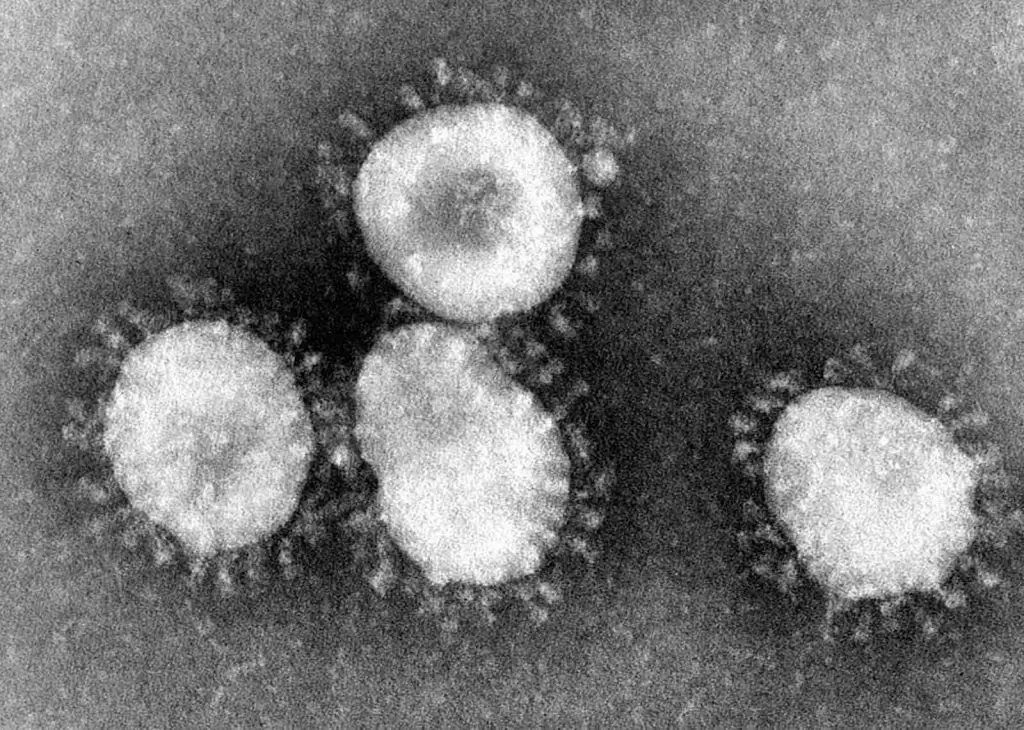
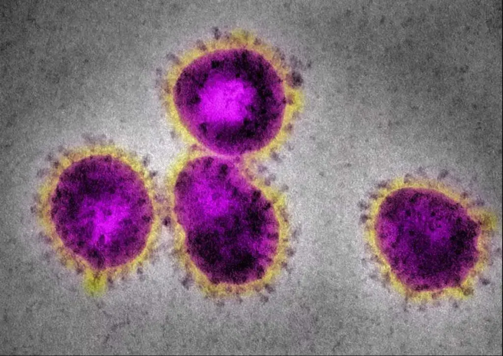
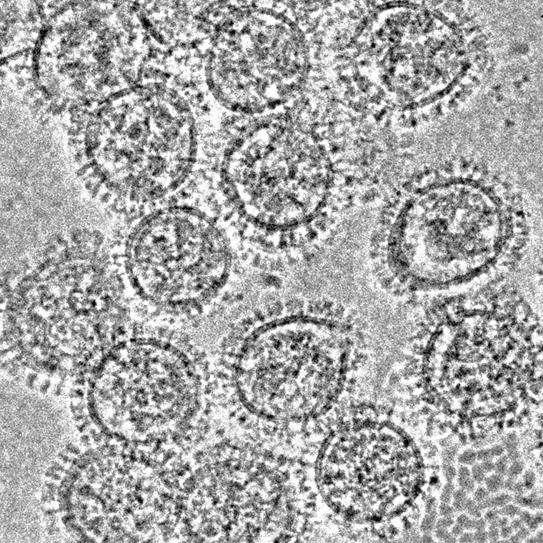
[In this figure] Coronaviruses have a halo or crown-like (corona) appearance.
The new virus, COVID-19, is one kind of coronaviruses. Original electron microscopic images are always black-and-white. The colors are artificially painted for better visualization (right). Photo credit: CDC.
By the way, the clinical diagnosis of COVID-19 is not by electron microscopes. It is done by reading the viruses’ genetic messages using a technique called Real-Time RT-PCR.
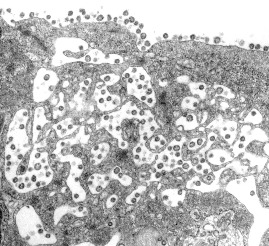
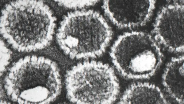
[In this figure] Coronaviruses are infecting a cell.
Many viruses have invaded the cell cytosol while some still adhere to the plasma membrane. Photo credit: CDC.
[In this figure] Herpes simplex virus or HSV. Photo credit: CDC/ Dr. Fred Murphy; Sylvia Whitfield.
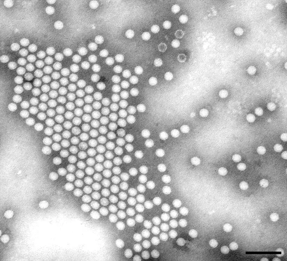
[In this figure] Polioviruses.
Polioviruses, which cause polio or infantile paralysis, are now rare due to the successful prevention of the polio vaccine. Photo credit: Joseph Esposito, CDC.
[In this figure] Influenza viruses cause flu. Photo credit: Ivan Kosik, Jonathan W. Yewdell, et al., NIAID.
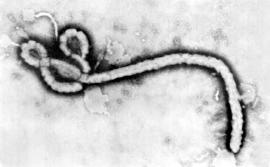
[In this figure] Ebola viruses.
Ebola viruses are very special viruses that look like flexible filaments with a consistent diameter of 80 nanometers, but they vary greatly in length and degree of twisting. Photo credit: Frederick A. Murphy, University of Texas Medical Branch, Galveston, Texas.
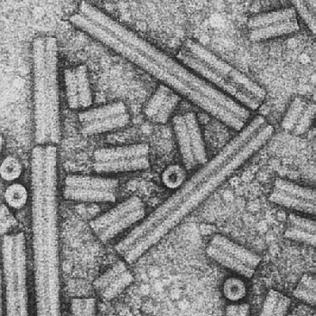
[In this figure] Plants also have their viruses.
Tobacco rattle viruses can infect tobacco and cause diseased leaves. Photo credit: John Antoniw, Rothamsted Experimental Station, UK.
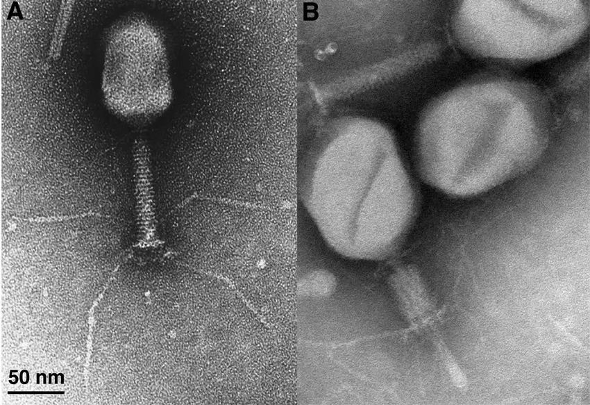
[In this figure] Bacteriophage T4.
Bacteriophage T4, a virus that infects a bacterium, looks like an Apollo Lunar Module in miniature. Source: Microbiology and Molecular Biology Reviews 67(1):86-156

[In this figure] The relative sizes of common human viruses. Source: https://viralzone.expasy.org/5216
Key takeaways
The size of viruses ranges from 20 to 400 nm, which is too small to be seen with an optical microscope. The resolution limit of an optical microscope is about 0.5 – 1 µm (500 nm – 1,000 nm). Therefore, we can not see viruses under the microscope. The electron microscope is required to study the structure of viruses.
Related posts
What Living Things You Can See Under a Light Microscope?
Advanced Microscopy on the Frontier of Science