

(update 6/15/2020)
This article covers
Why bacteria are difficult to see?
- The bacteria are small, typically 0.2-2 µm in diameter and 1-10 µm in their length.
- Most bacteria are colorless under a standard light microscope, so it is hard to see, not to mention identifying what kinds of bacteria they are. One way to improve this is to color them by staining. We discussed two types of staining in the post: simple stains and differential stains.
The size of bacteria
Most of the bacteria range from 0.2-2 µm in diameter. The length can range from 1-10 µm for filamentous or rod-shaped bacteria. The most well-known bacteria: E. coli, their average size is ~1.5 µm in diameter and 2-6 µm in length.
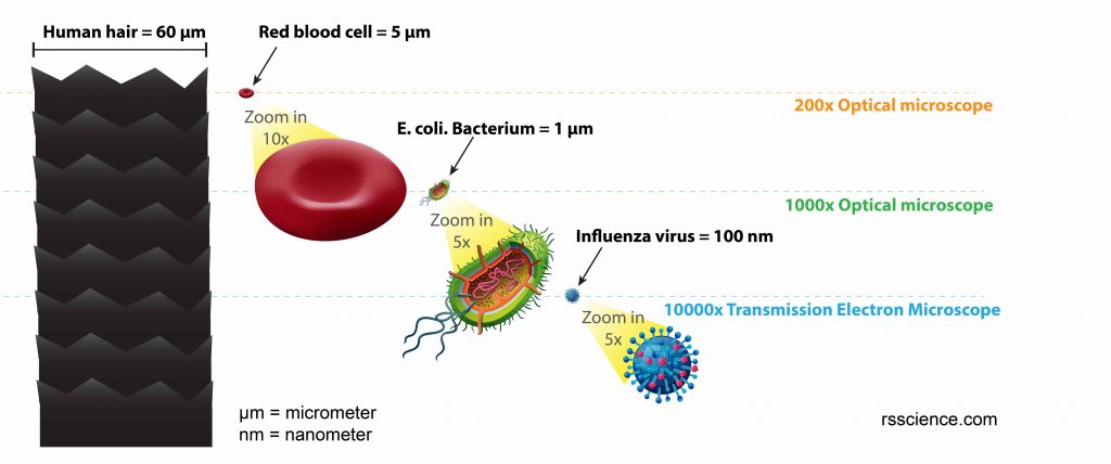
[In this figure] The size comparison between our hair (~ 60 µm) and E. coli (~1 µm). Notice how small the bacteria are. It requires 1000x magnification to see them well.
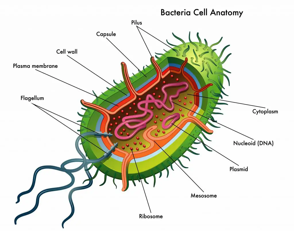
[In this figure] The anatomy of bacteria cell
One of the biggest bacteria in the world is called “Sulfur pearl of Namibia” (Thiomargarita namibiensis), a spherical shape bacterium found in the ocean sediment of Namibia. It is ~750 µm in diameter, slightly larger than a fruit fly’s eye so it is big enough to be seen with a naked eye. For more details, please check our post “Can a light microscope see bacteria?”.
[In this figure] Thiomargarita namibiensis. Credit: Wiki
Smear Preparation
Before staining, we need to prepare smears and fixed the sample. The fixing means attaching the microorganisms to the microscope slide and preserving their structure in their natural state with minimal distortion.
A smear is a thin film of microorganisms spread on the microscope slide. You can use an inoculation loop to pick up a colony or dip in the broth culture and then transfer some microorganisms to the surface of the microscope slide. The detailed steps are followed:
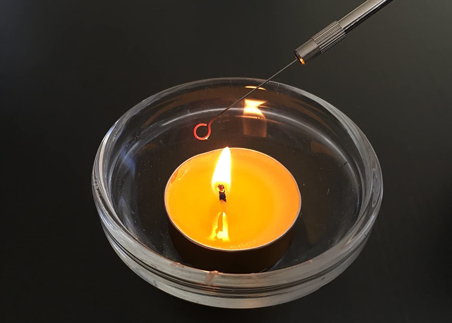
Before touching any sample, heat the inoculation loop until the color of the wire loop change and allow cooling (This step will clean and sterile the inoculation loop)
For the broth culture, place the inoculation loop into the broth to scoop a small amount of liquid, and gently spread the liquid on the surface of the microscope slide (size around a centimeter in diameter).

For the plate culture that forms colonies, you can put a drop of a saline solution on the microscope slide, then use an inoculation loop to pick up some microorganisms to stir on the drop of saline to form an emulsion.
Alternatively, if you don’t have bacteria colonies, you may try to take a little bit of yogurt, and spread it on the slide.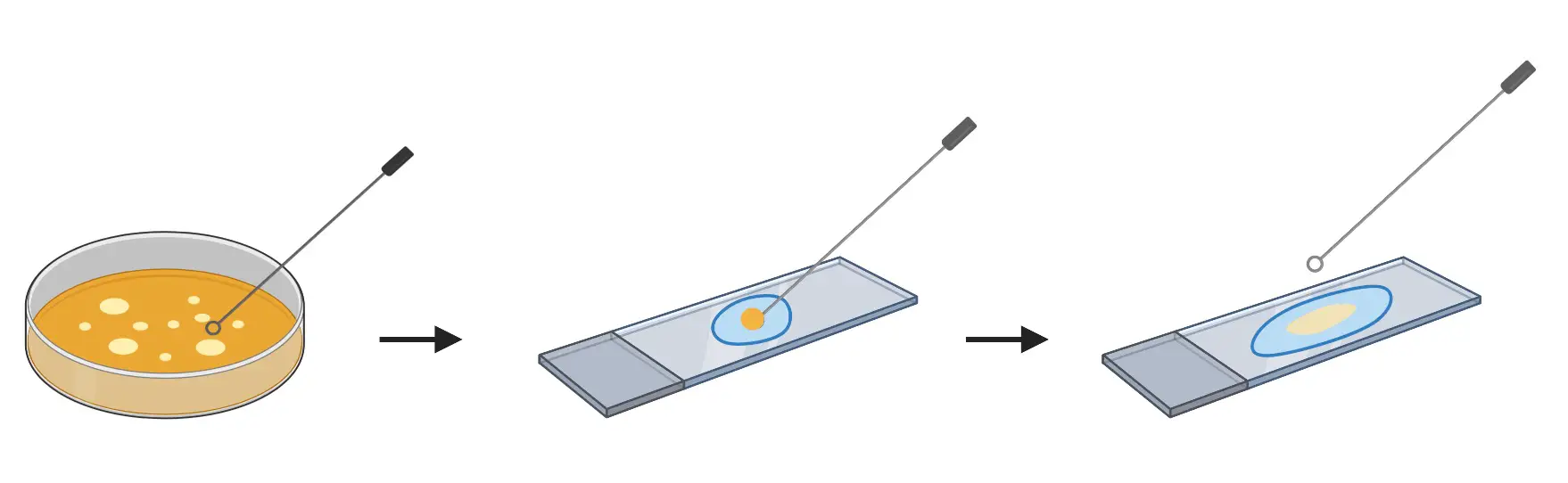
If you don’t have any bacteria culture, you may use the sterile cotton swab to gently scrape inside of your month to get some cheek cells and oral bacteria. We are able to see some oral bacteria and our cheek cells.
Let it air dry
Fixing
In most staining procedures, fixing is achieved by passing the slide through the flame of a Bunsen burner several times (2-3 times), smear side up. Do not overheat the slide.
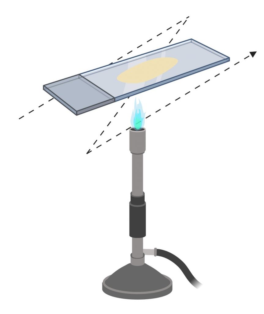
Alternatively, you can cover the slide with methyl alcohol for 1 min.
Then you can proceed with a simple stain or Gram stain.
Simple stains
A simple stain is using an aqueous or alcohol solution of a single dye. The primary purpose of this dye is to highlight the entire microorganisms, so the outline of cells and their basic structure are visible. The stain steps are
- Applying the stain to a fixed smear for a certain length of time
- Wash off the stain
- Let the slide dry
- Examine
Some of the common simple stains in the lab are methylene blue (aqueous), crystal violet (an alcohol solution) and safranin.
Below is an example of using methylene blue stain on cheek cells and oral bacteria.

[In this figure] Simple stain of the human cheek cells.
Differential stains- Gram stain
In contrast to simple stains, differential stains are used to distinguish the difference between bacteria. One of the most well-known differential stains is Gram stain, which differentiates gram-positive and gram-negative bacteria based on the difference in their cell wall structure.
The Gram stain was developed by the Danish bacteriologist Hans Christian Gram in 1884. It is one of the most useful and staining techniques in the Microbiology field. All bacteria can be classified into two large groups: gram-positive and gram-negative.
In the hospital, the Gram staining is a fast test to identify the type of infection (gram-positive or gram-negative), so the doctor can prescribe which antibiotic to use for treatment.)
The steps of Gram stain involved:
1. Primary stain (crystal violet)
Cover the heat-fixed smear with crystal violet for 1 min, then rinse with water very gently. You can use distilled or tap water. This purple stain passes its color to all cells, that’s why we called this step is primary stain.
2. Mordant (iodine)
Stain the smear with iodine for 1 min, then rinse with water very gently. The iodine is a mordant, which is to increase the affinity of a stain for the specimen. After iodine is washed off, both gram-positive and gram-negative bacteria appear dark purple.
3. Decolorization (alcohol wash)
The slide is washed with alcohol for about 10 seconds or until the alcohol running from the slide is clear. The alcohol removes the purple from the gram-negative cells. Do not wash with alcohol for too long. This is critical.
4. Counterstain (safranin)
The slide is stained with safranin for 1min, then rinse with water very gently until no color is coming from the water. Blot dry with absorbent paper. Place the slide on the microscope to view the sample.
The gram-positive bacteria will stain dark blue or purple. The gram-negative bacteria will stain pink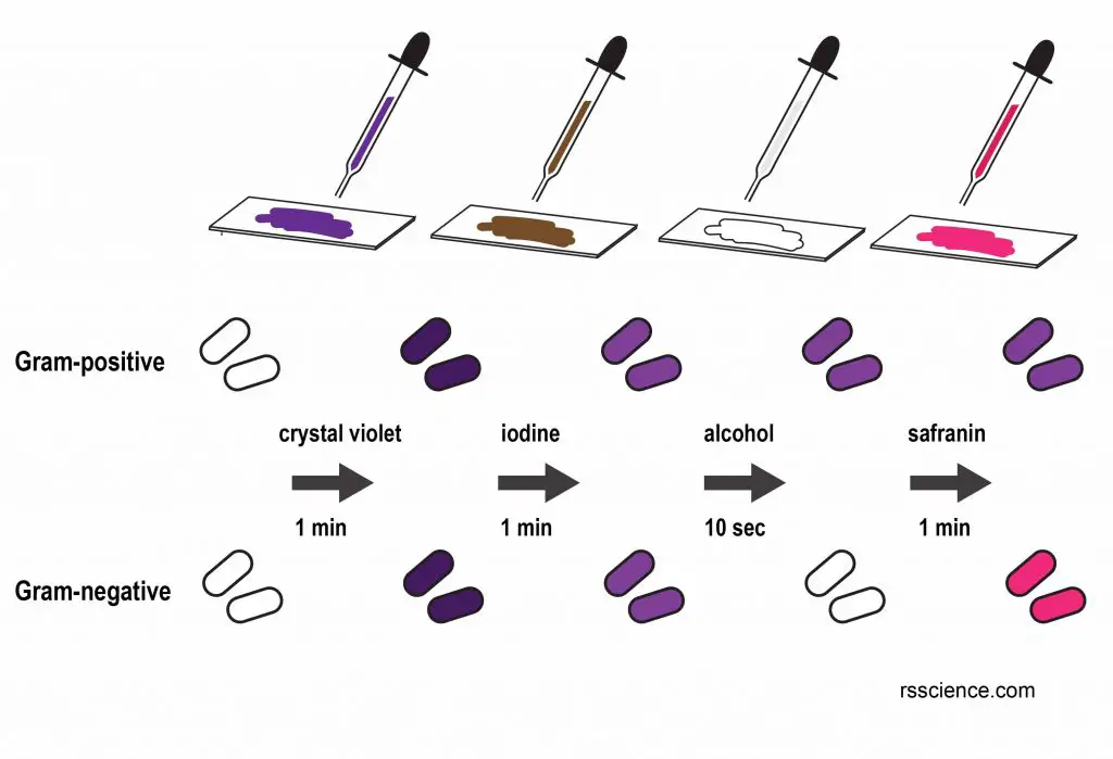
[In this figure] A procedure of Gram stain.
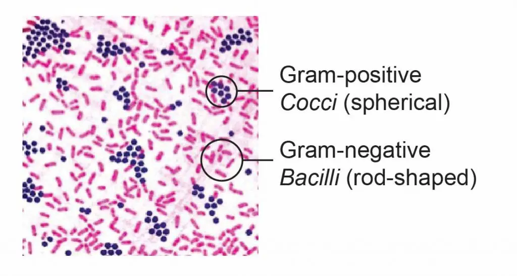
[In this figure] The gram-positive bacteria stain dark blue or purple while the gram-negative bacteria stain pink.
Why do bacteria stain differently after Gram stain?
This is because gram-positive and gram-negative bacteria have different cell wall structures.
The cell wall of the gram-positive bacteria contains many layers of peptidoglycan, forming a thick, rigid structure. In contrast, the gram-negative cell walls only contain a thin layer of peptidoglycan.
The cell wall of gram-negative bacteria consists of one or a few layers of peptidoglycan and an outer membrane. The outer membrane contains lipopolysaccharide, lipoproteins, and phospholipids. The function of the outer membrane is to serve as a barrier to certain antibiotics and digestive enzymes. It can also evade phagocytosis and host defense systems.
Although the outer membrane is an additional layer of barrier, it is not impermeable. There are proteins, called porins that form channels allowing nucleotides, disaccharides, peptides, amino acids, vitamin B12, and iron to pass through.
In addition, gram-negative bacteria contain periplasm, a fluid-filled space between the outer membrane and the plasma membrane. There are many degradative enzymes and transport proteins in periplasm.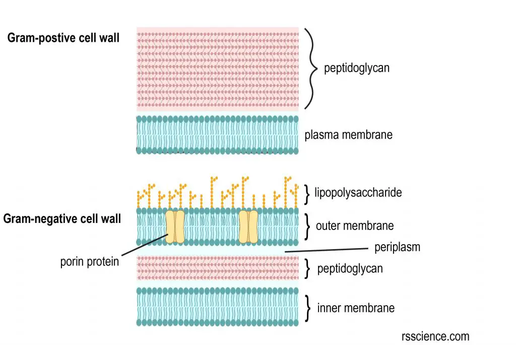
[In this figure] The cell wall structural difference between gram-positive and gram-negative bacteria.
Cell walls and the Gram stain mechanism
Now we know the cell wall difference between gram-positive bacteria and gram-negative bacteria. Let’s discuss the staining mechanism.
Crystal violet stains both types of bacteria purple because the dye enters the cytoplasm of both types of bacteria. When we applied iodine, it forms large crystals with the crystal violet that are too big to escape through the cell wall.
The application of alcohol in the following step dehydrates the peptidoglycan of gram-positive bacteria to make it impermeable to the crystal violet -iodine complex. However, gram-negative bacteria contain an outer membrane that consists of lipids. The alcohol dissolves the outer membrane and makes small holes, which allow crystal violet -iodine complex to diffuse away. This makes gram-negative bacteria colorless, thus can be stained as red when safranin is added.
In some cases, some gram-positive cells can give a gram-negative result. One possibility is that the cells are dead. Another possibility is that a few gram-positive genera show an increase of gram-negative cells as the culture ages. The best examples are Bacillus, Clostridium and Mycobacterium. They are gram-variable.
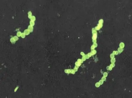
Example of Gram-positive cells
Staphylococcus and Streptococcus. 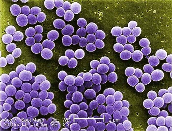
[In this figure] Under a high magnification of 10,000X, this digitally-colorized scanning electron microscopic (SEM) image shows a strain of Staphylococcus aureus bacteria taken from a vancomycin-intermediate resistant culture. Credit: CDC.
Example of Gram-negative cells
Escherichia, Salmonella, and Shigella.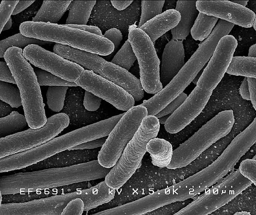
[In this figure] Escherichia coli: Scanning electron micrograph of Escherichia coli, grown in culture and adhered to a coverslip. Credit: wiki
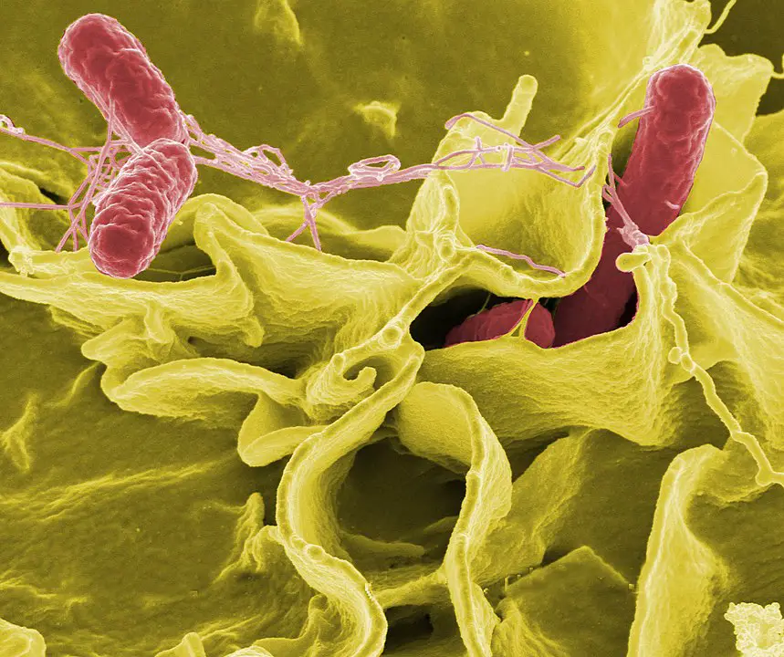
[In this figure] Color-enhanced scanning electron micrograph showing Salmonella Typhimurium (red) invading cultured human cells Credit: Rocky Mountain Laboratories. wiki
Bacteria shape and morphology
The bacteria can be grouped into three basic shapes, coccus (spherical, plural: cocci), bacillus (rod-shaped, plural: bacilli), and spiral.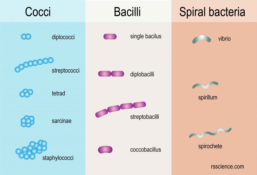
[In this figure] The bacteria shape and morphology.
Cocci
Cocci are usually round shapes but can be oval or elongated. The name of the cocci is based on how they reproduce and what they look like after dividing.
For example, cocci that remain in pairs after dividing are called diplococci and those that divide and remain attached together in a chain are called streptococci. Those that divide into two planes and remain in groups of four are called tetrads. Those that divide into three planes and remain in groups of eight are called sarcinae. Those that divide into multiple planes and remain in a cluster like a grape are called staphylococci. These shape characteristics are very useful for the identification of cocci.
Bacilli
Bacilli divide only across their short axis, so there are fewer groups of bacilli than cocci. Most bacilli are single rods. Those that remain in pairs after dividing are called dipobacilli. Similarly, those that remain together in a chainlike shape after dividing are called streptobacilli. Some bacilli are oval and look like cocci, so they are called coccobacilli.
Spiral bacteria
Spiral bacteria have one or more twists. Vibrios are bacteria that look like curved rods. Spirilla are bacteria that have a helical shape, like a corkscrew and rigid bodies. Another type of spiral bacteria is called spirochetes, which are also helical but with flexible bodies. Spirilla use flagella, a whiplike external appendage, to move while spirochetes use axial filaments to twist.
Most of the bacteria maintain in one single shape. However, there are also some bacteria that can change their shape based on environmental conditions. This will make identification more difficult.
Related posts
How to See Bacteria on Your Hand (Bacteria Handprint)
What Living Things You Can See Under a Light Microscope?
As a newcomer to microscopy, larger specimens are more interesting to observe. But this information about bacteria is essential for a more advanced student. And, germs are important! Especially staph germs.
Thanks for posting this.
Thank you very much for your kind words. I might have a simple way to see how much bacteria on your hands. Stay tuned 🙂
Good write-up, I’m regular visitor of one’s site, maintain up the excellent operate, and It’s going to be a regular visitor for a long time.