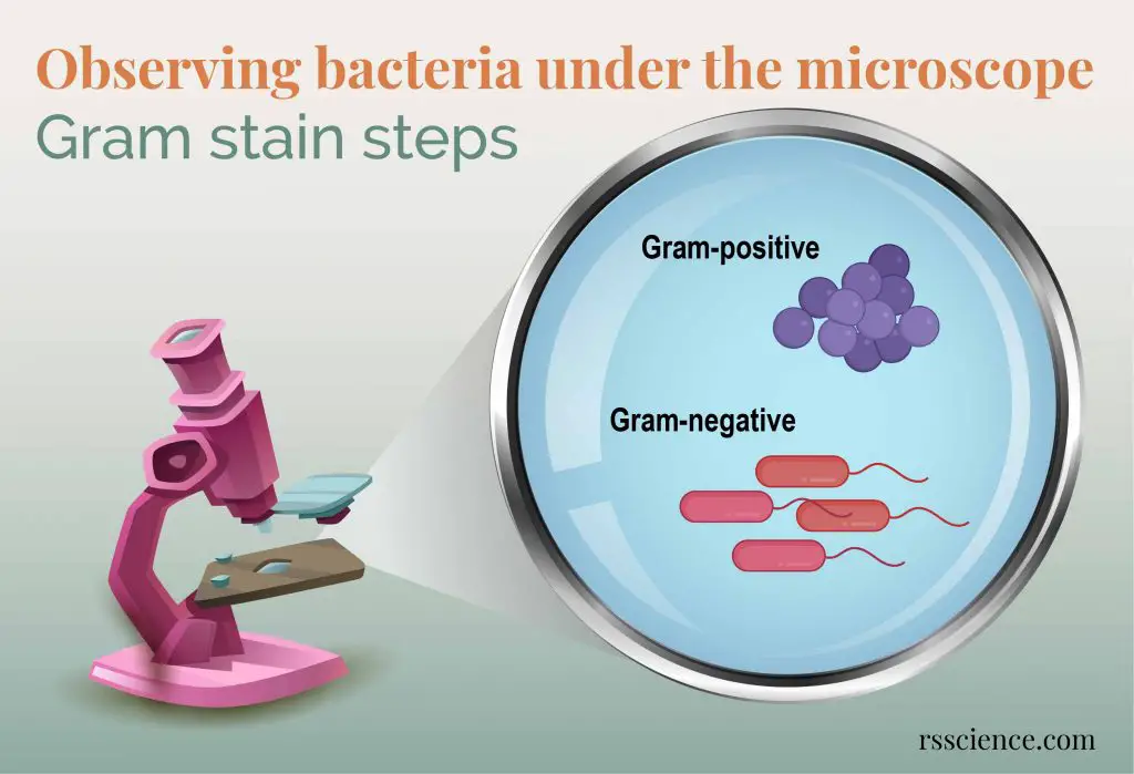
Why bacteria are difficult to see?
This is because:
1. The bacteria are small, typically 0.2-2 µm in diameter and 1-10 µm in their length.
2. Most bacteria are colorless under a standard light microscope, so it is hard to see.
What is a Gram stain?
What are Gram stain steps?
Why do bacteria stain differently after Gram stain?
This is because gram-positive and gram-negative bacteria have different cell wall structures.
Pages: 1 2


As a newcomer to microscopy, larger specimens are more interesting to observe. But this information about bacteria is essential for a more advanced student. And, germs are important! Especially staph germs.
Thanks for posting this.
Thank you very much for your kind words. I might have a simple way to see how much bacteria on your hands. Stay tuned 🙂
Good write-up, I’m regular visitor of one’s site, maintain up the excellent operate, and It’s going to be a regular visitor for a long time.