This is the second series of the article “Size Matters! – The Scale of Biology – Examples and Fun Facts”. If you are not sure what the Biological Scale is, check it out first.
Understanding the biological scale is important for several practical reasons, and for spiritual inspiration, too. Here are 6 reasons from my opinions:
This article covers
(1) A good comparison helps us understand biology better
In many branches of biology, such as molecular and cell biology, microbiology, and histology, we study some things that are invisible to naked eyes. Without a good scale, we could quickly lose our dimensions.
A good comparison can help us understand the relationships between biological components much intuitively.
Luckily, length scales in the human perspective also span about 5 orders of magnitude, with sizes ranging from 100 micrometers (width of a human hair) to 20 meters (height of 4 story building).
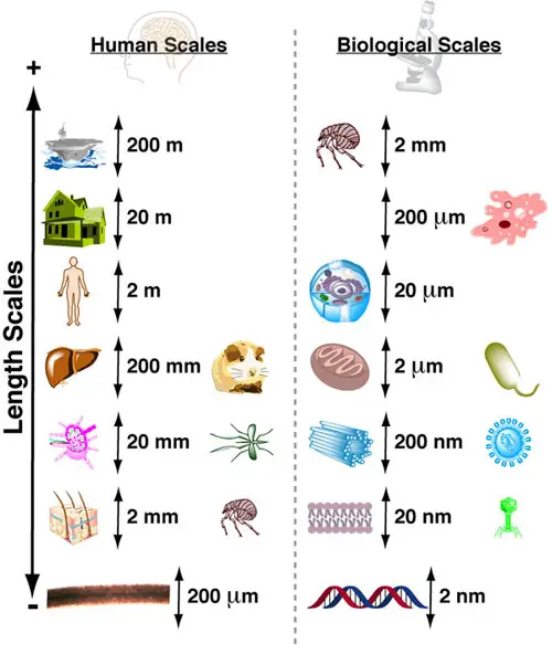
[In this image] An intuiting biological scale using human scales as references.
Photo source: Practically Science
By simply imagining the sizes of the objects in the Human Scales relative to you, you can intuit the relative sizes of biological organisms and molecules relative to a cell. For example, the relative sizes of rats to you are approximately similar to the relative sizes between bacteria and a white blood cell. A virus looks to a cell could be like a mosquito looks to you.
(2) Break the myths
By placing organisms on the same scale, you will immediately realize that we took many myths for granted.
Here is an example.
Are multicellular organisms bigger than simple, unicellular microbes?
The answer is No. For example, rotifers and tardigrades (water bears) are microscopic animals in the range of 100 – 500 µm. Their bodies are composed of hundreds to few thousand cells. They have fully functional organ systems like a complete digestive tract that includes both a mouth and anus.
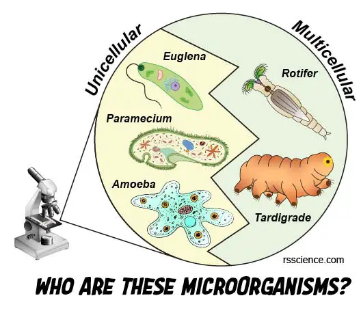
[In this figure] Many microorganisms fall in a size that ranges 100-500 μm.
However, they can be totally different. Some microorganisms, such as Euglena, Paramecium, and Amoeba, contain only one gigantic cell (called unicellular or single-celled) and are classified as protists. Others (like Rotifer and Tardigrade), however, contain thousands of cells and belong to multicellular animals or micro-animals.
On the opposite side, many single-celled protozoans also fall in this size range. Euglenas are typically 100-150 µm. Some paramecia can grow up to 300 µm. Amoebas can grow by engulfing more foods. They are typically 250-750 µm, and some big one can grow up to 1 mm long. Stentors are one of the largest unicellular microorganisms. Some Stentors can reach a size of up to 4 mm (4,000 micrometers) when stretched out. This is 10-20 times bigger than a rotifer!
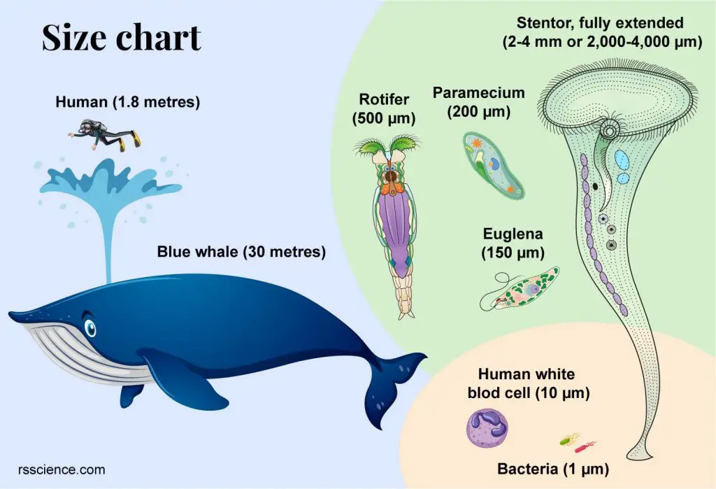
[In this image] In this relative size chart, you can see how big a stentor is (even it contains only one single cell). A rotifer looks to a stentor could be like a diver looks to a blue whale. Click here to learn more about stentors.
In the below case, the problem is not that the microorganism is too big; however, a multicellular animal could be much smaller than you can imagine.
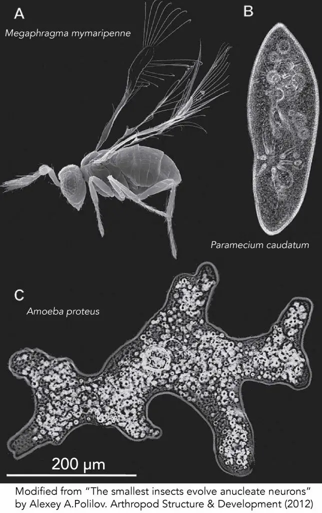
[In this image] Size of the smallest insect and two protozoans in comparison. (A) Megaphragma mymaripenne. (B) Paramecium caudatum. (C) Amoeba proteus. The scale bar is 200 μm. Megaphragma mymaripenne, a parasitic wasp, is the smallest known flying insect.
Let me ask you another question.
Are all bacteria very tiny (smaller than human cells)?
Surprisingly, the answer is No. There are some extremely big bacteria existing in nature as well. Thiomargarita namibiensis, a spherical shape bacterium found in the ocean sediment of Namibia, called “Sulfur pearl of Namibia” (Thiomargarita means sulfur pearl). It is ~750 µm in diameter (100 times bigger than our red blood cells), so it is big enough to be seen with a naked eye. 98% of its cell volume is vacuole, which is a nitrate reservoir used to fuel sulfide oxidation.
In addition, Epulopiscium spp., intestinal symbionts of certain marine surgeonfish, are the largest known heterotrophic bacteria (meaning they cannot produce their own food). These cigar-shaped cells are up to 600 µm long and 80 µm wide.
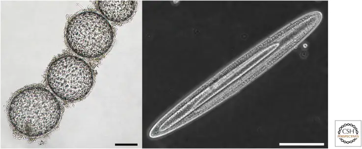
[In this figure] Giant bacteria.
On the left is a chain of Thiomargarita namibiensis cells. In this bright-field image, sulfur granules can be seen in the cytoplasm. The panel on the right shows an exceptionally large Epulopiscium cell with two large internal offspring. Scale bars, 100 µm.
Photo credit: Cold Spring Harb Perspect Biol. 2015 Jul; 7(7): a019216.
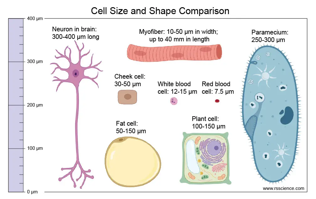
[In this figure] Different cell sizes and shapes comparison.
Human cells are relatively small. Some cells (like neurons and muscle cells) may be long. Fat cells (adipocytes) could be big because most of their cytoplasm is occupied by a huge oil droplet. On the other hand, plant cells and single-celled organisms (like paramecium or amoeba) are larger in size and require cytoplasmic streaming to distribute substances in these cells.
(3) Embarrass the excitement of extreme cases
By having the average biological scale in mind, you may find yourself more easily appreciating how extraordinary these extreme cases are.
For example, do you know what the largest cell on Earth is? It is an egg cell of ostrich. Ostrich’s egg could be about 150 mm (6 inches) in length by 125 mm (5 inches) in diameter and about 1.35 kg (3 pounds). On the opposite side, a human egg is only 0.12 mm. The difference in diameter is more than 100 times!
You may know the largest land animal is the African bush elephant, and the largest animal is the blue whale. If you are a fan of dinosaurs, you may also hear that the largest dinosaur is Argentinosaurus (40 meters), found in Argentina. But do you know what the largest living organism on Earth is?
The answer may surprise you. The largest living organism is a mushroom!
It is a honey fungus of the species Armillaria solidipes in the Malheur National Forest in the Blue Mountains of eastern Oregon, U.S. This fungal colony is spanning 8.9 km2 (2,200 acres) of area. Biologists found these mushrooms growing in this forest are in fact, a single organism collected by an underground network of tubular filaments (known by DNA testing). This organism is estimated to be 2,400 years old.
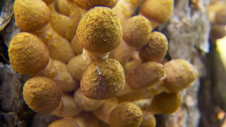
[In this image] The honey mushroom derives its common name from the amber-colored fruit that emerges in the fall.
Photo credit: OPB
(4) Know what you can see and what you cannot see
There are tons of misleading information out there on the internet. Seeing viruses under a pocket microscope is one example. If you know the scales of how tiny the viruses could be, you won’t be fool by these fake articles.
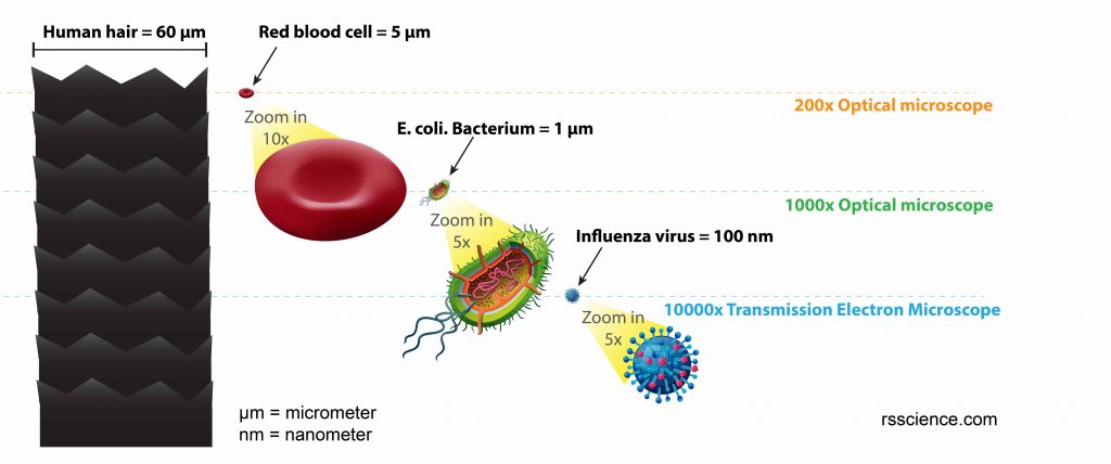
[In this figure] This relative size chart can give you an idea of how small the viruses are.
One influenza or “flu” virus particle is 600 times smaller than the diameter of human hair. 1 meter = 1,000 millimeters = 1,000,000 micrometers = 1,000,000,000 nanometer.
(5) Choose the right tools
Seeing is believing. This is totally true in studying biology. For this reason, you need the right tools. Depending on the subjects you like to study, you can choose from:
Optical microscopes (protists, cells, tissues, small organisms)
A microscope is an instrument used to see objects that are too small to be seen by the naked eye.
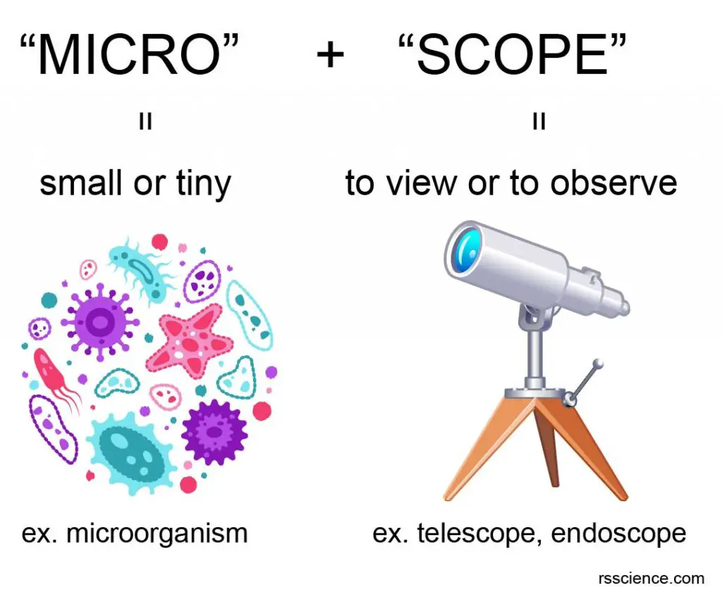
[In this figure] The name “microscope” came from two words – “micro” and “scope”.
“Micro” means small or tiny. “Scope” means to view or to observe. Therefore, a microscope can be understood as an instrument to see tiny things.
Basically, light microscopes use visible light to illuminate the specimen and to form the images. Based on the applications, light microscopes fall into two main categories: the compound microscope and the stereo microscope.
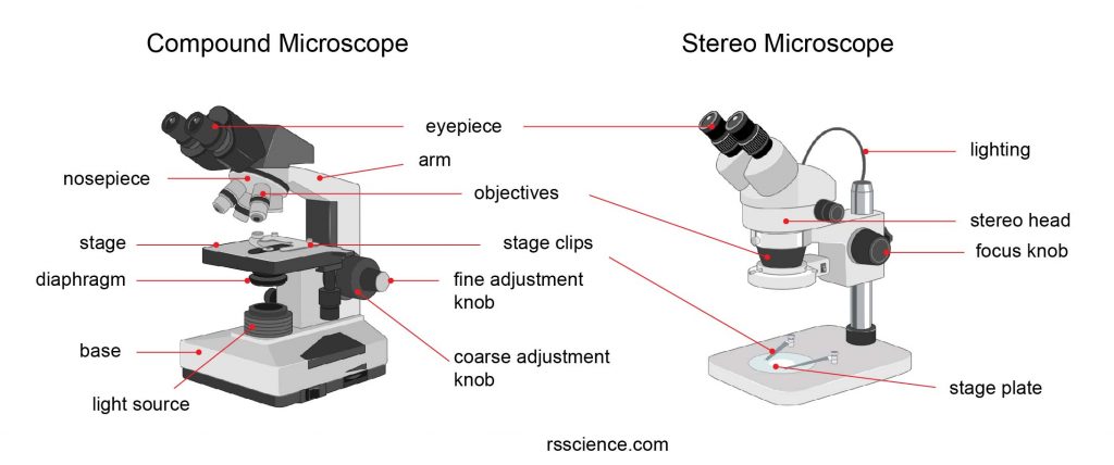
[In this figure] The structure of compound microscope vs stereo (dissecting) microscope.
Compound microscope
Compound microscopes have a higher magnification but only work well on thin specimens. Good examples are protozoa in the pond water, blood smear (blood cells), mouth swab (cheek cells), thin plant and tissue sections, pollens, cells, bacteria, semi-transparent water organisms (like water bear or algae), and premade slide sets. Images below are examples using a compound microscope.
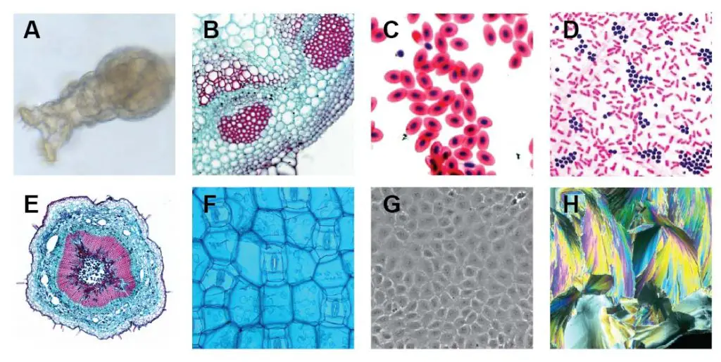
[In this figure] Compound microscope images examples.
A. Rotifer (bright field); B. Helianthus stem (thin stained section – bright field); C. Fish red blood cells (Stained – bright field); D. Bacteria (Gram’s Iodine stained – bright field); E. Loosen stem (thin stained section – bright field + image stitching); F. Rhoeo Discolor leaf (bright field + blue filter); G. Human cells cultivated in Petri dish (phase contrast); H. Citric acid micro-crystal (polarized light).
Typically, a compound microscope is supplied with 3-5 objective lenses that range from 4x to 100x (4x/10x/40x/63x/100x). Assuming you have 10x eyepieces and 100x objective, the total magnification of this combination is 1,000x (10×100 = 1000). By using special condensers, compound microscopes can also achieve darkfield, phase contrast, polarized light, or differential interference contrast (DIC) images for particular applications.
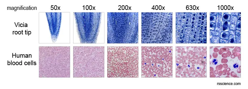
[In the figure] The low to high magnifications of Vicia root tip (top) and human blood cells (bottom).
Stereo microscope
You need a stereo microscope to view more substantial specimens such as insects, feathers, leaves, rocks, gems, coins, stamps, etc. Functionally, a stereo microscope is like a much powerful magnifying glass. Unlike a compound microscope that offers a flat image, stereo microscopes give the viewer a 3-dimensional image that you can see the texture of the specimen. Stereo microscopes have a lower magnification than compound microscopes.
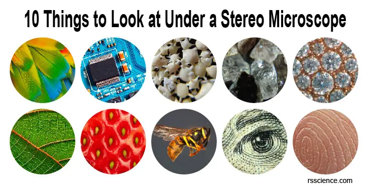
[In this image] Examples imaged under a stereo microscope.
(Top row) parrot feather, circuit board, star sands, crystals, and jewelry. (Bottom row) leaves, strawberries, bees, bills, and fingerprints.
Fluorescence microscopes and other advanced microscopies
Many biological subjects are transparent. In order to see these specimens, we may need to label them with fluorescent colors and use a fluorescence microscope. Several advanced microscopic techniques, which can see cellular structures in more detail, were developed in the last decades based on fluorescence microscopy.
[In this video] See amazing images of cells under a fluorescence microscope.
Learn more about the different types of microscopes.
The limitation of optical microscopes
Optical microscope does not have enough power to resolve things as tiny as a virus. It is because of the limitation of visible light. More specifically, the resolution is limited by the wavelength of the visible light that illuminates the specimen.
The wavelength of the visible light ranges from about 400 (purple light) to 700 (red light) nanometers. Generally speaking, you cannot see the stuff clearly if its size is smaller than half of the wavelength of the visible light, which is about 0.2 – 0.35µm. Therefore, anything smaller than 0.35 µm is not viable using an optical microscope. In summary, 1 µm (the size of a bacterium and mitochondrion) is about the limitation of the “common” optical microscope.
Electron microscopes (viruses, bacteria, cell organelles)
In order to see smaller things, scientists have to find an illumination with a wavelength much smaller than visible light.
According to modern physics, an electron has both properties as a particle and a wave. As a result, an electron has a wavelength (called De Broglie wavelength) depending on its energy (or speed). For example, if an electron is speeded up in a vacuum tube to the speed of 1,000,000 meters per second (equal to 2.2 million miles per hour!), the De Broglie wavelength we get is around one-tenth of a nanometer, which is about the size of an atom.
This is why we can use electron microscopes to probe the structure of atoms in a crystal directly. Therefore, seeing viruses with an electron microscope is a piece of cake.
There are two types of electron microscopes. The transmission electron microscope (TEM) is used for thin specimens (like tissue sections) because electrons can pass through the specimens to generate a projection image. On the other hand, the scanning electron microscope (SEM) can see the surface of a specimen.
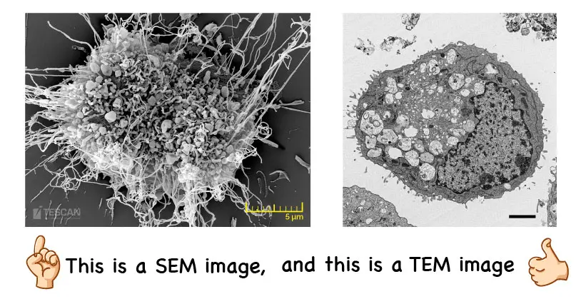
[In this figure] The examples of SEM and TEM images.
The left image was taken by an SEM. In contrast, the right image was taken by a TEM.
Learn more about electron microscope images.
X-ray Crystallography (DNA and proteins)
Using X-ray diffraction, scientists can figure out how atoms arrange in a crystal material. By making a tiny crystal of proteins or DNA, biologists can use the same principle to decipher the structures of biomolecules down to the atomic level.
Other than X-ray, nuclear magnetic resonance (NMR) spectroscopy and cryo-EM are also used to study the structures of proteins.
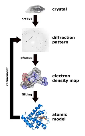
[In this image] Workflow for solving the structure of a protein by X-ray crystallography.
Photo credit: wiki
A summary of what equipment you need to see the biological subjects
| Magnification | Instrument | Example |
| 1x | Naked eye | Hair (approx. 0.1 mm) |
| 2x – 5x | Magnifying glass | Plant and insect in detail |
| 10x – 20x | Stereo microscope | Insect’s compound eyes |
| 50x | Compound microscope | Daphnia, Rotifers, Water bears, Stentors |
| 100x | Compound microscope | Paramecium, Amoeba, Vorticella, Volvox |
| 200x | Compound microscope | Pollen, Euglena, small green algae, Diatom |
| 400x | Compound microscope | Cheek cells, Onion skin cells |
| 800x – 1,500x | Compound microscope | Red blood cells (8µm), bacteria (1µm), condensed chromosomes in nuclei |
| 2,000x – 1,000,000x | Electron microscope | Objects smaller than 1μm, such as a virus (100 nm) and DNA (2 nm) |
| > 1,000,000x | X-ray Crystallography | Protein and DNA structures at the atomic level (1 Å) |
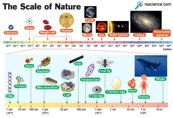
(6) Knowing how small we are
On the scale of a galaxy, let alone the universe, we’re smaller than we can readily comprehend. However, we do have the capacity to wonder, question, explore, investigate, and imagine. Take a moment to think about the enormity of what is beyond your vision. Everything will look different!