
This article covers
What are Flagella and Cilia? – quick overview
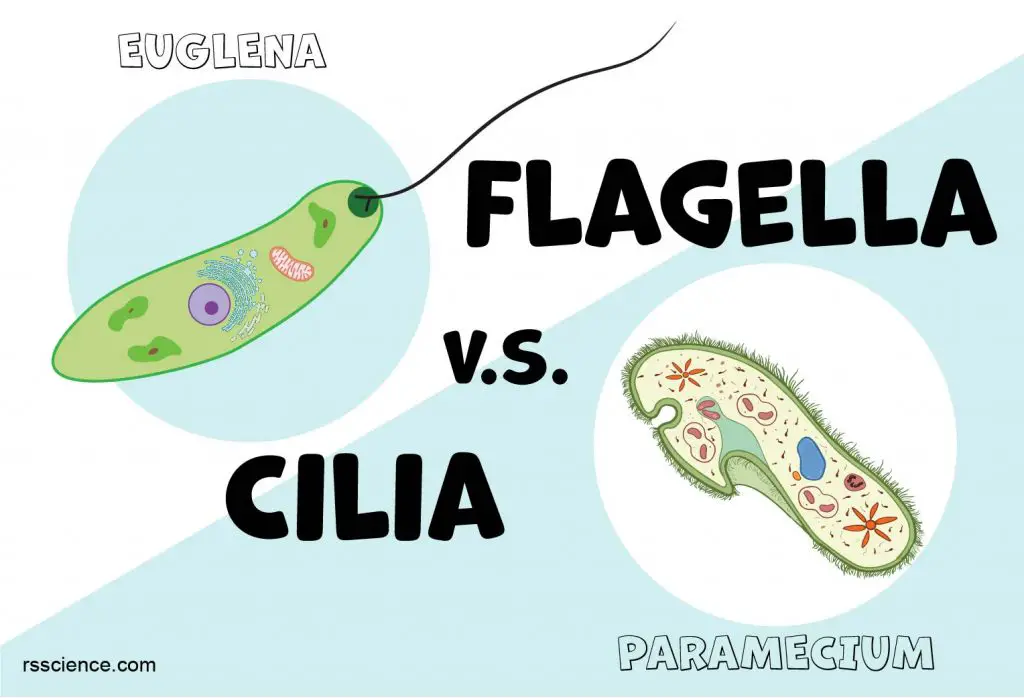
Flagella (singular: flagellum) and cilia (singular: cilium) are two types of cellular structures that allow movement in most microorganisms and animal cells, but not in high plant cells. Both flagella and cilia look like beating, hair-like appendages growing on the surface of cells. Usually, flagella are much longer than cilia. However, one cell may have thousands of cilia, but only one or two flagella.
[In this video] Microorganism moving methods: Euglena by flagella, Amoeba by pseudopodia, and Paramecium by cilia.
In single-celled microorganisms, cilia and flagella are fundamental units of motion. For example, the cilia covering a Paramecium‘s skin help it swim by waving these tiny hairs. One long, whip-like flagellum on the head of a Euglena can drive the cell moving like a propeller.
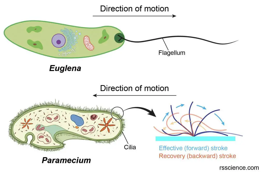
[In this image] The cell structures include cilia and flagella in Paramecium and Euglena.
Note that the direction of Euglena’s movement leads by the position of its flagellum. On the other hand, the sperm’s flagellum functions like a tail that propels the cell to move forward like a submarine.
In multicellular organisms, cilia and flagella also play vital roles. For example, our respiratory tract is lined with many cilia that keep inhaled dust, smog, and potentially harmful microorganisms from entering the lungs. Male reproductive cells, sperms, use their tail-like flagella to move toward the egg.
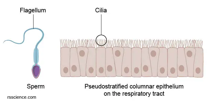
[In this figure] Cellular structures that allow the movement of animal cells: Flagellum (the tail of sperm) and Cilia (the waving hairs on the surface of airway cells).
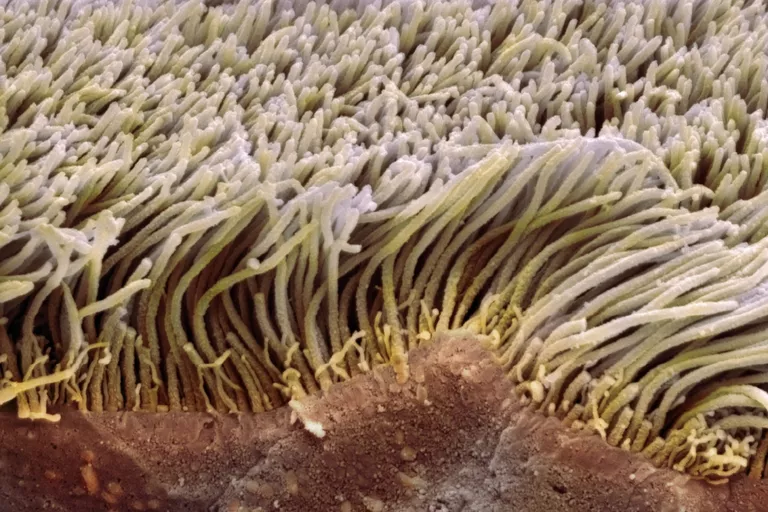
[In this image] Scanning electron micrograph (SEM) of ciliated epithelial cells.
Image source: Steve Gschmeissner/Science Photo Library
Part 1. Flagellum
A Flagellum (plural: flagella) is a long whip-like structure that protrudes from the cell body. Usually, a cell has one or several (less than 5-10) flagella. The primary function of a flagellum is for cell movement. But some specialized flagella can act as a sensory organelle (like insects’ antenna), being sensitive to chemicals and temperatures outside the cell.
Who has flagella?
Cells that have flagella are called “flagellates”. Flagella can be found among the three Domains of life – Bacteria, Archaea, and Eukaryotes. All three kinds of flagella can be used for swimming, but they differ greatly in structure, protein composition, and mechanism of propulsion.
Examples of flagellates:
1. Helicobacter pylori is a bacterium that causes an ulcer. Helicobacter pylori have multiple flagella to propel itself through the mucus lining to reach the stomach.
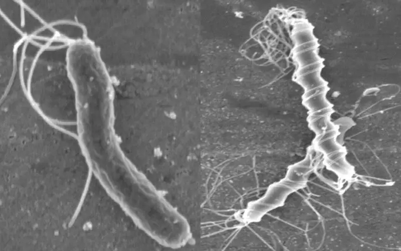
[In this image] Several flagella on one cell of Helicobacter pylori (left) and Helicobacter felis (right).
Image source: Electron Micrographs courtesy of Lucy Thompson, school of Biotechnology & Biomolecular Sciences, University of New South Wales
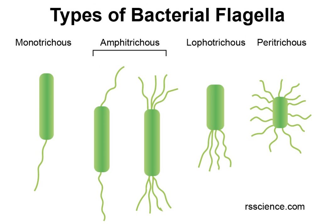
[In this image] Depending on the number and location of bacterial flagella, they can be classified as shown in this image.
2. The flagella of archaea have a special name, archaellum, to emphasize their difference from bacterial flagella.
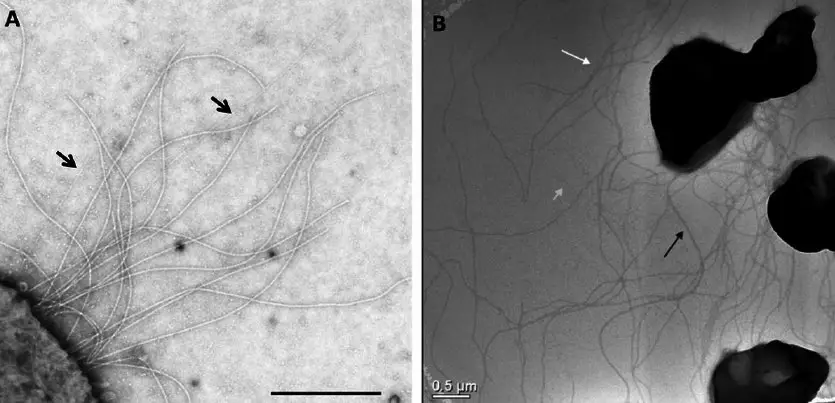
[In this image] Electron micrograph of archaea (Left: M. maripaludis and right: S. acidocaldarius) showing numerous archaella.
Image source: Jarrell KF., et. al., Life, 2013
3. A sperm cell is an example of a eukaryotic flagellate cell. It uses its flagellum to propel itself through the female reproductive tract.
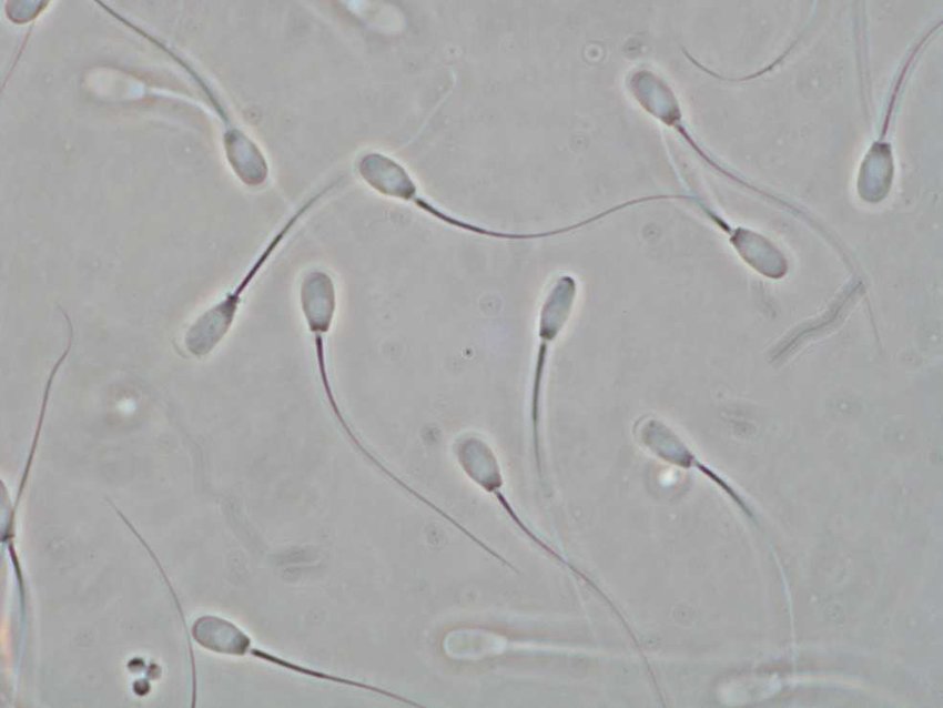
[In this image] Boar sperm cells with long tail-like flagella under the phase-contrast microscope.
Image source: Computational Modeling of Objects Represented in Images, Proc. CompImage Symposium 2006
The structure of a flagellum and how it works
Bacterial and Archaeal flagella
Bacterial flagella are helical filaments, each with a rotary motor at its base, which can turn clockwise or counterclockwise. There is a segment called “hook” between the motor and the filament, which functions as a flexible joint and transmits the rotating force from the motor to the flagellum. The bacterial movement can divide into two kinds:
- Run – resulting from a counterclockwise rotation of the flagellum
- Tumbling – resulting from a clockwise rotation of the flagellum
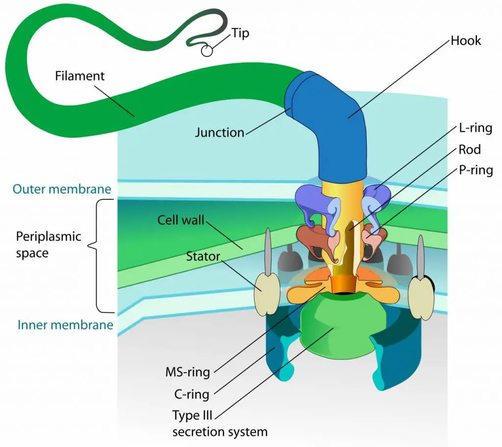
[In this image] The detailed structure of a bacterial flagellum.
The bacterial flagellum is a long, slender projection from the cell body. It rotates like a propeller when the bacterium swims.
Image source: wiki
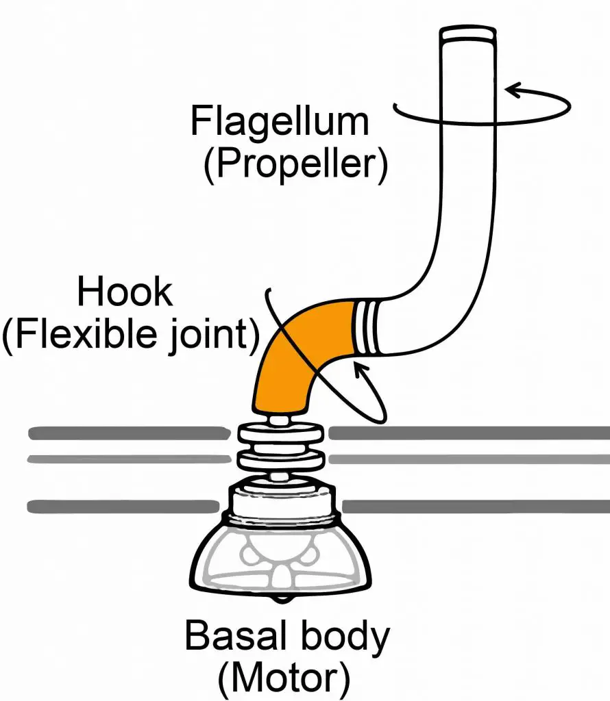
[In this image] The hook acts as a dynamic joint, transmitting torque from inside the cell to rotate the outer tail, allowing bacteria to move.
Image source: Dr. Satoshi Shibata
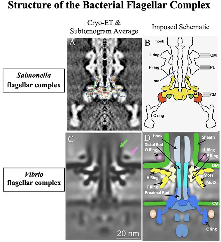
[In this image] Bacterial flagella are one of the most delicate biological micro-machines.
Examples showing in this image are the flagellar base complex of two kinds of bacteria – Salmonella enterica and Vibrio alginolyticus. Scientists used advanced cryo-electron tomography (Cryo-ET) to see how these fantastic micro-machines works.
Image source: Echazarreta M. A. and Klose K.E., Front. Cell. Infect. Microbiol., 2019
[In this video] See bacteria moving under the microscopes.
Archaeal flagella (archaella) are similar to bacterial flagella and also work as a rotary motor. However, the proteins to make archaeal flagella are different from bacterial flagella.
Note: Bacteria have another type of long hair on their cell, called pili (singular: pilus). Pili are thin, rigid fibers made of proteins that protrude from the cell walls. Pili help bacteria to attach to specific surfaces or other cells. Some bacteria can transfer DNA from one bacterial cell to another by their special pili. However, pili, cilia, and flagella are different structures.
Eukaryotic flagella
Eukaryotic cells (those of animal, plant, and protist cells) have their flagella in a very different way. One major difference is that prokaryotic (bacterial and archaeal) flagella run in a rotary movement, while eukaryotic flagella run in a bending motion (that lash back and forth).
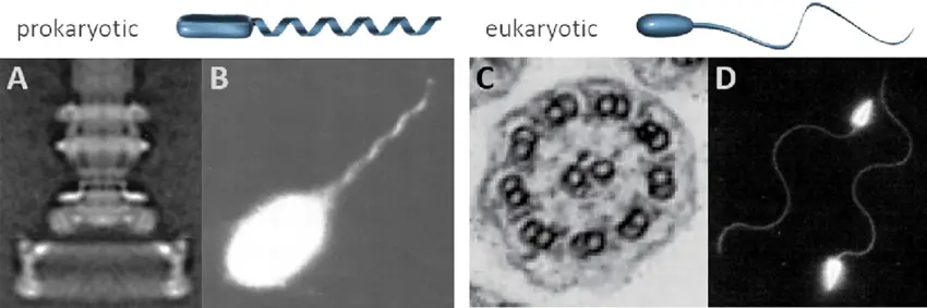
[In this image] Prokaryotic (helical) and eukaryotic (whiplash) flagella are fundamentally different. You can see the rotary motor of the bacterial flagellum (A) with a base width of about 45 nm. (B) A bacterium, S. typhimurium, swims with a polar bundle of flagella. Conversely, (C) eukaryotic flagella are powered by microtubule bundles (cross-section diameter ~200 nm). An example of eukaryotic flagella is swimming sperm cells in (D).
Image source: Schwarz L., et. al., Applied Physics Reviews, 2017
Structurally, eukaryotic flagella and cilia are indistinguishable. Both possess a central bundle of filament proteins, called axoneme. Each axoneme contains nine pairs of microtubules (doublet) forming the outside of a ring and two central microtubules, known as “9+2”. Microtubules are held together by cross-linking proteins.
In the flagella/cilia, there are motor proteins, called Dynein, sitting across each paired microtubule fiber. Dynein uses ATP as energy to crawl along the microtubules. When dynein proteins move upward on one side but downward on the other side, the flagellum/cillum bends. The repeat of bending-relaxing cycles makes flagella/cilia act like oars, beating back and forth to create movement.
Usually, flagella are much longer. One cell may have only one or two flagella on its head or tail regions. On the other hand, cilia are shorter in length but greater in number, typically covering a significant area of the cell surface.
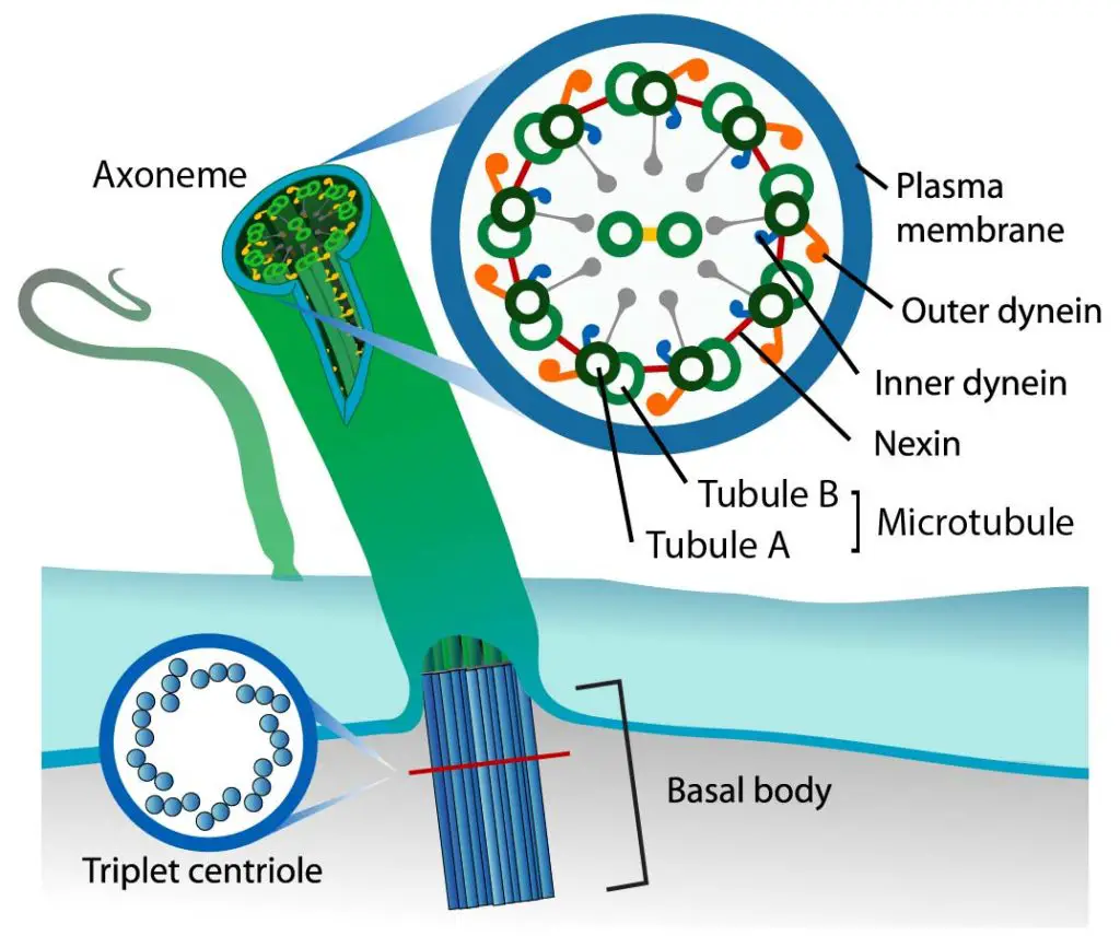
[In this figure] The organization of eukaryotic flagella and cilia.
Each flagellum /cilium contains nine pairs of microtubules forming the outside of a ring and two central microtubules. This structure is known as an axoneme. Microtubules are held together by cross-linking proteins. There are motor proteins, called dynein, sitting across each paired microtubule fiber.
Photo credit: modified from LadyofHats on wiki.
Click here to learn what are microtubules and other cytoskeleton proteins.
Case study – Euglena
Euglena (Greek: eu = true, glene = eye-ball) is a genus of single-cell eukaryotes, and they can be found in freshwater ponds and ditches. Euglena shares some characteristics of both plants and animals. For example, euglena contains chloroplasts; as a result, they can make their own food, a characteristic of plants. In contrast, euglena can also move using its flagella and consume food through phagocytosis, which are characteristics of animals. Euglena also lacks a cell wall.
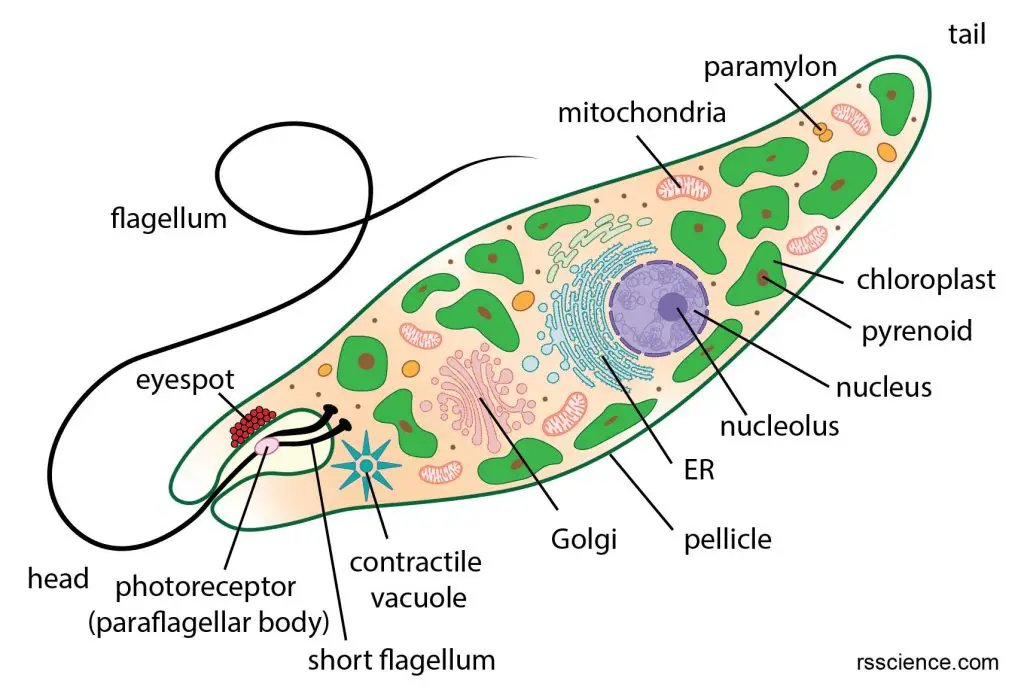
[In this image] Euglena anatomy and its organelles, including two flagella.
One of the two flagella is long and can be seen under a light microscope, but the other one is very short without protruding from the cells. The function of flagella is to help euglena swim. Learn more about the structure of euglena here.
Case study – Volvox
Volvox is a genus of green algae. Volvoxes are free-floating single-cellular algae but typically stay together as spherical colonies (or balls) of 500-50,000 cells. They can live in various freshwater habitats, including ponds, pools, and ditches. Under a microscope, volvoxes look like green marbles, slowly rotating, making them one of the most adorable microscopic organisms.
These volvox cells sitting on the surface of the sphere bear two flagella. These flagella face the side of the surrounding water and beat to propel the whole colony through the water. This is why a volvox moves like a rolling ball.
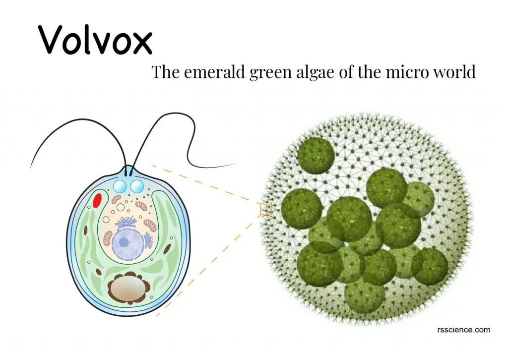
[In this image] Volvox is a cluster of many single-cell green algae. Each cell sitting on the surface has two flagella. Learn more about Volvox.
Case study – Sperm cells
The only human cells with flagella are male gametes – that is, sperm cells. The sperm cell (also called spermatozoon) is the male reproductive cell of animals, which bears a haploid set of chromosomes within the nucleus. The sperm comprises a head, neck, middle piece, and tail. Its nucleus locates in the head part. The rest of the sperm cell is basically a powerful flagellum propeller.
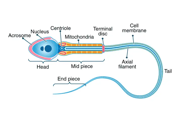
[In this image] The structure of a sperm cell.
Except for the head part, the sperm cell is built for its flagellum. The neck of a sperm cell hosts a centriole, which is the root of microtubule bundles. These microtubule bundles assemble into an axoneme and elongate into a long tail. A sheath covers the axial filaments as protection in the tail part. In the middle piece, the axial filament is surrounded by many mitochondria, which provide the energy for the flagellum movement.
Image source: BYJU’S
Case study – Flagellated lifecycle stages
Many organisms do not have flagella in mature forms. However, they develop specialized flagellate forms during a particular stage of their lifecycle. For example, sperms are male reproductive cells of animals, many of these flagellated lifecycle stages are associated with reproduction. Examples are zoospores (motile spores use a flagellum to move) and male gametes (male reproductive cells) for most green/brown algae, some fungi, lower plants (liverworts, hornworts, and mosses), cycads, and ginkgo.
Part 2. Cilium
Cilium (plural: cilia) is a structure only found on eukaryotic cells. It is in the shape of a slender protuberance that projects from the cell body. There are look-alike structures (i.e., pili and fimbriae) on bacteria, but they are not cilia.
Many microorganisms have cilia. Examples include Paramecium, Stentor, Vorticella, Lacrymaria, Coleps, and Dileptus. They are classified as a group called “ciliates” by sharing this common characteristic.
[In this video] A video collection of several common ciliates under the microscope.
Larger eukaryotes, such as animals, have cilia as well. Cilia are usually present on a cell’s surface in large numbers and beat in coordinated waves. In humans, cilia are found on the epithelial cells lining the respiratory tract. These cilia move constantly to clean sweeping mucus and dirt out of the lungs.
The structure of a cilium and how it works
Cilia look like many tiny hairs growing on the surface of cells. Their roots are cytoskeleton-like structures called the basal body sitting under the cell membrane. Microtubule bundles are extending from the basal body to form the axial core of the cilia. As we mentioned above, both eukaryotic cilia and flagella consist of a “9+2” arrangement of microtubule bundles. The movement is created by the bending of microtubule bundles through the motor proteins Dynein.
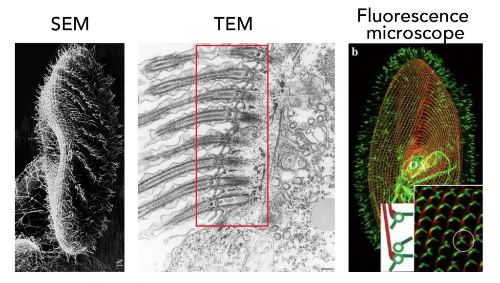
[In this figure] Scientists used advanced microscopes to answer their questions of paramecium’s cilia.
Left: Scanning electron microscopy shows us the morphology of cilia (Credit: Judith L. Van Houten). Middle: Transmission electron microscopy gives us the transverse section image of cilia in detail (Credit: Richard Allen). Right: Fluorescence microscope shows us how cilia anchor on the cell’s surface.
Microtubules are protein fibers inside the cells with multiple functions. Microtubules can also serve as an intercellular highway for the transportation of molecules and organelles. During cell division, microtubule fibers projected from two centrosomes pull chromosomes apart into new nuclei.
Two types of cilia
There are two major types of cilia: motile and non-motile cilia. Motile cilia move by consuming energy to create constant movement – for the mobility of single cells (i.e., Paramecium) or the transportation of fluid outside the cells (i.e., airway epithelium).
Non-motile cilia are also called primary cilia which serve as a sensory organelle. Many cells have at least one primary cilium per cell. For example, our olfactory neurons in our noses possess several non-motile cilia which function as a cellular antenna to sense odor molecules.
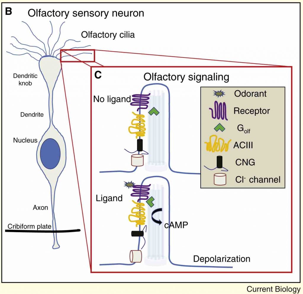
[In this image] Illustration of primary cilia on olfactory sensory neuron.
The olfactory epithelium contains olfactory neurons with multiple non-motile cilia extensions (olfactory cilia). The cilia trap odor molecules as they pass. Information about the odor molecules is then transmitted from the odorant receptors to the olfactory bulb in the brain.
Image source: Berbari N.F., et. al., Current Biology, 2009
Case study – Paramecium
Paramecium (pair-ah-me-see-um; plural, Paramecia) is a unicellular (single-celled) living organism with a shape resembling a slipper. Paramecium is naturally found in aquatic habitats. You need a microscope to see the paramecia because they are only 50 to 300 µm (micrometers) in length.
Paramecium’s skin is covered by many tiny cilia like fine hairs on our bodies. One Paramecium cell can have 5000 – 6000 cilia. Each cilium is very tiny – approximately 0.25 μm in diameter and up to 20 μm in length. Paramecium’s cilia can be classified into two types: oral cilia and body cilia. Oral cilia are present on the surface of the oral groove. They help collect food materials. Body cilia are on the body surface and facilitate its locomotion. They act like microscopic oars to move the organism in one direction.
Body cilia are arranged in longitudinal rows (along the head-to-tail axis) with a fairly uniform length throughout the cell. There are also a few longer cilia present at the posterior end of the cell (quite obvious in P. caudatum). These form caudal tuft of cilia (hence the name caudatum).
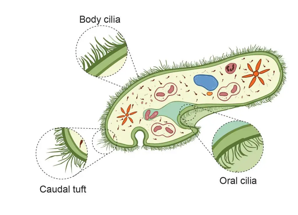
[In this figure] Different types of cilia on paramecium cell – oral cilia, body cilia, and caudal tuft.
If Paramecium’s cilia just wave back and forth in the same way, the cells can’t go anywhere. The forward and backward strokes have to be in different phases to create a meaningful propulsive force.
[In this video] Motion of Paramecium’s cilia recorded by a high-speed video camera under the microscope.
To answer this question, Scientists used a microscope with a high-speed video camera to capture how cilia beat to propel the entire body of paramecium. By analyzing the high-speed video frame by frame, scientists found that the paramecium swims in a way similar to how we swim in the front crawl stroke.
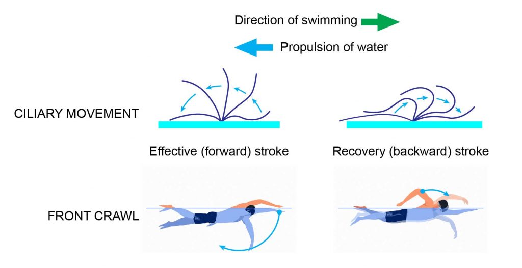
[In this figure] Stroke pattern of cilia on a Paramecium.
The movement of cilia can be divided into Effective (forward) and Recovery (backward) strokes. Two kinds of strokes alternately repeat to propel the body of paramecium as we swim in the front crawl style.
Learn more about how Paramecium’s cilia do the wave below
Case study – Stentor
Stentor (also known as the “Trumpet Animalcule”) is a genus of the ciliate. They have a horn-shaped body with cilia uniformly covering the most surface area. Some species of Stentors are among the biggest known unicellular organisms on earth, which can grow up to 2 mm (0.08 inches) long.
The most notable part of a Stentor is the “crown” of cilia surrounding the trumpet “bell”. These cilia can wave back and forth in a coordinated way to create a current or vortex that pulls food closer. Adjacent to the oral opening, the fusion of several cilia to form “super cilia” or membranelles. These giant cilia form a spiral that leads into the stentor’s mouth. Every little while, the stentor will close up the cilia crown and contract, bringing the food down into the body.
[In this video] Under the microscope, you can see these longer cilia forming the “crown” of Stentor’s trumpet. There are also many shorter cilia covering the entire Stentor’s body for free swimming.
Case study – Respiratory epithelium
Respiratory epithelium (or airway epithelium) is a type of ciliated columnar epithelium found lining most of the respiratory tract. Its functions serve to moisten and protect the airways.
Each ciliated epithelial cell has around 200 cilia that beat constantly at a rate of between 10 and 20 times per second. The direction of their beat removes microbes and debris up and out of the airways.
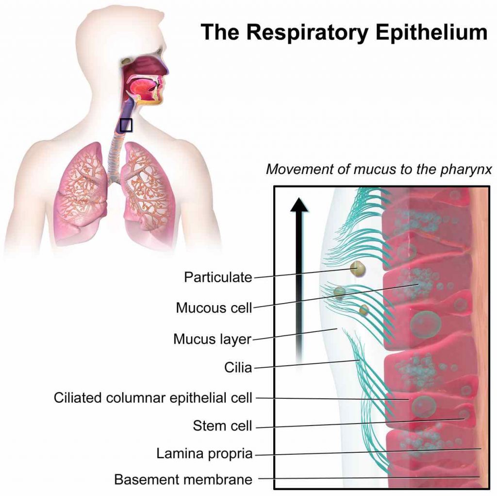
[In this image] Respiratory epithelium protects our lungs by the mucous layer and cilia-propelling clearance.
Image source: wiki
Part 3. What are the differences between Flagella and Cilia?
This table summarized the major differences between Flagella and Cilia.
| Character | Flagella | Cilia |
| Definition | Flagella are long, thread-like cell structures present on the surface of a living cell. | Cilia are short, hair-like cell structures extending from the surface of a living cell. |
| Presence | Flagella are found in both prokaryotic and eukaryotic cells. | Only found in Eukaryotic cells. |
| Length | Long; up to 150 µm in length. | Short; 5-10 µm in length. |
| Position | Usually, located at both or one of the ends of a cell. | Present throughout the surface of cells. |
| Density | Few (less than 10) per cell. | Many (hundreds to thousands) per cell. |
| Motion | Rotary movement in prokaryotes whereas bending movement in eukaryotes; both generate a propeller-like motion. | Fast waving motion by beating back and forth. |
| Beating | Flagella beat independent of each other. | Cilia beat in coordination or one after the other. |
| Functions | Mainly in locomotion only. | Locomotion, transportation, feeding, sensing, circulation, etc. |
| Structure | Prokaryotic: helical filament proteins with a rotary motor base. Eukaryotic: basal body, microtubule bundles (9+2), motor proteins. | Same as flagella: basal body, microtubule bundles (9+2), motor proteins. |
| Examples | Flagellate bacteria, like Helicobacter pylori, Salmonella; some Archaea. Euglena, Dinoflagellates, Green algae Male gametes, Sperm cells | Protozoan Ciliates (Paramecium, Stentors, etc.) Respiratory epithelium in human airway |
Summary
1. Flagella and cilia are two types of organelles that help cells to move.
2. Flagella are long, thread-like cell structures that can be found in both prokaryotic and eukaryotic cells. However, their structures and protein compositions are different.
3. Cilia are short, hair-like cell structures extending from the surface of eukaryotic cells.
4. Eukaryotic flagella and cilia are the same in their structures. They both consist of microtubule bundles as the core.