
This article covers
What is epithelium? A quick overview
Epithelial tissue or Epithelium (plural = epithelia) is a protective, continuous sheet of compactly packed cells. Epithelium covers all internal and external surfaces of our body, and lines body cavities and hollow organs. Epithelium also forms major tissues in all glands.
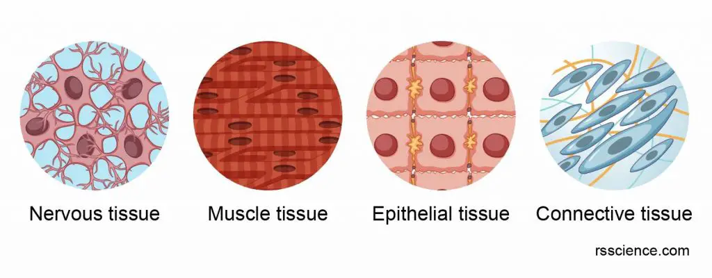
[In this image] Epithelium is one of the four basic types of animal tissue, along with connective tissue, muscle tissue, and nervous tissue.
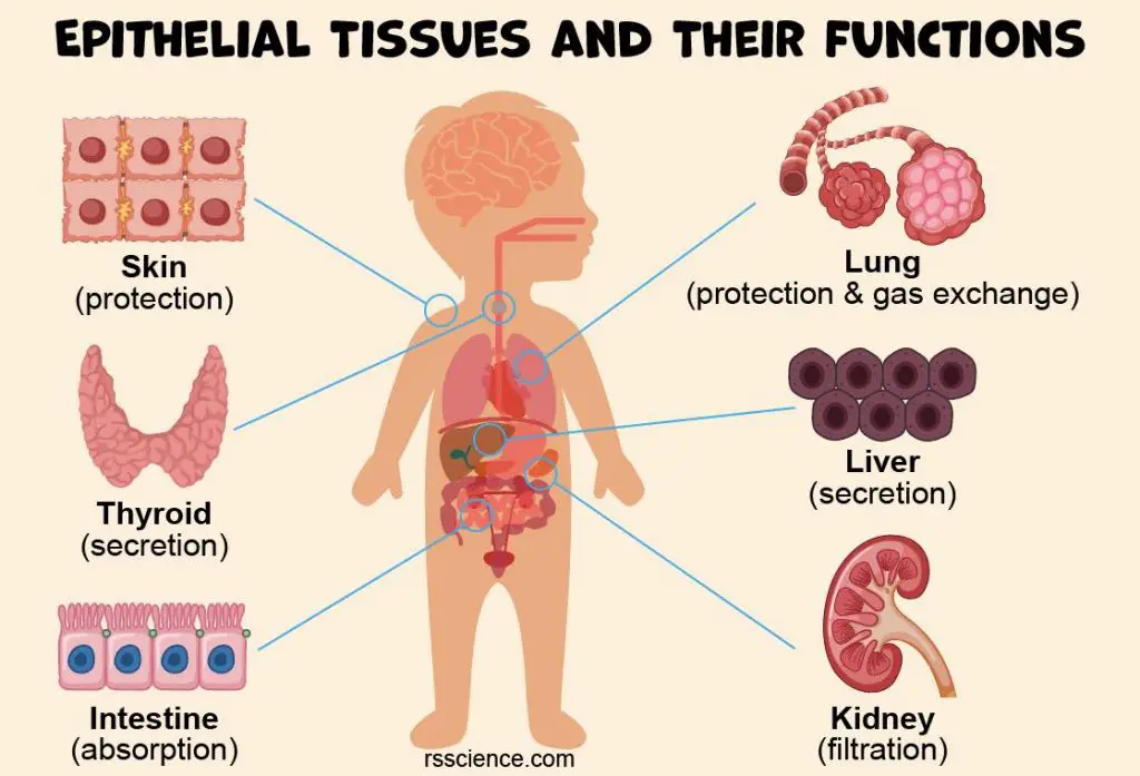
The function of the epithelium varies depending on where it’s located in your body. Examples include protection, secretion, sensation, and absorption.
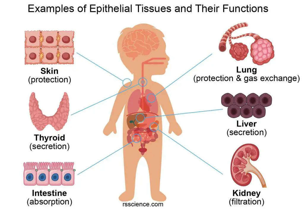
[In this image] Examples of epithelial tissues and their functions.
To help you have a quick idea of what epithelial tissues can do, here are some examples:
- Epithelium forms the outer layer of your skin (called epidermis).
- Specialized epithelium forms secretory organs like sweat glands and thyroid glands.
- Epithelial cells line the airway of our respiratory tract, waving their hair-like cilia to prevent dust and pathogens from entering our lungs.
- Epithelial cells line the inner surface of our digestive tract. In the intestines, these epithelial cells absorb nutrients from the digested food.
Characteristics of epithelial tissue
Epithelial cell sheets and cellularity
Epithelial tissue is formed by a tightly packed continuous layer of epithelial cells. There is little intercellular material or matrix present between epithelial cells. Instead, epithelial cells stay very close to each other, leaving no space between neighbors. Epithelial tissue has high cellularity (Epithelial tissue mainly comprised cells).
In comparison, cells in the connective tissues are separated from each other and embedded in a matrix. Thus, the connective tissue is low cellularity.
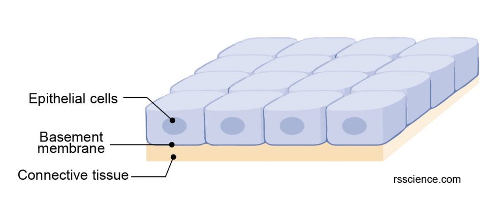
[In this image] Epithelial tissues are tightly packed cell sheets, in which epithelial cells adhere firmly to each other. These cell sheets may consist of only a single layer of cells or a stack of multilayered cells.
Intercellular adhesion and other junctions
Epithelial cells use specialized junctions to link adjacent cells tightly together. These junctions include:
- Tight junctions – provide a strong bonding between neighboring cells and prevent leakage across tissues
- Adherens junctions – link the cytoskeleton from two adjusted cells together and transmit mechanical forces
- Gap junctions – facilitate the movement of ions and molecules across the tissue
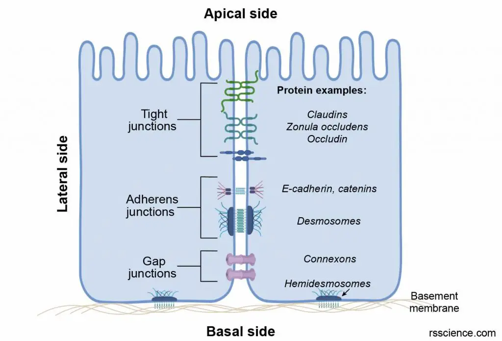
[In this image] Cell junctions link epithelial cells into tissues.
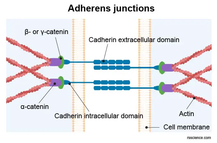
[In this image] Adherens junctions link actin cytoskeleton from two adjacent cells together.
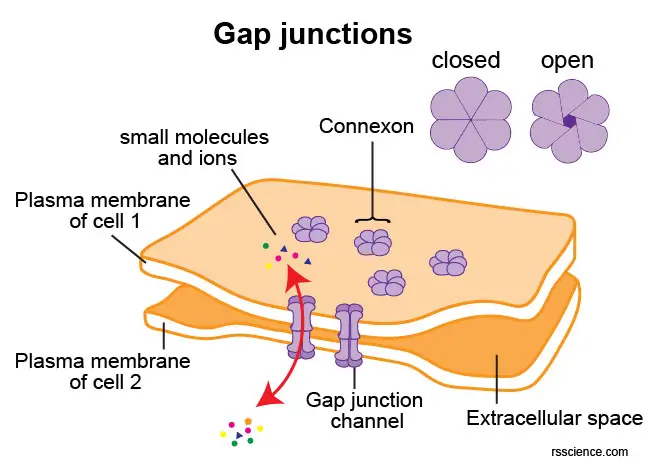
[In this image] Gap junctions serve as channels for the exchange of small molecules between cells.
Polarity
To maintain the integrity of epithelial barriers, epithelial cells tend to be in a cube or cuboid shape. As we know, a cube has six faces or sides; each face of an epithelial cell is different. This intrinsic asymmetry observed in cells is called Cell polarity.
The apical face of the epithelial tissue is exposed to either the external environment or the body fluid. The lateral faces are the four sides closely linked to the neighboring cells by intercellular junctions. The basal face is attached to the underneath connective tissues by the basement membrane.
Basement membrane
The basement membrane separates the epithelia from the underlying connective tissues. The basement membrane can be further divided into two parts. The basal lamina (close to the epithelium) consists of fibers and polysaccharides secreted by epithelial cells. The reticular lamina (close to the connective tissue) is rich in collagen proteins produced by cells of connective tissues, also known as the fibroblasts.
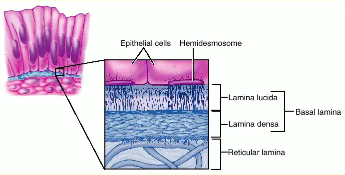
[In this image] Most epithelial cells are separated from the connective tissue by a sheet of extracellular material called basement membrane. It is formed by the association of two layers: Basal lamina and Reticular lamina.
Image credit: Socratic Q&A
Avascular and innervated
Blood vessels don’t grow into the epithelial tissues therefore called avascular. Instead, epithelial cells receive their nutrients from capillaries in the underlying connective tissues. The exchange of chemicals between epithelial and connective tissues is accomplished through diffusion.
Although blood vessels do not penetrate epithelial tissues, nerve endings do; that is, epithelia are innervated, meaning they have their own supply of nerves.
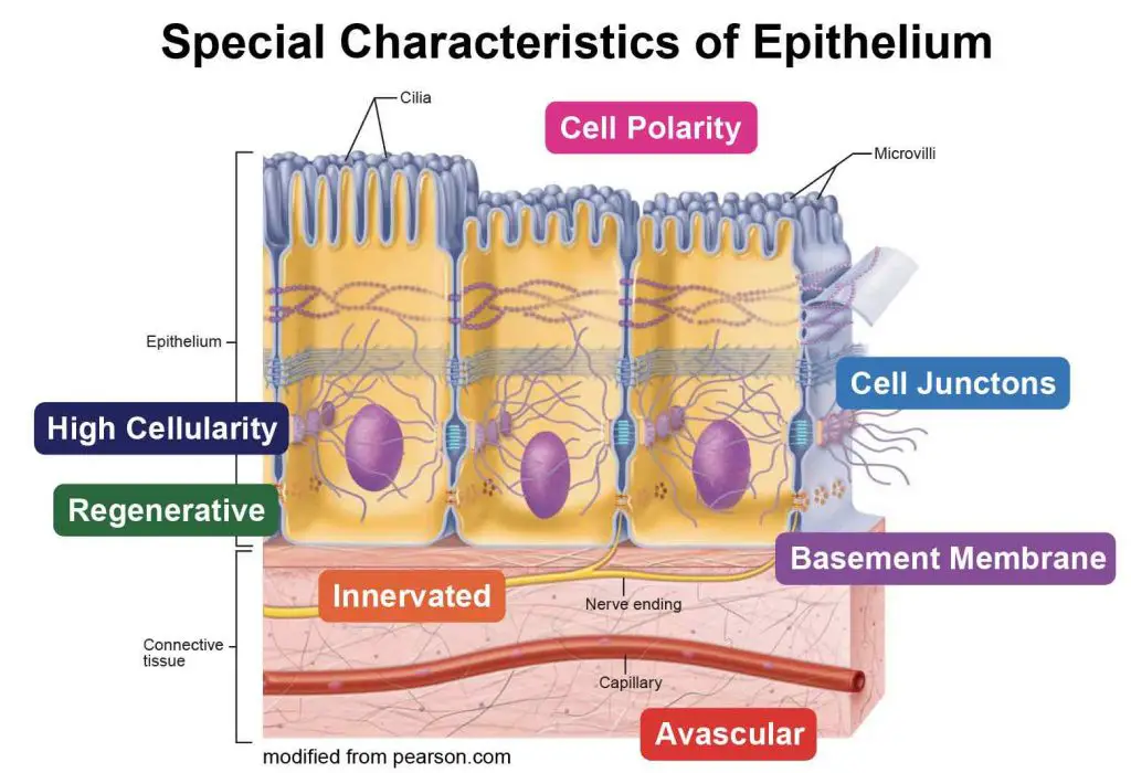
[In this image] A summary of key characteristics of epithelial tissues.
Regeneration
As epithelia line all the external and internal surfaces to protect our body, epithelial cells are constantly exposed to hostile substances such as bacteria, acids, and smoke. Fortunately, our epithelial tissues display a high regenerative capacity. As long as epithelial cells receive adequate nutrition, they can quickly replace dead cells by cell division to catch up with a high turnover.
[In this video] Quick proliferation of human kidney epithelial cells in a culture dish.
Development
Epithelial tissues can derive from all three embryological germ layers:
from ectoderm (e.g., the epidermis);
from endoderm (e.g., the lining of the digestive tract);
from mesoderm (e.g., the inner linings of body cavities).
Sometimes, endothelium and mesothelium (both derived from mesoderm) are also listed as epithelium. However, many scientists do not count endothelium and mesothelium as “true” epithelium.
The structure of epithelial cells
Like all other cells, epithelial cells are enclosed within a cell membrane. Each cell has one cell nucleus and several organelles, including mitochondria, endoplasmic reticulum, ribosomes, Golgi apparatus, peroxisomes, and lysosomes.
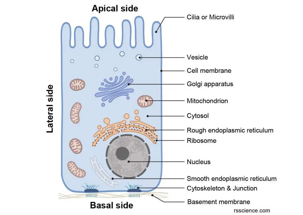
[In this image] The anatomy of epithelial cells.
Epithelial cells have all the common cell organelles like other animal cells. Some epithelial cells have hair-like protrusions (cilia or microvilli) on their apical sides.
Some epithelial cells have unique characteristics on the cell surface that help them perform certain functions.
Cilia
Cilia are tiny, hair-like structures on the surface of some epithelial cells. Ciliated cells usually have hundreds of cilia on their surfaces. Their cilia can wave in a coordinated way to sweep small objects away from the epithelium-lined surface.
For example, epithelial cells lining our airway have cilia that trap dust, bacteria, and other substances that you breathe in and move them toward your nostrils so that they don’t go into your lungs. Another example of cells with cilia are the epithelial cells that line the fallopian tubes that help move an egg from an ovary to the uterus.
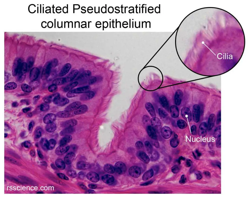
[In this image] Tiny hair-like cilia are observed on the apical sides of trachea epithelial cells under a high magnification light microscope. Ciliated epithelium lines most parts of our upper airway.
Microvilli
Microvilli are tiny finger-like structures on the surface of intestinal epithelial cells. Unlike cilia, microvilli don’t move. However, they can increase the cell’s surface area so that it can absorb substances efficiently. Each small intestine epithelial cell may have thousands of microvilli that absorb nutrients from the food you eat and protect your body from intestinal bacteria.
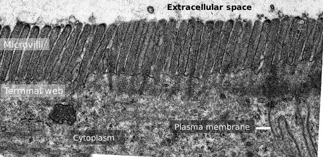
[In this image] Transmission electron microscopy image of small intestine epithelium surface covered by the dense microvilli.
Image credit: Atlas of plant and animal histology
Stereocilia
Stereocilia are specialized microvilli that resemble cilia and project from the surface of auditory and vestibular sensory cells. For example, stereocilia are needed on the epithelial tissue in your inner ear for hearing and balance.
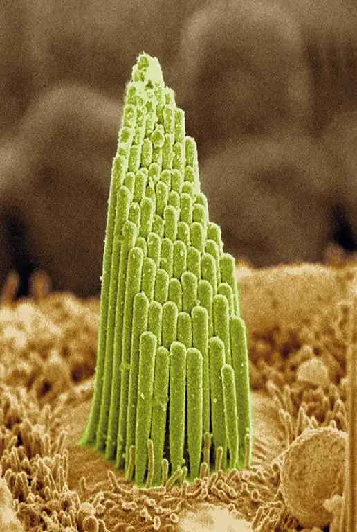
[In this image] Colored scanning electron microscopy image of a bunch of stereocilia on the inner ear epithelium.
Image credit: Estereocilios
Types of epithelia
In general, epithelial tissues are classified by the number of layers, the cell shape, and the function of the cells. Here is just a quick summary of common types of epithelial tissues. See “Classification and Types of Epithelial Tissues” for more detailed explanations and examples.
Arrangement
Three types of epithelial tissue arrangements – Epithelial tissues could form by cells assembling in Simple (only one layer), Stratified (multiple layers), and Pseudostratified (one layer of cells with different sizes resulting in a multilayered appearance) arrangements.
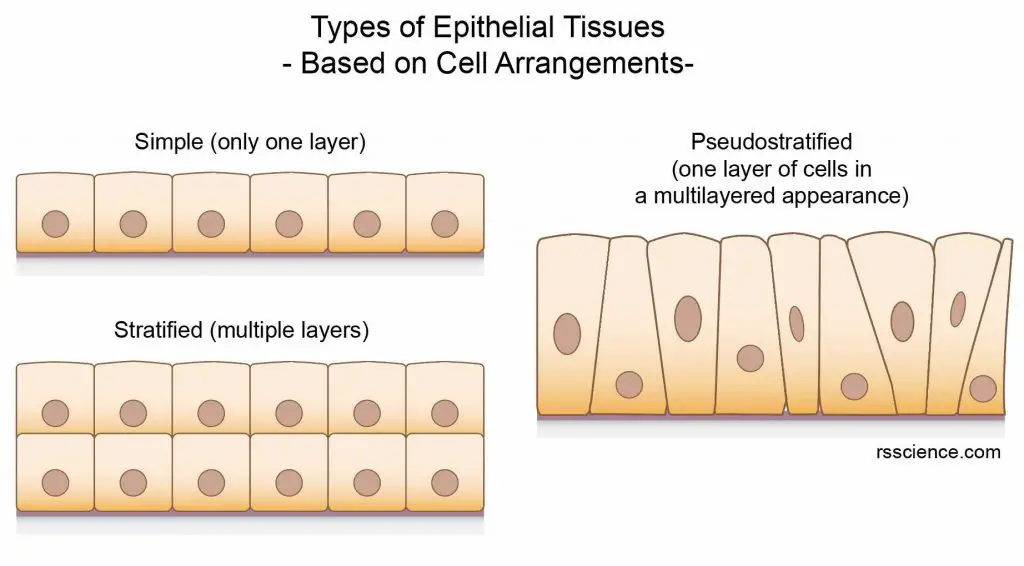
[In this image] Schematics of simple, stratified, and pseudostratified arrangements of epithelial cells.
Shape
Three types of epithelial cells based on their shapes – Epithelium may consist of cells that are Squamous (flat and scale-like), Cuboidal (cube-like), and Columnar (taller, column-like) in appearance.
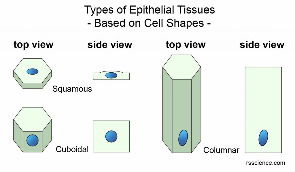
[In this image] Schematics of Squamous, Cuboidal, and Columnar shapes of epithelial cells.
Function
Some unique epithelial tissues are categorized by their special functions, including Transitional (can become flattened when stretched), Glandular (can secrete substances and form the glands), Mucous (can produce mucus), Serous (lines the body cavities), and Olfactory (involves in the sense of smell).
Given the different shapes and arrangements of epithelial cells, there can be several types of epithelial tissue in combination. Examples are Simple squamous epithelium (a single, flat layer of cells forming the alveoli of lungs), Simple columnar epithelium (a single, thick layer of cells lining the stomach and intestines), Stratified squamous epithelium (multilayered cells forming the outer layer of skin), Stratified cuboidal epithelium (forming the ducts of salivary and sweat glands), and Pseudostratified columnar epithelium (lining our upper respiratory tract and usually has a lot of cilia).
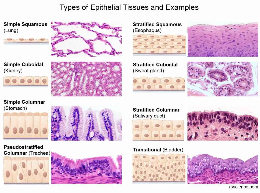
[In this image] A summary of eight types of epithelial tissues and examples. To learn more, see “Classification and Types of Epithelial Tissues”.
Functions of epithelium – What does the epithelium do?
Epithelium plays many important functions that are vital to our life. Since epithelial cells are found throughout our body, they adapt to different functions based on their location and organs’ needs.
[In this image] Epithelial tissues pick up specialized functions to fit the needs of different organs in our body.
Epithelium can have one or a combination of the following functions:
Protection
As epithelial tissues cover the entire body surface, their primary function is serving as the first line of defense against all kinds of pathogens, injury, and chemicals. For example, the outer layer of our skin is a thick epithelial tissue called the epidermis. It protects your body from harm, keeps your body hydrated, produces new skin cells, and contains melanin, which determines your skin color.
Epithelium also protects our respiratory tract. Cilia presented by epithelial cells in the nose or upper respiratory tract help trap the dust particles and prevent them from entering the lungs.
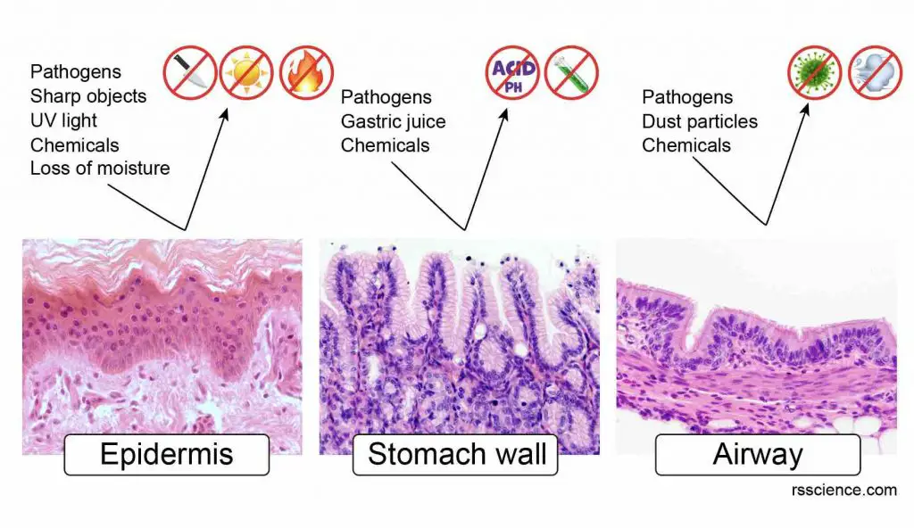
[In this image] Examples of epithelial tissues that function as a protective barrier are the epidermis (skin), stomach wall epithelium, and ciliated airway epithelium.
Secretion
Epithelial tissues in various exocrine and endocrine glands (i.e., thyroid, salivary glands, sweat glands, mammary glands) can secrete hormones, enzymes, saliva, mucus, sweat, milk, fluids, etc. Major organs like the liver and pancreas are also secretory organs. For glands that release substances through secretory ducts, these ducts are also made of epithelial tissues.
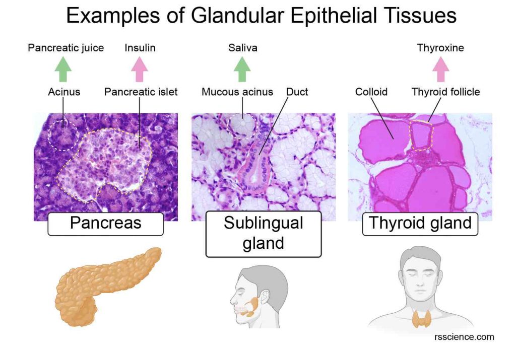
[In this image] Examples of epithelial tissues that secrete essential substances for our body: pancreatic juice and insulin from pancreas; saliva from sublingual gland; and thyroxine from thyroid gland.
Examples of epithelial tissues that secrete essential substances for our body:
1. Pancreas has both exocrine and endocrine functions. Acinar epithelium (exocrine) secrets the pancreatic juice through the pancreatic duct into the intestines to help food digestion. Pancreatic islets (endocrine) secrete insulin directly into the bloodstream to regulate blood sugar levels.
2. Sublingual glands are a pair of salivary glands (exocrine) situated underneath the tongue. They produce saliva in the mouth.
3. Thyroid (endocrine) is a butterfly-shaped gland that sits low on the front of the neck. It secretes several thyroid hormones into the bloodstream.
Lubrication
Mesothelium is a layer of epithelium that lines our body cavities, such as the peritoneum (abdomen cavity), pleura (lung cavity), and pericardium (heart cavity). Mesothelium also surrounds the internal organs. Mesothelium secretes a lubricant film called serous fluid, which prevents the frictions between organs and body walls during movement, breathing, or heart beating.
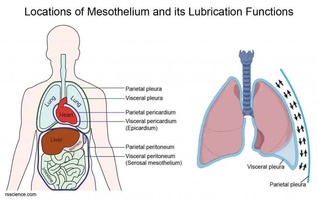
[In this image] Mesothelium covers the body wall and the organs.
(Left) Mesothelial cells lining the cavities/body walls in these subdivisions are called parietal mesothelia, while visceral mesothelia cover the organs. (Right) The size and location of our lungs change during each breath. For this reason, the lubricant layer between parietal pleura (mesothelia lining the chest cavity) and visceral pleura (mesothelia covering the lungs) is critical to reduce the fraction and prevent injury.
Absorption
The epithelial lining of the digestive tract absorbs water and nutrients. For example, the intestine epithelial cells forming villi can increase the surface area of cells and absorb nutrients from the food we eat.
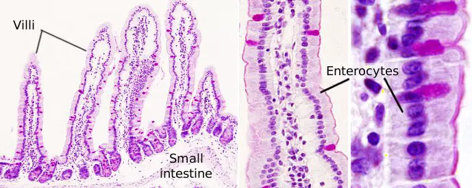
[In this image] Villi arrangement of small intestine epithelium.
Image source: Atlas of plant and animal histology
Exchange of substances
Epithelial tissues control the exchange of substances between the body and the external environment. For example, the oxygen/carbon dioxide exchange in our lungs happens across the thin epithelial layer of alveoli.
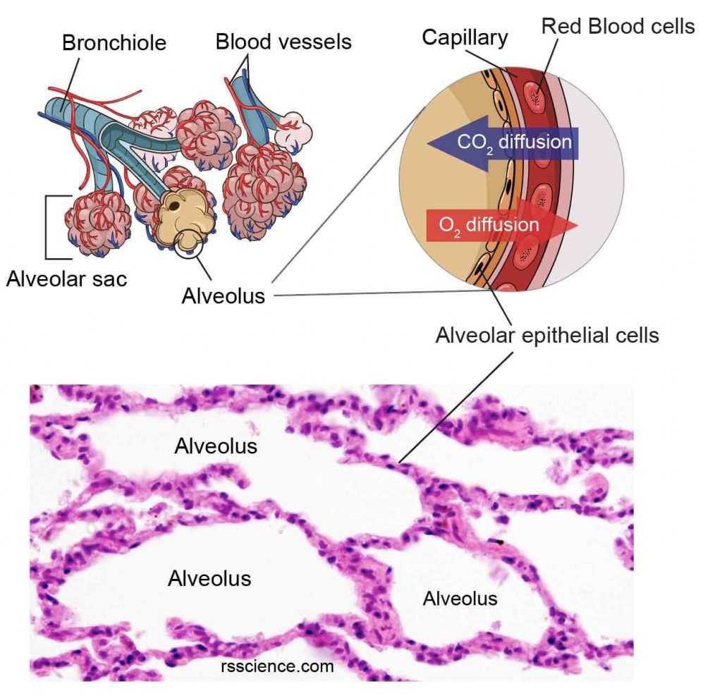
[In this image] Simple squamous epithelium lines the surface of alveoli (singular: alveolus) and serves as the site for gas exchange.
Filtration and excretion
Excretion is the removal of waste from our bodies. The epithelial cells in the kidneys form renal tubules to filter and clean the blood. These cells filters waste from the blood into the fluid (which becomes urine) as it flows through the tubules. They also reabsorb and return needed water, electrolytes, and nutrients (such as glucose and amino acids) back to the blood.
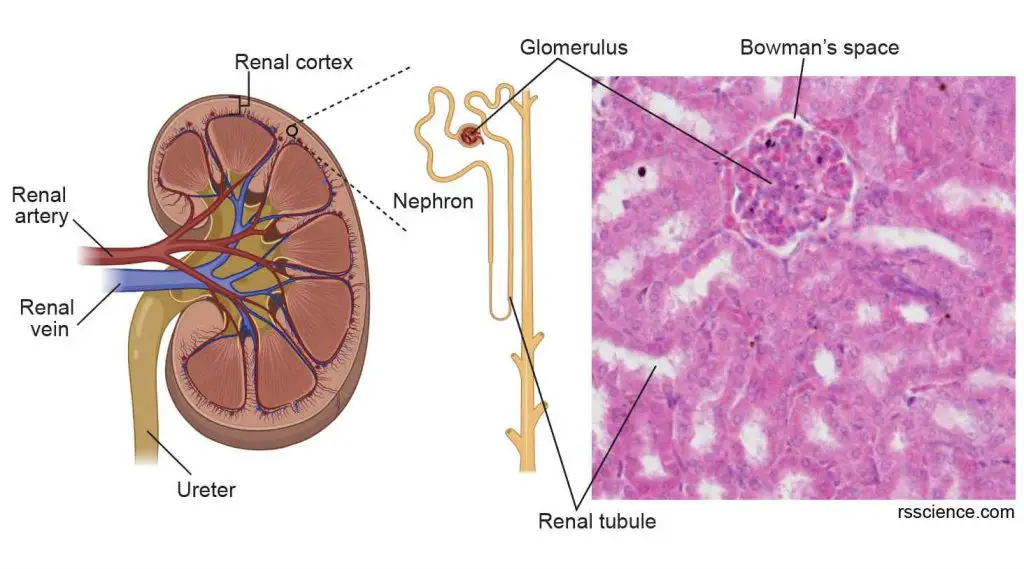
[In this image] Nephron is the kidney’s microscopic structural and functional unit. A nephron consists of a tuft of capillaries, called the glomerulus, and renal tubules made of tubule epithelial cells.
Sensation
Sensory nerve endings are present in the epithelial tissues of the skin, nose, ears, taste bud, etc. These receptors can receive outside sensory stimuli and transmit signals to the brain. For example, the stereocilia on the surface of the ear epithelial tissues are essential for hearing and balance. In addition, your taste buds sit in the stratified squamous epithelium of your tongue.
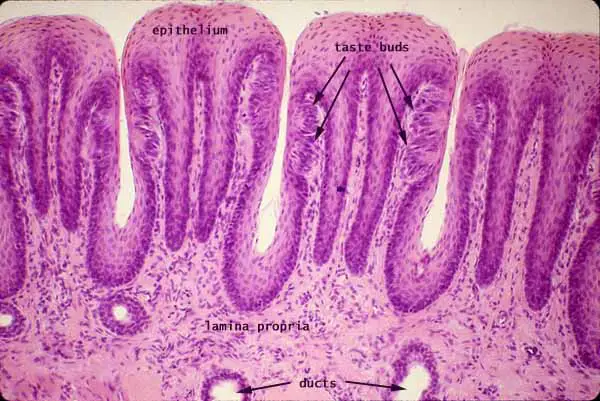
[In this image] Examples of epithelial tissues that host sensory nerves are the tongue epithelium with taste buds.
Image source: Histology at SIU
Diseases – What conditions affect epithelial tissue?
Cancer
One of the biggest concerns with epithelial tissue is the potential for malignancy development (a bad side of their robust regeneration capability). The types of cancer that initiate from epithelial tissues are called carcinomas.
Carcinomas are the most common type of cancer. They make up about 85 out of every 100 cancers in the USA. Based on their origins, carcinomas include squamous cell carcinoma, adenocarcinoma, transitional cell carcinoma, and basal cell carcinoma, etc.
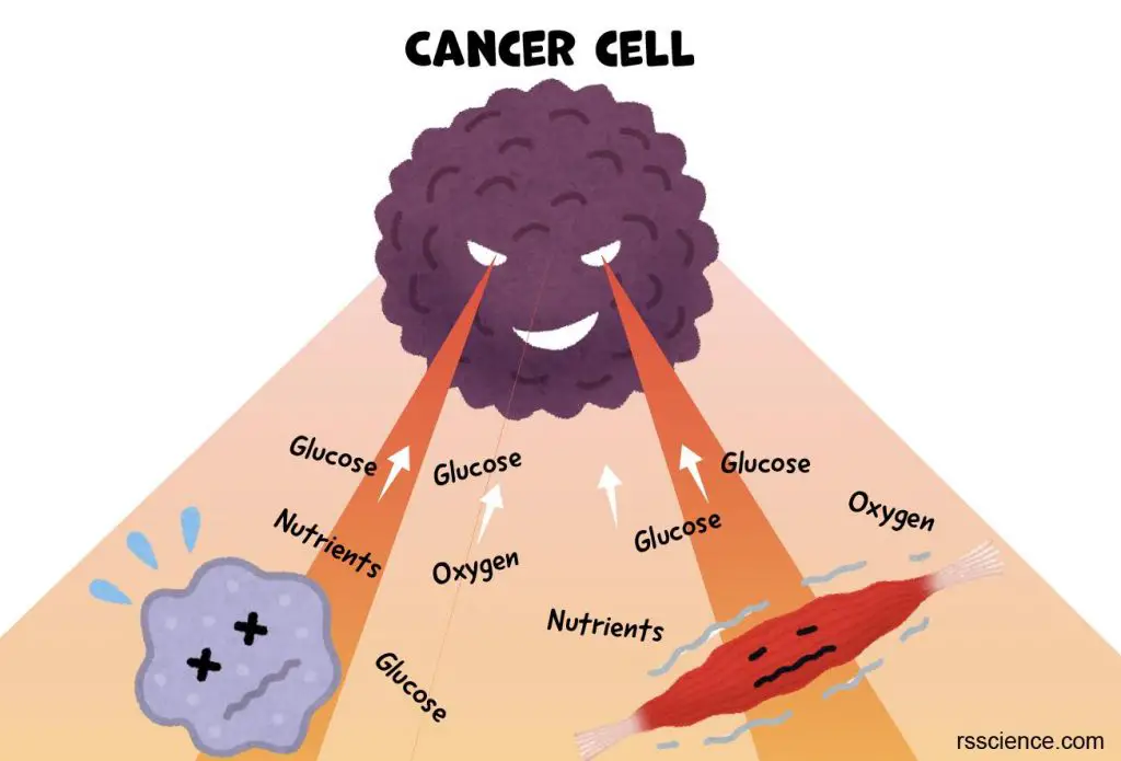
[In this image] Cancer cells plunder the nutrients from surrounding cells and tissues to sustain the rapid growth of tumors.
In addition to cancer, various organs can develope disease. Examples are asthma (airways), celiac disease (intestines), vertigo (ears), dermatitis (skin), etc.
Look at epithelial cells under a microscope
Project 1 – Look at your cheek cells
The tissue that lines the inside of our mouth is known as the basal mucosa. This protective layer is composed of stratified squamous epithelial cells or cheek cells. These cells divide approximately every 24 hours and are constantly shed from the surface of our mouth. Quick cell turnover makes sure any damage to this protective barrier is repaired as soon as possible.
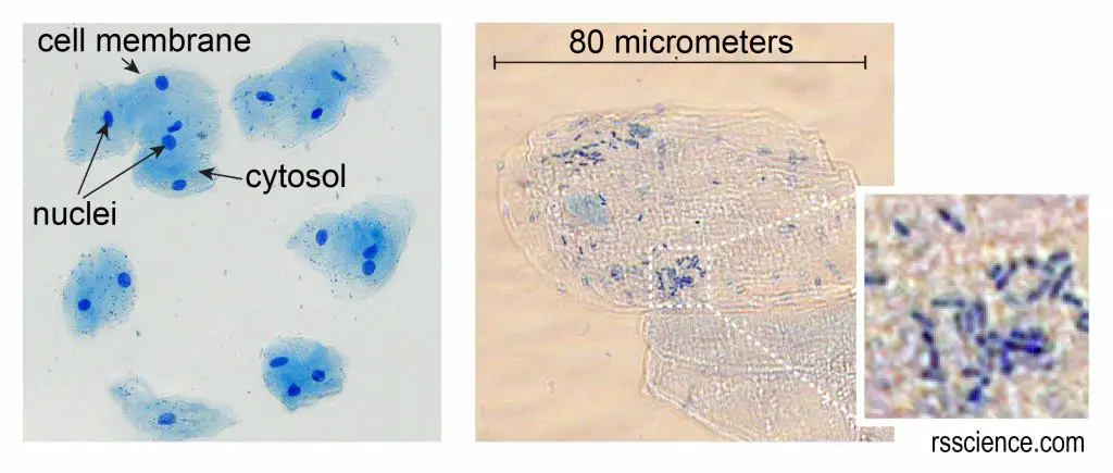
[In this image] Cheek cells (epithelial cells) form a protective barrier lining your mouth. You can easily obtain your cheek cells for your microscope project by scraping the inside of the mouth using a clean cotton swab. See more details here.
Project 2 – Study premade histological slide sets
All colorful pictures in this article are taken from my collection of professional slides for histological study.
Histology is the branch of biology that studies biological tissues’ microscopic anatomy. In order to see the detail of individual cells in the tissues or organs, the specimens have to be cut into very thin sections. To do so, a professional microtome and special chemical treatments are required. Below is the typical process to prepare professional microscopic slides:
(1) The tissue is grossly cut into a suitable piece and placed in a cassette. Soft animal tissues require chemical fixation (including formalin and alcohol) to preserve the cell structures.
(2) The specimen is then embedded in a wax-like material called paraffin to become a hard, solid block.
(3) A professional rotary microtome.
(4) Carefully place the specimen block and the sectioning blade.
(5) By rotating the drive wheel, the blade will trim a thin section of the specimen block like the peeler in the kitchen. Each section can be as thin as 3 micrometers (1/30 the thickness of your hair!) Series of sections can be cut like a ribbon. The ribbon will then be moved to a water bath for flotation. The good sections will be selected and transferred to the glass slide.
(6) Depending on the purpose of the specimen, the slides can be stained with specific stains, then washed, dried, and permanently mounted with coverslips.
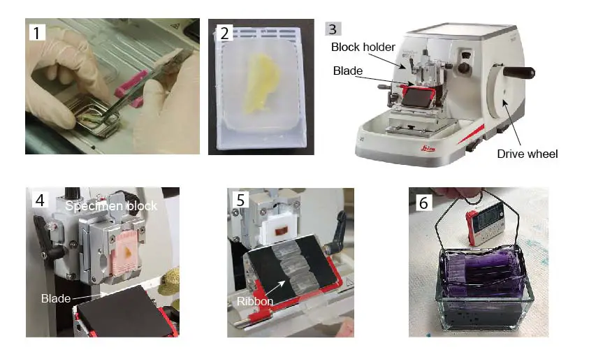
[In this image] The steps of histological slides preparation.
Hematoxylin and eosin stain (or H&E stain) is one of the most common stains used in histology. The hematoxylin stains cell nuclei a purplish blue, and eosin stains the matrix and cytoplasm pink. Most pictures of epithelial tissues we saw in this article are done by H&E stain.
None of us can have these professional microtomes and special chemicals at home. But don’t worry, you can purchase premade slide sets. There are all kinds of slide sets on Amazon.com, from 10 to 200 slides per set with different contents.
You can check out our 25 Microscope Prepared Slide Set, which includes 6 slides of human histology (blood, trachea, stomach, liver, pancreas, and brain).

Summary
1. Epithelial tissue or Epithelium (plural = epithelia) is a protective, continuous sheet of compactly packed cells. Epithelium covers all internal and external surfaces of our body, and lines body cavities and hollow organs.
2. Characteristics of epithelial tissue include cell sheets and cellularity, cell junction, polarity, basement membrane, high regeneration, nerve innervation and lack of blood vessels.
3. Epithelial cells have all the common cell organelles like other animal cells. Some epithelial cells have hair-like protrusions (cilia or microvilli) on their apical sides.
4. Epithelial tissues are classified by the number of layers and by the shape or the function of the cells. Epithelial tissues could form by cells assembling in Simple (only one layer), Stratified (multiple layers), and Pseudostratified (one layer of cells with different sizes resulting in a multilayered appearance). Cell shape includes Squamous (flat and scale-like), Cuboidal (cube-like), and Columnar (taller, column-like).
5. There are 8 types of epithelial tissue: simple squamous, simple cuboidal, simple columnar, pseudostratified columnar, stratified squamous, stratified cuboidal, stratified columnar, and transitional epithelia.
6. Major functions of epithelia are protection (skin), secretion (thyroid), absorption (intestine), gas exchange (lung), secretion (liver), and filtration (kidney).
7. Carcinomas are the most common type of cancer, which initiated from epithelial tissue (a bad side of their robust regeneration capability).
8. How to look at epithelial cells under a microscope? 1. Look at your cheek cells. 2. Study premade histological slide sets.
Extended Reads
Classification and Types of Epithelial Tissues