
This article covers
What is epithelium?
Epithelial tissue or Epithelium (plural = epithelia) is a protective, continuous sheet of compactly packed cells. Epithelium covers all internal and external surfaces of our body and lines body cavities and hollow organs. The epithelium has a variety of functions depending on where it’s located in your body, including protection, secretion, sensation, and absorption.
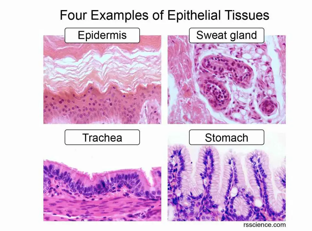
[In this image] Four examples of epithelial tissues with distinct cell morphology and functions.
This article will dive deeper into the classification of epithelial tissues and give you a few examples of each type.
We have an overview article on Epithelium – covering its definition, characteristics, cell structures, types, and functions. If you like to have a quick understanding of epithelial tissues in general, click here to see it.
How to classify epithelial tissues?
In general, epithelial tissues are classified by the number of cell layers, the cell shapes, and the functions of the cells.
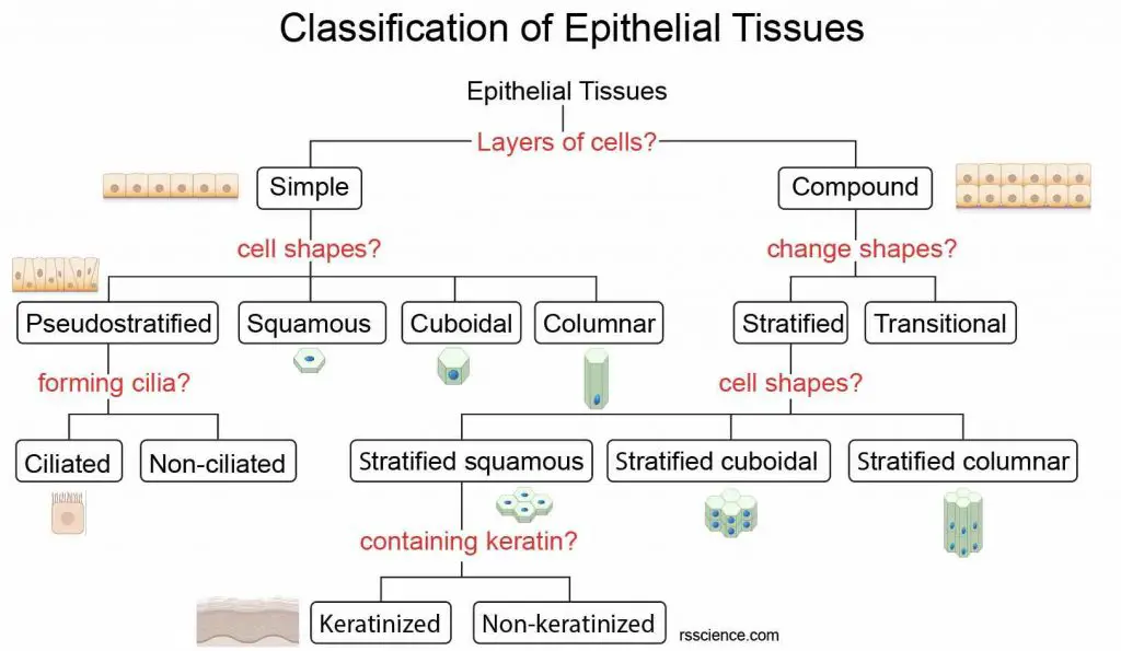
[In this image] A hierarchical classification system of epithelia.
The structure of tissue provides clues to its name and location. Epithelial tissues are grouped by the number of their layers (simple vs. compound/stratified), by the cell shapes (squamous, cuboidal, or columnar), and by the presence of specialized structures (i.e., cilia or keratin). See below to learn what these terms mean.
Classification by cell shapes of epithelial cells
There are three principal shapes of epithelial cells: squamous, cuboidal, and columnar.
- Squamous epithelium: Squamous epithelial cells are flat and scale-like in appearance. They can be found in the smooth lining of the mouth, esophagus, and the alveoli of the lungs.
- Cuboidal epithelium: Cuboidal epithelial cells are cube-like in appearance, meaning they have equal height and width. They usually form ducts of secretory organs and glands.
- Columnar epithelium: Columnar epithelial cells are taller than their width, showing a column shape in appearance. Columnar epithelia can be further classified into ciliated columnar epithelium (lining airway surface) and glandular columnar epithelium (forming glands).
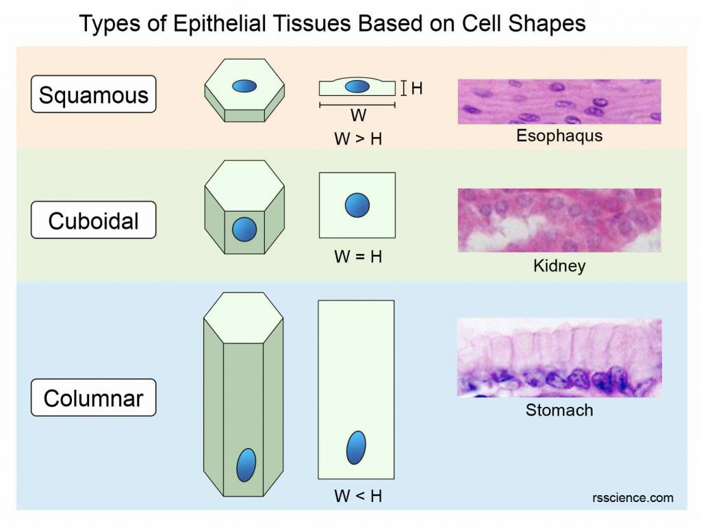
[In this image] Schematics and examples of Squamous, Cuboidal, and Columnar shapes of epithelial cells. W: width, H: height of cells.
Classification by the arrangement of epithelial layers
Epithelial tissue can also vary based on how the cells are arranged.
- Simple epithelium forms by only a single layer of cells. In general, the thinness of the epithelial barrier allows them to perform functions like absorption and filtration. Examples are cells of the bronchi, alveoli, kidney tubules, uterus, body cavities, stomach, and small intestine.
- Stratified (or compound) epithelium is made up of more than one layer of cells. Multilayered structures allow the stratified epithelia to withstand mechanical or chemical insult. Even when some cells are damaged and detached from the surface of the stratified epithelium, the underlying tissues are still protected without exposure.
Usually, the cells sitting in the basal layer can divide and generate new epithelial cells to replenish the loss of surface cells. Examples of stratified epithelia are the lining of the skin, mouth, esophagus, sweat glands, salivary glands, and mammary glands.
- Pseudostratified epithelium is made up of closely packed cells that appear to be arranged in layers because they’re different sizes, but it’s actually just one layer of cells. Examples are ciliated epithelial cells lining the trachea and upper respiratory tract.
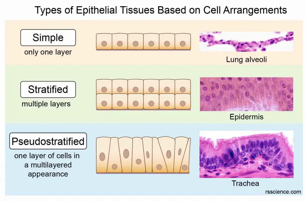
[In this image] Schematics and examples of Simple, Stratified, and Pseudostratified arrangements of epithelial cells.
Classification based on specialized functions
Some unique epithelial tissues are categorized and named by the particular functions they have, including:
- Transitional epithelium is made up of multilayers of cells that become flattened when stretched, depending on the amount of tension on the epithelial tissues. Examples are the urothelium, which allows our bladder to expand.
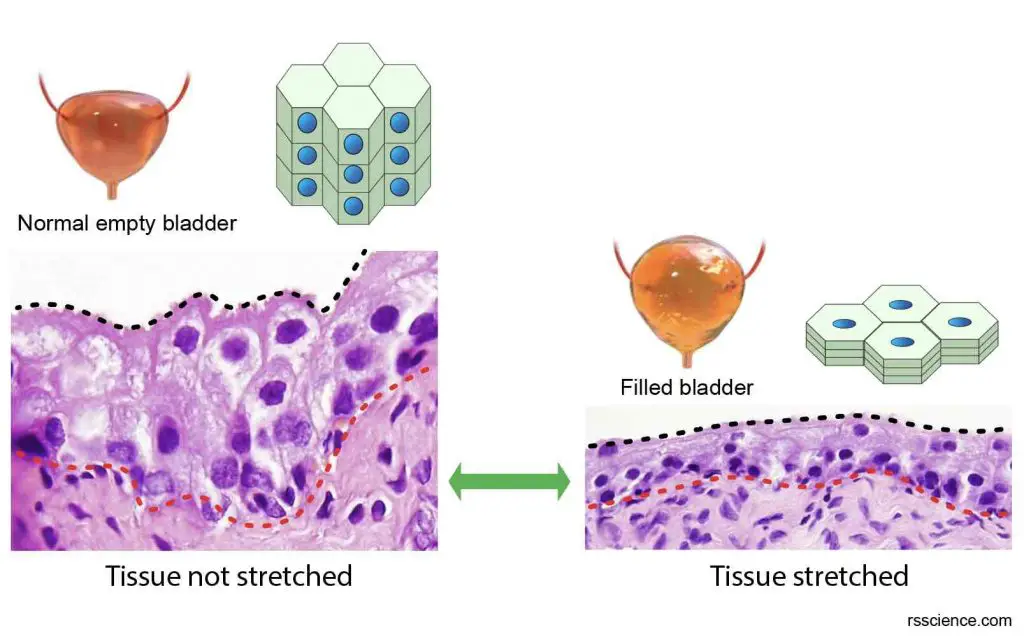
[In this image] Transitional epithelium can undergo “transitions” in shape. Examples are epithelial cells lining the lumen surface of our bladder. They can become flattened when stretched to expand the volume of the bladder to accommodate more urine.
Image source: modified from Atlas of plant and animal histology
- Glandular epithelium forms various glands and is specialized to secrete substances. They can produce hormones, enzymes, saliva, mucus, sweat, milk, body fluids, etc.
Glands are classified as endocrine (internal secretion) or exocrine (external secretion), depending on where they release their product. Exocrine glands secrete their product into a duct. Examples of exocrine glands are salivary glands, sweat glands, and mammary glands.
Endocrine glands are ductless glands, and they release their product directly into the blood or intestinal fluid. Examples of endocrine glands are the thyroid, pineal gland, pituitary gland, ovaries, testes, and adrenal glands. The liver and pancreas contain both exocrine and endocrine tissues.
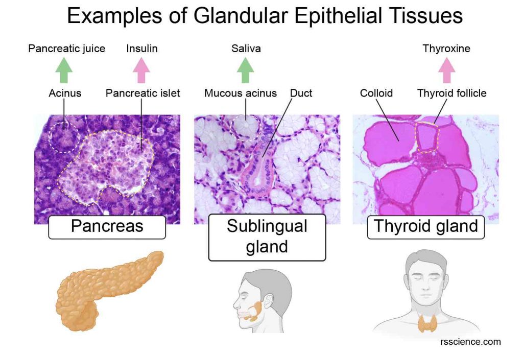
[In this image] Examples of epithelial tissues that secrete essential substances for our body.
1. Pancreas has both exocrine and endocrine functions. Pancreatic islets (endocrine) secrete insulin directly into the bloodstream to regulate blood sugar levels. Acinar epithelium (exocrine) secrets the pancreatic juice through the pancreatic duct into the intestines to help food digestion.
2. Sublingual glands are a pair of salivary glands (exocrine) situated underneath the tongue. They produce saliva in the mouth.
3. Thyroid (endocrine) is a butterfly-shaped gland that sits low on the front of the neck. It secretes several thyroid hormones into the bloodstream.
- Mucous epithelium is also known as mucosa. Mucous epithelium lines our respiratory and digestive tracts, which constantly interact with the outside world. Mucous epithelial tissues secrete gel-like mucus. The mucus helps in lubrication, protection, and easy movement of materials. It prevents tissues from drying.
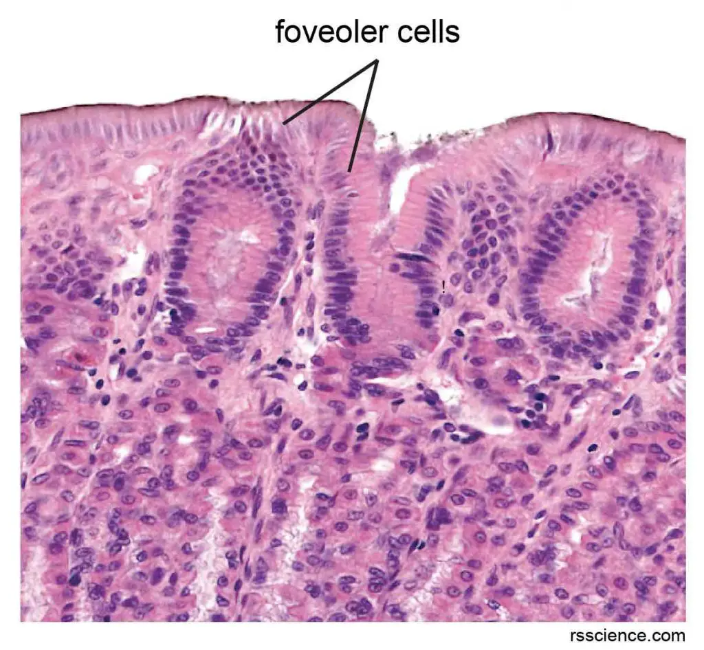
[In this image] Microscopic section of gastric mucosa, which forms a protective barrier of our stomach. Foveolar cells can be seen at the top of the image lining the surface. They are the mucus-producing cells of the stomach.
- Serous epithelium lines the body cavities, such as the peritoneum (abdomen cavity), pleura (lung cavity), and pericardium (heart cavity). It also surrounds the internal organs like the heart and lungs. These cells secrete a lubricant fluid that prevents the frictions between organs and body walls during movement, breathing, or heart beating.
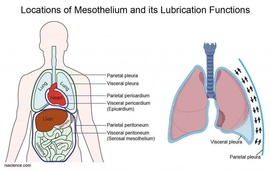
[In this image] Mesothelium covers the body wall and the organs.
(Left) Mesothelial cells lining the cavities/body walls in these subdivisions are called parietal mesothelia, while visceral mesothelia cover the organs. (Right) The size and location of our lungs change during each breath. For this reason, the lubricant layer between the parietal pleura (mesothelia lining the chest cavity) and visceral pleura (mesothelia covering the lungs) is critical to reduce the fraction and prevent injury.
- Olfactory epithelium is found inside the nasal cavity. It contains olfactory receptor cells and involves in the sense of smell. Olfactory receptor cells do so by specialized cilia extensions. The cilia trap odor molecules you breathe in as they pass across the epithelial surface. Stimuli triggered by these molecules are transmitted from the receptors to the olfactory bulb in your brain, where your brain interprets the smell.
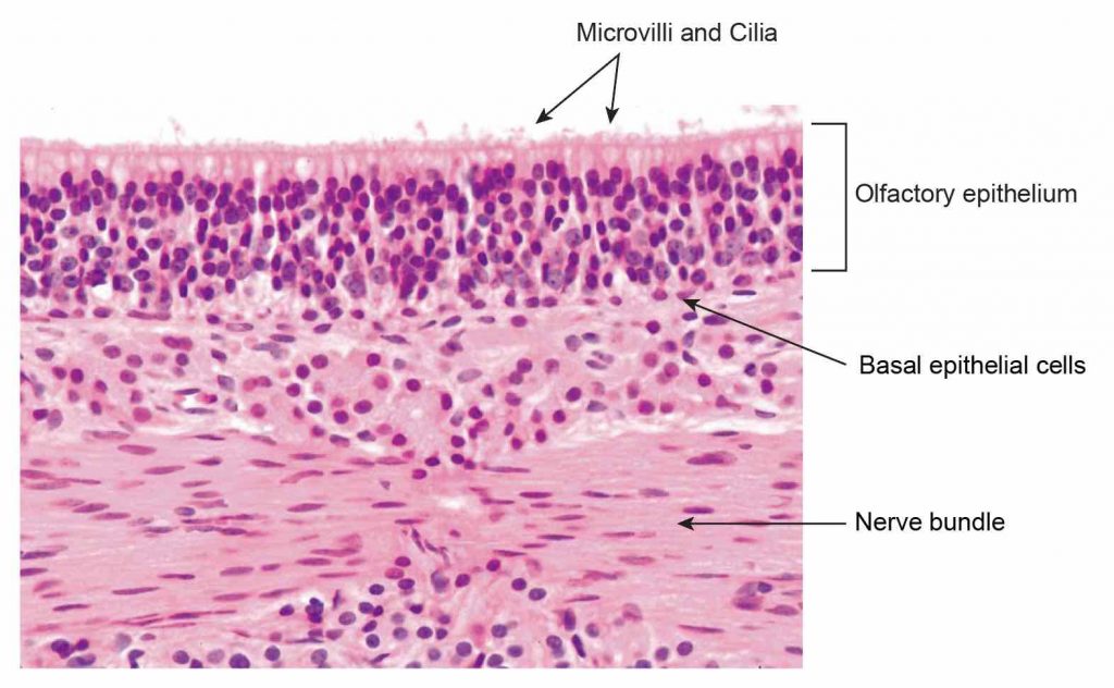
[In this image] Microscopic section showing the olfactory epithelium with a meshwork of microvilli and olfactory cilia on the surface. You can also see a nerve bundle under the olfactory epithelium.
Image source: modified from Histology@Yale
Eight common types of epithelial tissues
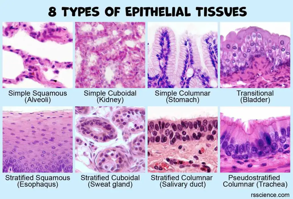
Given the different shapes and arrangements of epithelial cells, there can be 8 types of epithelial tissue in combination, including:
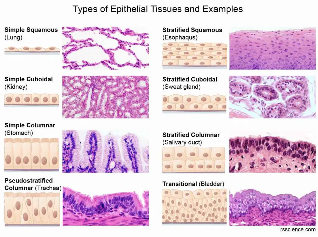
[In this image] A summary of 8 common types of epithelial tissues and their examples.
(1) Simple squamous epithelium
Simple squamous epithelium is a single layer of flattened cells. When viewed from above, these cells closely fit, resembling a tiled floor. Simple squamous epithelial cells have disc-shaped central nuclei. When viewed in the lateral section, they resemble fried eggs seen from the side.
Simple squamous epithelium is thin and often permeable. This property makes simple squamous epithelium ideal to form a membrane for diffusion or filtration, wherever small molecules must pass through quickly. For example, the epithelium (also called Endothelium) lines the walls of capillaries (tiny blood vessels). The exceptional thinness of epithelial walls allows the efficient exchange of nutrients and wastes between the bloodstream and surrounding tissue cells.
Simple squamous epithelium also forms the thin walls of alveoli (singular: alveolus) and serves as the site for gas exchange in the lungs.
Some simple squamous epithelium serves as a lubrication layer. For example, specialized epithelium called mesothelium lines the cavities of the body and the surface of organs. This lubricant layer between body walls and organs is critical to reduce the fraction and prevent injury (i.e., during each breath and heart beating).
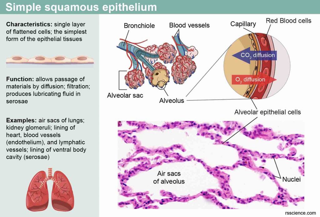
[In this image] The essential information of simple squamous epithelium. An example of simple squamous epithelium is the epithelial lining of the air sacs, where gas exchange occurs in the lungs.
(2) Simple cuboidal epithelium
Simple cuboidal epithelium consists of a single layer of cube-shaped cells. This type of epithelium is ideal for forming the secretory cells of glands and the walls of the smallest ducts of glands. In the kidneys, simple cuboidal epithelium forms the walls of many tubules of nephrons, which is the basic units to perform the kidney’s filtration function.
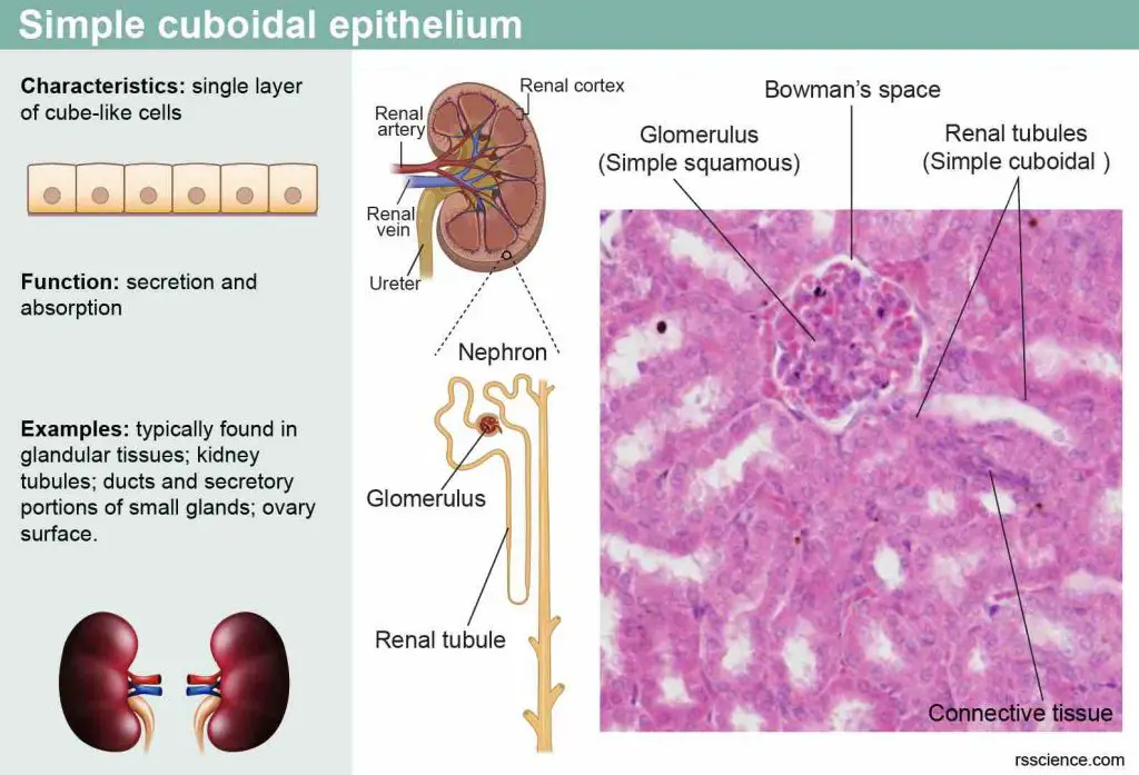
[In this image] An example of simple cuboidal epithelium is the epithelial lining of renal tubules. The nephron is the functional unit of the kidney. It is composed of a renal corpuscle and a renal tubule. The nephron is responsible for removing waste from the blood. There are about 1,000,000 nephrons in each human kidney.
(3) Simple columnar epithelium
Simple columnar epithelium is a single layer of tall cells aligned like soldiers in a row. For this reason, this type of epithelium provides a good protection function while maintaining the ability to absorb nutrients.
Our digestive tract is lined by simple columnar epithelium from the stomach to the rectum. The structure of simple columnar epithelium is ideal for the active movement of molecules, namely in absorption (i.e., uptaking nutrients by small intestine epithelia) and secretion (i.e., producing gastric juice by stomach epithelia).
Simple columnar epithelium is thin enough to allow large numbers of molecules to pass through it quickly, yet thick enough to house the specialized cells needed to perform the complex processes of molecular transport. To increase the efficiency of absorption, some cells have microvilli on their apical side to enlarge their surface area.
Some simple columnar epithelia have cilia on their cell surface. These cilia look like tiny whiplike hairs and can beat rhythmically to move substances across certain body surfaces. For example, the inside of the uterine tube is covered by simple ciliated columnar epithelium. Its cilia help move the ovum to the uterus.
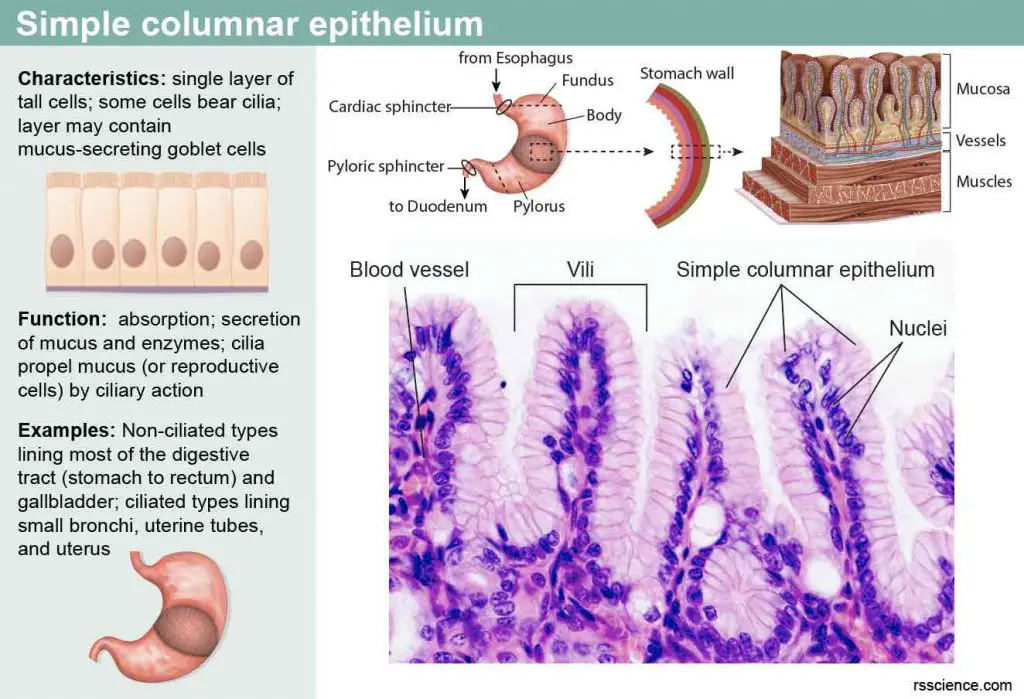
[In this image] An example of simple columnar epithelium is the surface of the stomach. In the microscopic section, you can see a single layer of aligned tall cells with their nuclei near the cells’ bottom. Goblet cells, which synthesize and secrete mucus, sit inside the stomach epithelial walls. These mucins help neutralize the acids produced by the stomach. They also help in lubricating the epithelium for the easier passage of food.
(4) Pseudostratified columnar epithelium
“Pseudo” means “false”. This name suggests that the pseudostratified columnar epithelium is not a real multilayered stratified epithelium. In fact, the cells of pseudostratified columnar epithelium are varied in height. All its cells rest on the basement membrane, but only the tall cells reach the surface of the epithelium. The short cells are hidden under these tall cells. These short cells are progenitors and continuously give rise to tall cells. The cell nuclei lie at several different levels, giving a false impression that this epithelium is stratified.
Pseudostratified columnar epithelium, like simple columnar epithelium, functions in secretion and absorption. This type of epithelium lines our upper respiratory tract and usually has a lot of cilia. Their cilia can propel dust-trapping mucus out of the lungs.
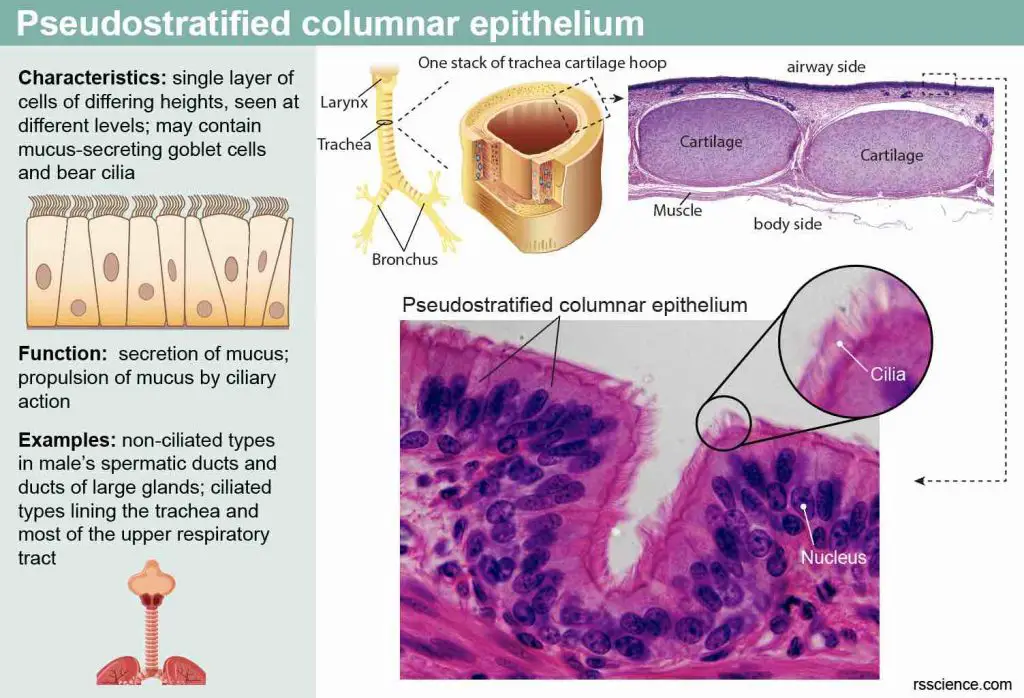
[In this image] An example of pseudostratified columnar epithelium is the cells lining the surface of the trachea. The high magnification of the microscopic image shows many hair-like cilia on the surface of epithelial cells.
(5) Stratified squamous epithelium
Stratified epithelium regenerates from below; that is, the basal cells divide and push upward to replace the older surface cells. For this reason, stratified epithelia are more durable than simple epithelia, and their major (but not only) role is protection.
As you might expect, the stratified squamous epithelium consists of many cell layers whose surface cells are squamous. In the deeper layers, the undifferentiated cells could look cuboidal or columnar.
Stratified squamous epithelium is the thickest and best adapted for protection among all the epithelial types. It covers the outer surfaces of our body, forming the epidermis of the skin and the inner lining of the mouth, esophagus, and vagina. This type of epithelium can protect us against microbes from invading underlying tissue and/or protection against water loss.
The epidermis of the skin is keratinized, meaning that its surface cells contain a tough, protective protein called keratin. The other stratified squamous epithelia of the body lack keratin and are non-keratinized.
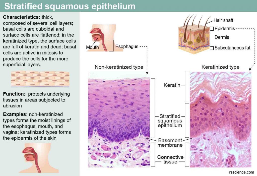
[In this image] Two examples of stratified squamous epithelia.
(Left) The tissue lining the surface of the esophagus is a non-keratinized stratified squamous epithelium. The mature cells near the apical surface are flattened, while the dividing cells (which can replenish the damaged cells) closer to the basement membrane are more cuboidal. (Right) The epithelial tissues forming the epidermis are keratinized. The tough keratin fibers provide additional protection to the underlying tissues.
(6) Stratified cuboidal epithelium
Stratified cuboidal epithelium generally consists of two layers of cuboidal cells. This type of epithelium is not as common. You can find them located in the excretory ducts of some glands, for example, sweat glands, mammary glands, and salivary glands.
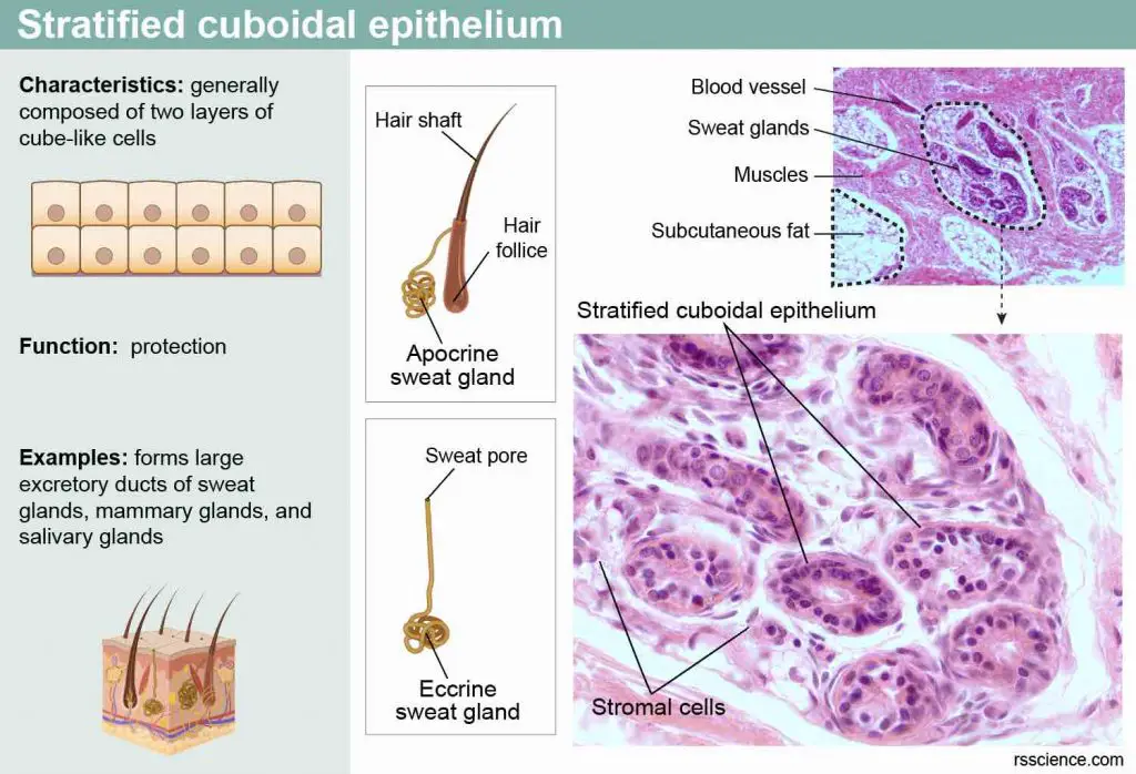
[In this image] Stratified cuboidal epithelium is a rare type of tissue.
One example is the excretory ducts of sweat glands. There are two types of sweat glands. Eccrine sweat glands are the major sweat glands of the human body, found in virtually all skin, with the highest density in palm and soles. They have their opening pores on the surface of the skin. On the other hand, apocrine sweat glands are associated with the hair follicles and found only in certain body locations, like the armpits.
(7) Stratified columnar epithelium
This type of epithelium is not as common and is seen in the large excretory ducts of our salivary glands. Stratified columnar epithelium is also found in small amounts in the male urethra and the mucous membrane (conjunctiva) lining your eyelids.
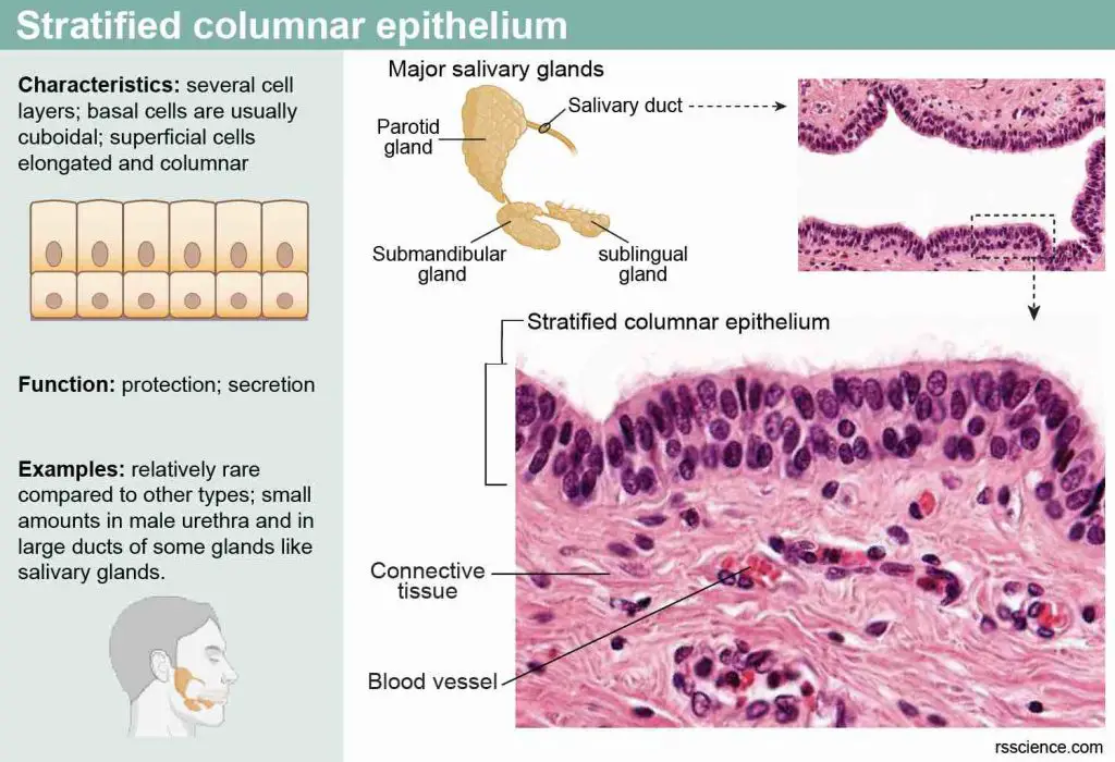
[In this image] Stratified columnar epithelium is rare to find. One example is the biggest duct of our salivary glands. Humans have three paired major salivary glands (parotid, submandibular, and sublingual) and hundreds of minor salivary glands.
(8) Transitional epithelium
Transitional epithelium is one type of stratified epithelia that can change its cellular arrangement.
Transitional epithelium lines the inside of the hollow urinary organs, like the urinary bladder. Our bladders stretch as they fill with urine. As the transitional epithelium stretches, it thins from about six cell layers to three, and its apical cells unfold and flatten. When relaxed, portions of the apical surface invaginate into the cell. Thus, this epithelium undergoes “transitions” in shape. It also forms an impermeable barrier that keeps urine from passing through the bladder wall.
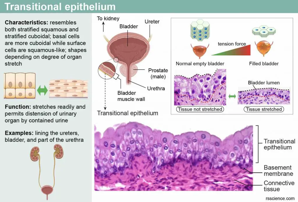
[In this image] An example of transitional epithelium is the lining of the urinary bladder. The arrangement of these epithelial cells can transform depending on the size of the bladder. When the bladder is empty, the transitional epithelium is relaxed and resembles a stratified cuboidal type. Once the bladder is filled with urine, the epithelium stretches, making the surface cells more squamous-like.
Summary of eight types of epithelial tissues
| Cells | Location | Function | Example |
| Simple squamous epithelium | Air sacs of lung alveoli, lumens of the heart, blood vessels, and lymphatic vessels (endothelium), lining of body cavities (mesothelium) | Allows materials to pass through diffusion and filtration; secrets lubricating substance | Air sacs of lung alveoli 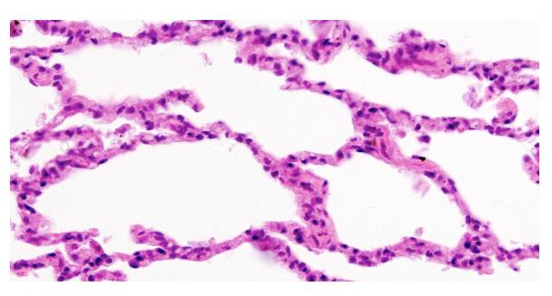 |
| Simple cuboidal epithelium | Secretory cells of glands, small ducts of glands, renal tubules in nephrons of kidneys | Secretion and absorption | Renal tubules of kidney 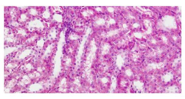 |
Simple columnar epithelium 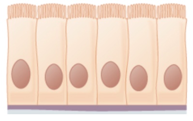 | Ciliated types are in bronchi, uterine tubes, and uterus; non-ciliated types are in the digestive tract (from the stomach to the rectum) | Secretion (mucous and enzymes) and absorption | Stomach wall 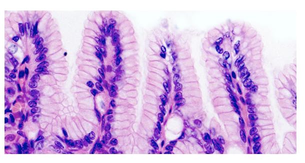 |
Pseudostratified columnar epithelium 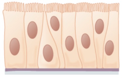 | Ciliated tissues line the upper respiratory tract and the trachea | Secretes mucus; waving the cilia to move dust-trapping mucus out of the lungs | Airway side of trachea 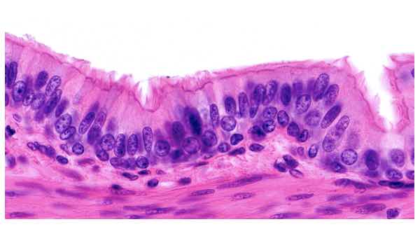 |
Stratified squamous epithelium 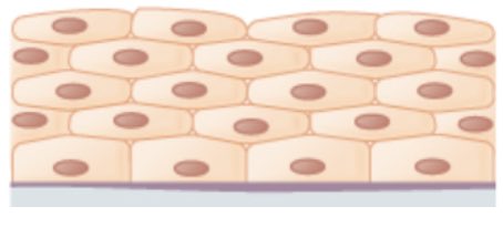 | Keratinized types form the epidermis of the skin; non-keratinized types line the mouth, esophagus, and vagina | Protection against abrasion, infection, and loss of water | Esophagus 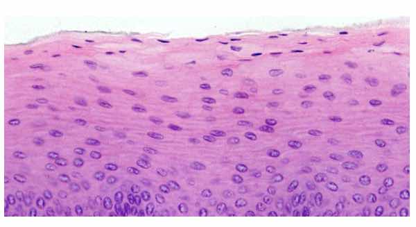 |
Stratified cuboidal epithelium 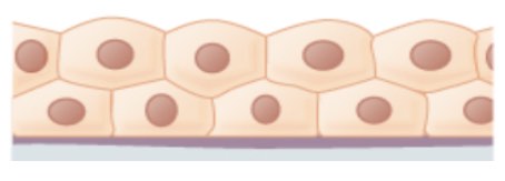 | Sweat glands, mammary glands, and salivary glands | Protection; forming excretory ducts of glands | Excretory ducts of sweat glands  |
Stratified columnar epithelium 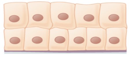 | Rare, the male urethra and the ducts of some glands | Secretion and protection | Large excretory ducts of salivary glands 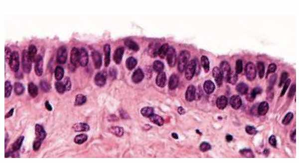 |
Transitional epithelium 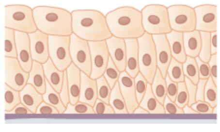 | Lines the bladder, urethra, and the ureters | Allows the urinary organs to expand and stretch | Urinary bladder 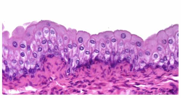 |