
This article covers
Why microscope is important in biology?
The microscope is a very important tool in a biological laboratory. Many cellular structures are too tiny to see by naked eyes. Examining specimens under a good microscope enables us to study these cellular structures and investigate their biological functions.
In this article, we will show you that you can study plant biology and anatomy using a premade slide set.
Overview of a flowing plant
A plant is made up of several different parts. The five main parts are the roots, the leaves, the stem, the flower, and the seed. Each part has its unique job to keep the whole plant healthy. By looking at the microscopic structures of different parts of the plant parts, we can learn how the plant function at the cellular level.
The Roots
The function of the roots is to absorb water and minerals from the soil. The roots also anchor the plant in the ground. The way of roots growing deep into the ground is through the elongation of the root tips.
In this premade slide of Vicia pea’s root, you can see the active cell division at the tip of a growing root. Under high magnification, you can even identify cells undergoing mitosis, and different phases of mitosis, prophase, metaphase, anaphase, and telophase.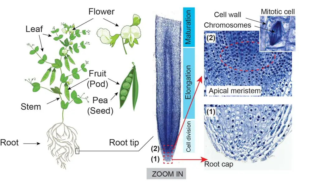
[In this figure]
Left: The anatomy of a typical flowering plant, including flower, fruit (pod), leaf, stem, and root.
Right: The microscopic image of the longitudinal section of the Vicia pea’s root tip. The specimen was stained with Methylene blue, a dye that can highlight the cell wall and nucleus (containing DNA).
With higher magnification, you can see regions of (1) root cap that protects the root tip, and (2) apical meristem, which contains actively dividing cells near the end of the root tip. The highly active mitosis area is highlighted with a red dash line. Within that area, you can easily find cells undergoing different phases of mitosis, prophase, metaphase, anaphase, and telophase.
(Modified from the guidebook of Rs’ Science – 25 Microscope Prepared Slide Set)
The Stem – Xylem and Phloem
The function of the stem is to support the plant above ground and to transports the water and nutrients from the roots to the leaves. The critical structure in the stem is the vascular system. The vascular system consists of Xylem and Phloem. The xylem is responsible for transporting water upward from the roots. The phloem carries nutrients like sugars around the plant (both upward and downward directions).
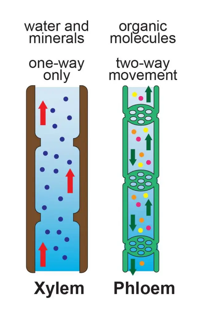
[In this figure] Illustration of Xylem and phloem.
Xylem and phloem are both transport vessels that combine to form a vascular bundle in higher order plants. Xylene transport water unidirectionally from the roots. Phloem carries nutrients made from photosynthesis (typical from the leaves) to the parts of the plant where need nutrients.
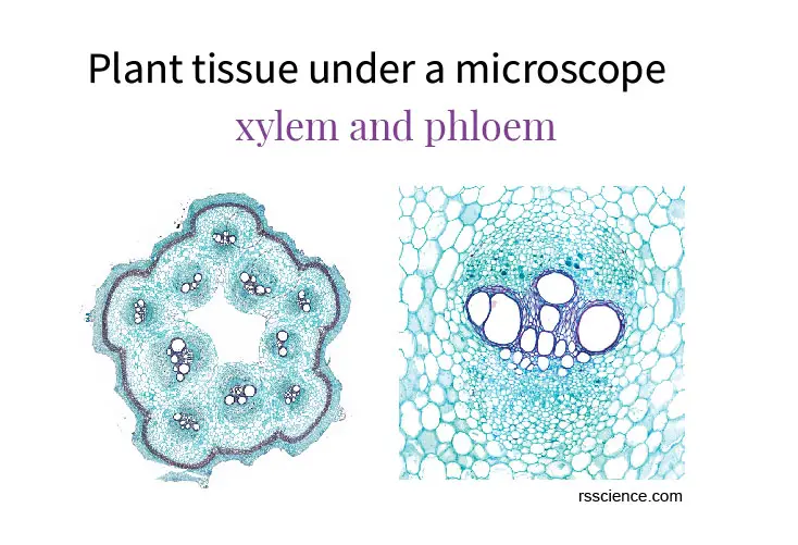
By looking at the cross-section slide of the pumpkin’ stem, you can easily identify the vascular bundles in a ring arrangement. Each vascular bundle includes two types of vascular tissues – Xylem and Phloem.
The xylem is responsible for keeping a plant hydrated by transporting water upward from the roots. Xylem cells are dead, elongated, and hollow. The phloem carries important sugars, organic compounds, and minerals around a plant (both directions). The phloem is made from cells called sieve tube members. Vascular bundles are enclosed inside the ground tissue and protected by the epidermis layer.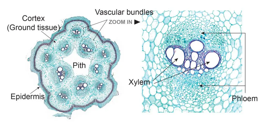
[In this figure] Vascular bundle distribution of a pumpkins’ vine.
The cross-section of a pumpkin’s vine shows the typical vascular bundle distribution in a ring arrangement with pith in the center. Each vascular bundle includes the xylem (stained with dark blue) in the middle surrounded by phloem.
The Leaves
The function of the leaves is to collect energy from the sunlight and convert the energy into sugars for the plant. This process is called photosynthesis, which requires special organelles – Chloroplast. By looking at the slide of the rice leaf, you can see the vascular system extending from the stem into the leaves as a continuous pipe network.
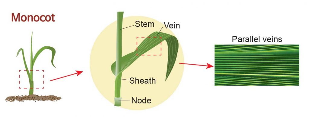
[In this figure] A monocot plant with leaves characterized by their parallel veins.
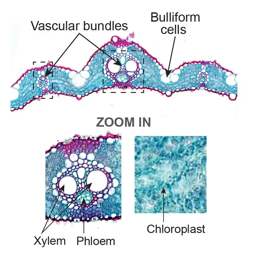
[In this figure] The microscopic image of the cross-section of rice leaf.
When you zoom in to have a closer view, you will see vascular bundles set inside the veins. Leaf cells with many chloroplasts can absorb the sunlight and perform photosynthesis. You can also see some large, bubble-shaped cells called Bulliform cells. Bulliform cells can regulate the water evaporation from the leaves.
The Flowers
The flowers are the reproductive parts of plants. The flowers often have brightly colored petals to attract pollinators. In the center of a flower, there are female parts called pistils and male parts called the stamen. In this slide of the lily flower, you can see the pollen grains inside the pollen sac of the anther (the structure at the tip of the stamen). Mature pollen grains will be released and carried by wind or insects to pistils.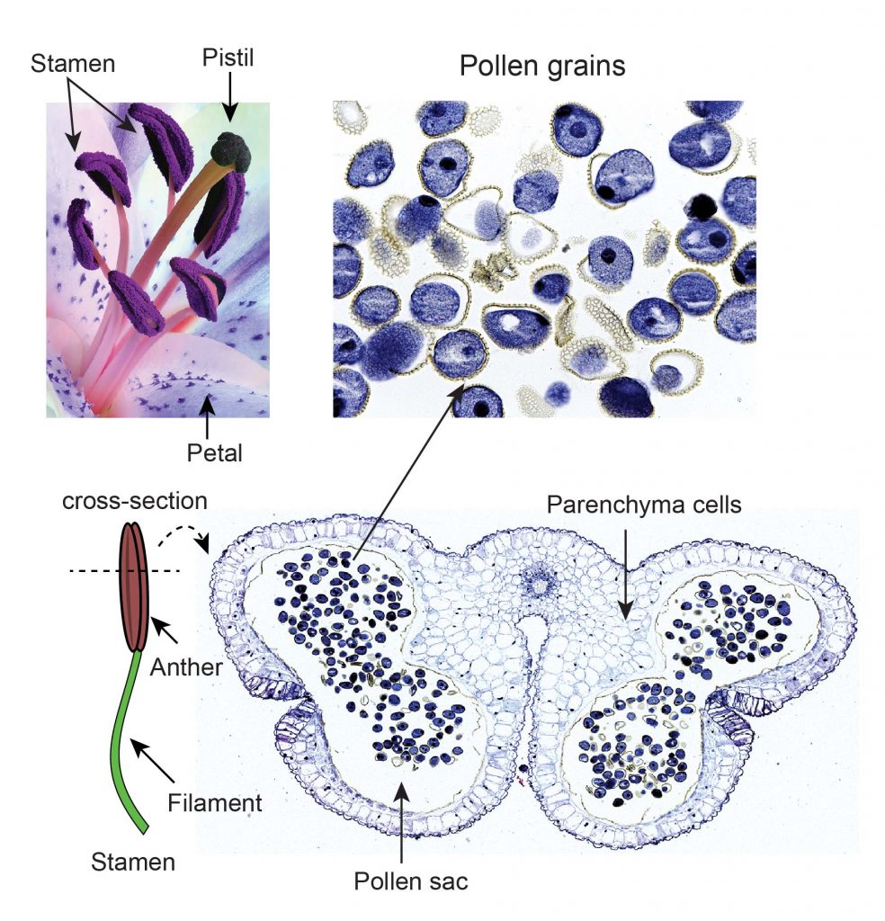
[In this figure] The anatomy of lily flowers.
The lily flowers contain a pistil, several stamens, and petals. The cross-section of a lily anther shows the pollen sac containing many pollen grains inside. Pollen grains are very beautiful and delicate viewing by a higher magnification.
The Seeds
Fertilized flowers will develop fruits and seeds. The seeds can grow into new plants if the environment is favorable. By looking at the slide of a corn kernel, you can see the tiny embryonic plant enclosed in a protective outer covering. The seeds also store plenty of nutrients like starch reserved for the growth of new plants.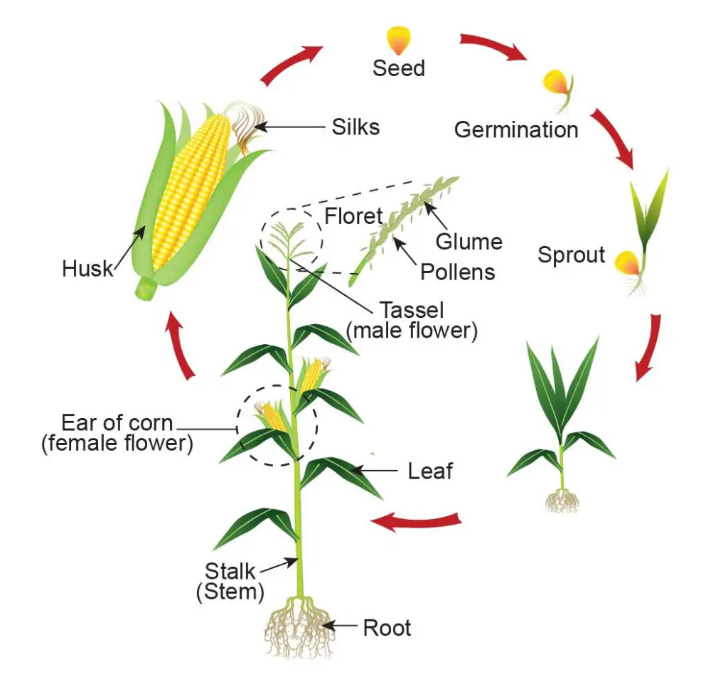
[In this figure] The life cycle of the corn plant.
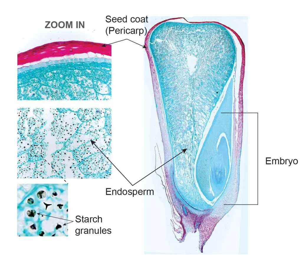
[In this figure] A longitudinal microscopic section of corn seed showing the seed coat, endosperm, and embryo.
The endosperm stores the energy in starch granules, which are stained with black color with iodine. The embryo can give rise to a new plant after seed germination.
I hope you enjoy learning plant biology and plant anatomy, and if you have premade slide sets on your hands, please take a look. Biology is amazing.