
(update 6/07/2020)
This article covers
What is a hemocytometer
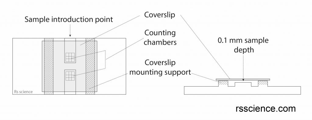
The hemocytometer is a counting chamber device originally designed for counting blood cells (the name “hemo” means blood). Hemocytometer is like a thick specimen slide with a grid engraved on it. When the coverslip is placed on the hemocytometer, the coverslip held the sample at a specific height (typically 0.1 mm).
The grid also has specific dimensions, therefore, the volume in the chamber can be calculated. Since you can count the number of cells in the area of grids and the volume is known, the concentration of cells in the sample can be determined easily. Below is an illustration of a hemocytometer.
How to use a hemocytometer to count cells
- Preparing the sample
Mixing the sample well before taking the samples. Avoid clumps.
- Making appropriate dilutions
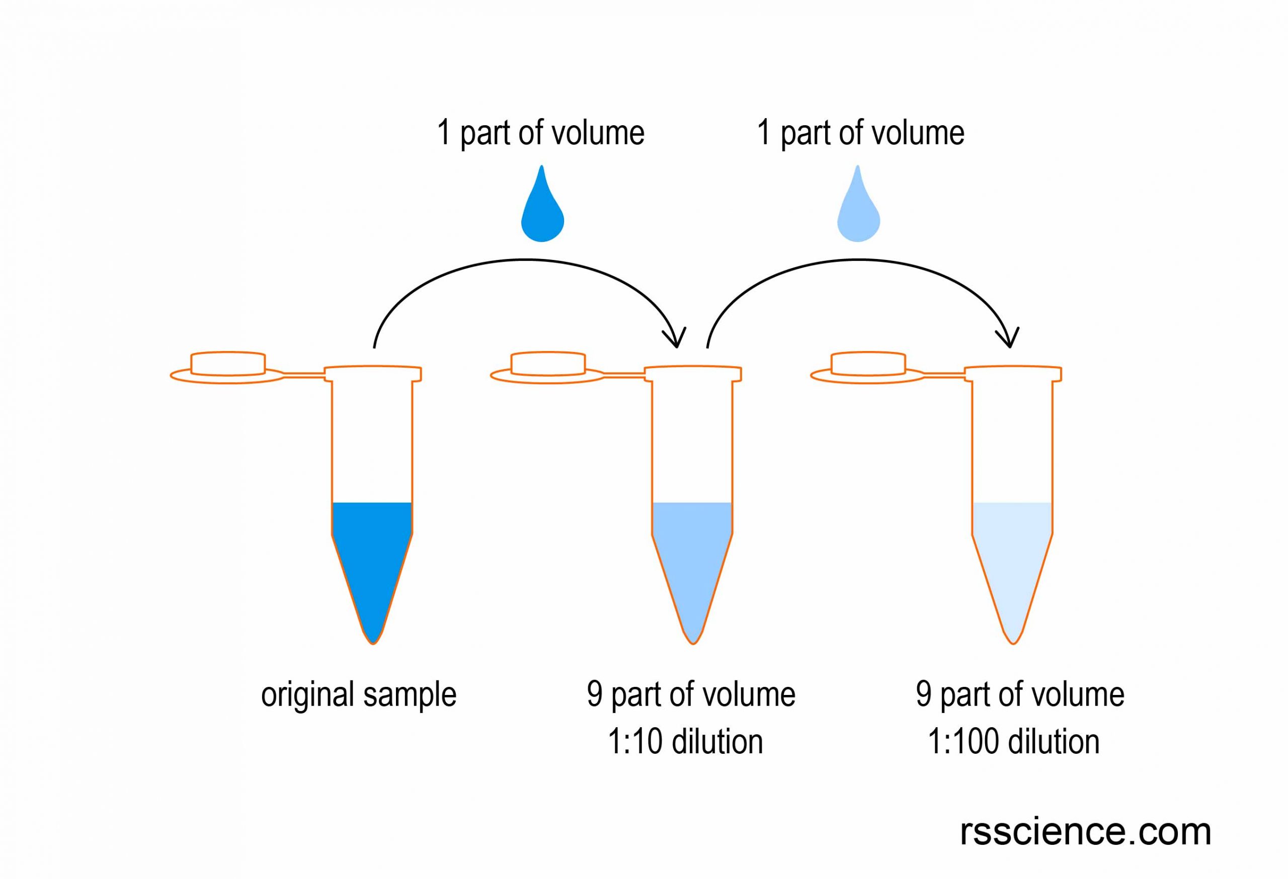
The concentration of cells should not be too high or too low. If the cell concentration is too high, it is hard to count cells visually under the microscope. If the cell concentration is too low, you need to count more squares and perhaps more times to avoid the statistical errors. If the cell concentration is too high, try to do 1:10, 1:100, 1:1000 serial dilutions (see the illustration below).
- Introducing cell suspension
Place the coverslip over the counting surface. Introduce the cell suspension (10 μL or one drop) from the edge of the coverslip with a fine tip transfer pipette. The capillary force will drag the liquid to fill the area under the coverslip. Enough liquid should be introduced so that the counting surface is just covered.
- Counting the cells
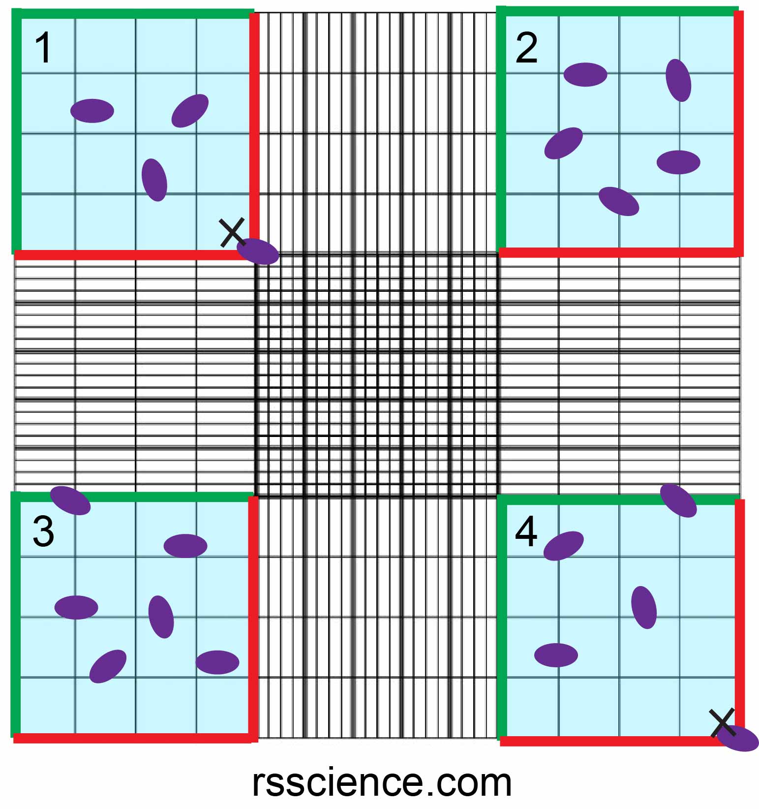
Using a microscope, focus on the grid lines of the counting area with a 5-10x objective. Count the cells in one set of 16 squares (1×1 mm square area; the blue area). You should set a counting rule.
For example, count the cells on the top and left lines of a square (marked by the green line), but do not count the cells on the bottom and right lines of a square (marked by the red line). Move the hemocytometer to the next set of 16 corner squares and continue to count until all 4 sets of 16 squares are counted. Take the picture below as an example, the cell numbers of 4 sets of 16 squares are 3, 5, 6, 4, respectively. Therefore, the average cell number of this counting is (3+5+6+4)/4 = 4.5
- Calculating the cell concentration
To calculate the number of cells per mL: Take the average cell count from each of the sets of 16 corner squares. Multiply by 10,000. The final value is the number of cells/mL in the original cell suspension if there is no dilution. In this case, the average cell number you calculated above is 4.5, then multiply by 10,000. Therefore, 4.5*10,000 = 45,000. The concentration is 45,000 cells/mL. If this is a 10 fold dilution, you will need to multiply by 10; bring it back to 450,000 cells/mL.
You can also see a demo video here.
How to use a hemocytometer to determine cell viability
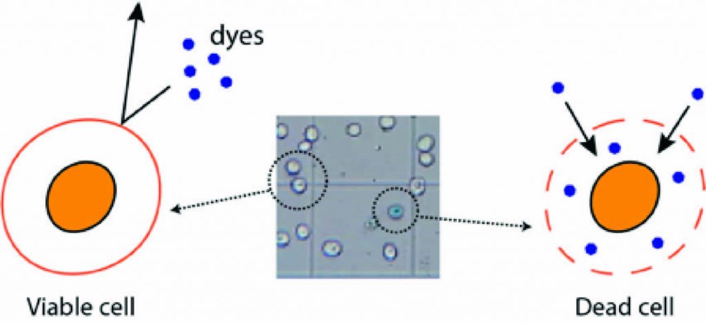
Trypan blue is a dye used to distinguish between live and dead cells. It is a vital stain that is not absorbed by healthy viable cells, but stains cells with a damaged cell membrane. Therefore, only dead cells can be stained as blue and can be seen under the microscope. This method is referred to as the dye exclusion method.
To determine cell viability, we use the same method as cell counting. The only difference is that you need to mix the cell suspension with Trypan blue dye before introducing cell suspension to the hemocytometer. Let’s start!
1. Making appropriate dilutions
Like what we discussed above, appropriate dilutions will make your counting life easier. Too many or too few cells is not good for counting.
2. Mixing equal volume of cell suspension and that of Trypan blue dye
The concentration of Trypan blue is 0.4% (w/w). The working concentration is 0.2%; that is the reason why we mix an equal volume of cell suspension with an equal volume of 0.4% Trypan blue. Remember that your cell concentration now is diluted 2 fold!
3. Introducing cell suspension
Place the coverslip over the counting surface. Introduce the cell suspension (10 μL or one drop) from the edge of the coverslip with a fine tip transfer pipette. Wait for 1 min allowing cells to settle.
4. Counting the cell viability
Using a microscope, focus on the grid lines of the counting area with a 5-10x objective. Now you can see some cells are transparent, shiny, with round and smooth shape. These are healthy and live cells. You can also see some cells are blue inside, with a really rough outline in their shape. These are unhealthy and dead cells. After you are confident to distinguish them, you can start counting the number of live cells and dead cells.
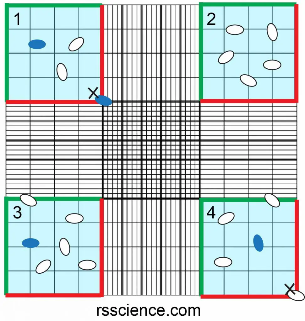
Let’s take an example of the picture below.
The counts of live cells (white cells) are (2, 5, 5, 3) in 4 light blue squares.
The average counts of live cells are (2+5+5+3)/4 = 3.75.
The counts of dead cells (blue cells) are (1, 0, 1, 1) in 4 light blue squares.
The average counts of dead cells is (1+0+1+1)/4 = 0.75
Therefore the cell viability is calculated as:
[live cells / (live cells + dead cells)]: 3.75 / (3.75+0.75) = 0.83, which is 83%.
You can also calculate the number of live cells and dead cells. In this case, the number of live cells is 3.75*2 (dilution factor)*10,000 = 75,000 cells/mL.
The number of dead cells is 0.75*2 (dilution factor)*10,000 = 15,000 cells/mL.
Hemocytometer applications
Estimate the cell size
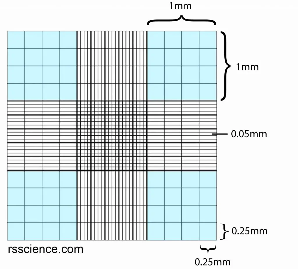
Because the grid has a specific dimension, some lines are 0.25 mm in between; some lines are 0.05 mm in between (see the image below). Therefore, you can easily estimate the size of the cells. If you want to determine the cell size more precisely, you can take a picture of the image under the microscope. Then, use softwares such as image J, where you can tell the software “a fixed length is equal to 1 mm”. It will convert and calculate the cell size for you.
Perform a blood cell count
The hemocytometer initially was designed for the blood count. Our blood consists of Red blood cells, White blood cells, and Platelets. The cell number and the composition of each cell type can tell a doctor your body health condition. For example, low in Red blood cell count causes feelings of fatigue and weakness (anemia). An increase in the number of White blood cells may indicate infection, allergy, inflammation or other diseases. Therefore, a hemocytometer is a very important tool in the medical test lab because of its ability to count the number of cells and the composition of each cell type.
Perform cell count for cell culture
When I grow cells in the lab, I need to change the cell medium every couple of day (depends on how fast the cells grow). After adherent cells grow to occupy 95% of the surface of a dish, it is time to “passage” cells to another dish. What we do is to detach cells, make a homogenous suspension, and count the number of cells. The number of cells is very important for cell culture. Cells may not grow very well or die if you have too few cells in a dish. Hence, the ability to count the cell number is a fundamental skill for cell culture.
Where to buy a hemocytometer
Amazon is a good place to start. You can find a variety of hemocytometers with different price ranges and qualities at Amazon. Rs’ Science also offers an “all in one kit” which comes with a hemocytometer, coverslip, Trypan Blue and Methylene Blue to evaluate the cell viability and sample vials, fine tip transfer pipettes, lens paper booklet. All the stuff you need to get started is in one kit.
Related posts
10 Everyday Things You Should Look at Under a Microscope
Lesson 7: Grow Your Own Yeasts & Use a Hemocytometer to “Count Cell Density”