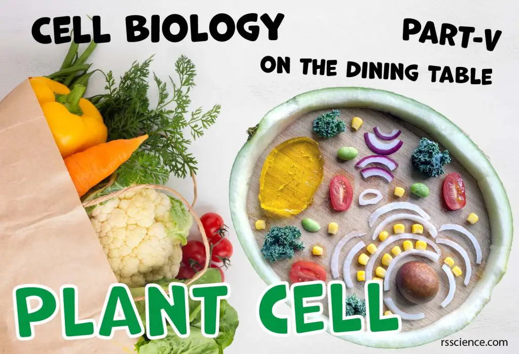
Cells are building blocks of life. Building a cell model should deepen your understanding of the cell, each organelle’s role, and its function. Some organelles are constantly present in the cell. Some organelles are temporary and only present when cells perform a particular process such as mitosis. If you want to learn more and see how we make an edible model of each organelle, check the following posts as well.
- Animal Cell Model Part I – cell membrane, cytosol, nucleus, and mitochondria.
- Animal Cell Model Part II – endoplasmic reticulum, ribosome, Golgi apparatus, peroxisome, and lysosomes.
- Animal Cell Model Part III – two types of temporary organelles involving in eating behaviors, autophagosomes, and endosomes.
- Animal Cell Model Part IV – two types of temporary organelles only appearing during mitosis, centrosomes, and chromosomes.
- Plant Cell Model Part V – cell wall, vacuole, and chloroplast.
- Cell Organelles and their Functions – overview of each organelle.
This article covers
The difference between plant cells and animal cells
Plant cells and animal cells have many in common. They both have essential organelles including a nucleus, mitochondria, cell membrane, ribosomes, endoplasmic reticulum (ER), Golgi apparatus, lysosomes, peroxisomes, and cytoskeleton.
In addition to these common features, plant cells have three unique cellular structures or organelles that animal cells do not have – Cell wall, Vacuole, and Chloroplast.
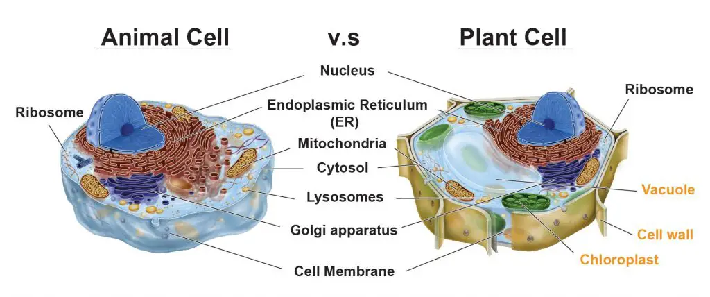
[In this figure] The cell anatomy of animal and plant cells.
The animal cell and plant cell share many organelles in common, such as a nucleus, ER, cytosol, lysosomes, Golgi apparatus, cell membrane, and ribosomes. The organelles that are unique for plant cells are Vacuole, Cell wall, and Chloroplast (shown in orange text).
In this article, I will briefly introduce these three organelles and explain their specific roles in plant cell biology. At the same time, I am going to build my plant cell model using food items that have scientific meanings related to each organelle.
The receipt to make an edible plant cell model
Before we start, I want to show you my shopping list for the ingredients to make a plant cell model.
A big watermelon, a box of cherry tomato, a can of corn kernels, a pack of Jell-O, a handful of kale, one avocado, one red, and one yellow onion.
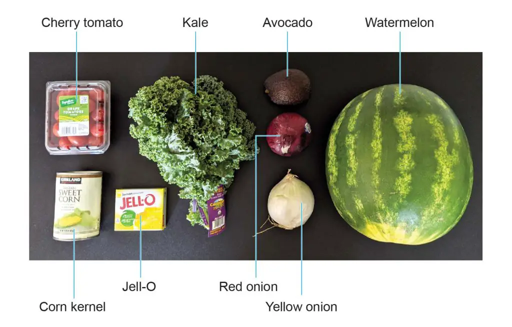
[in this figure] My recipes of an edible plant cell model.
Before I tell you the story, would you like to guess why I need these materials and what kinds of organelles they stand for?
Cell wall – where the name “Cell” came from
The cell wall is a unique structure that only exists in plant cells, but not in animal cells. The cell wall is made of cellulose and encloses the cell just outside the cell membrane. It provides the cell with both structural support and protection.
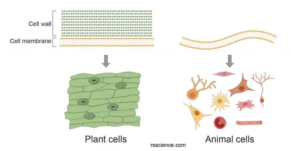
[in this figure] The difference of cell membrane between plant and animal cells.
The cell wall provides additional protective layers outside the cell membrane. Plant cells can maintain the cell shape and support the growth due to the strength of cell walls. On the other hand, animal cells only have a cell membrane. The flexibility of the cell membrane allows animal cells to move and change their cell shapes.
The composition of cell wall – cellulose
Cellulose is a polymer type of sugars. Cellulose can not dissolve in water and is resistant to many chemicals. When you eat vegetables, you can not even digest and break down their cellulose for energy. Cows and other herbivores have special bacteria in their stomachs to digest the cellulose into simple sugars.

[in this figure] The chemical structure of cellulose (a linear polymer of D-glucose units).
The hydrogen bonds (dashed) within and between cellulose molecules provide the strength of cellulose fibers.
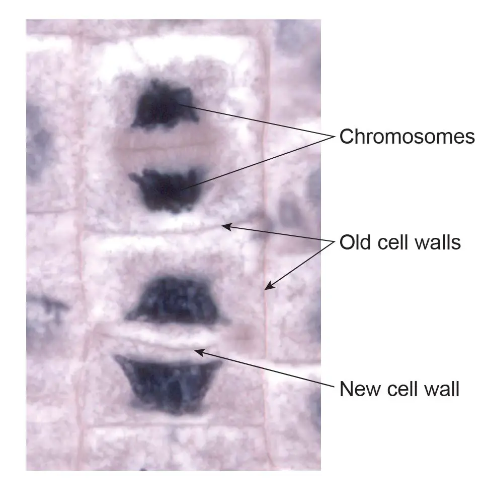
[in this figure] Microscopic image of dividing onion root cells.
The formation of new cell walls (also called phragmoplast) separates two new daughter cells at the late telophase.
Original image: Dr. phil.nat Thomas Geier, Fachgebiet Botanik der Forschungsanstalt Geisenheim.
The function of cell wall
Cell wall is tough and sometimes rigid. You can think of an animal cell like a water balloon, which is flexible to change its shape. In contrast, a plant cell is like a water balloon squeezed in a cardboard box. The cardboard box is so fit that the balloon and cardboard almost stick together. The balloon is protected from the outside world by a structure that provides protection and support.
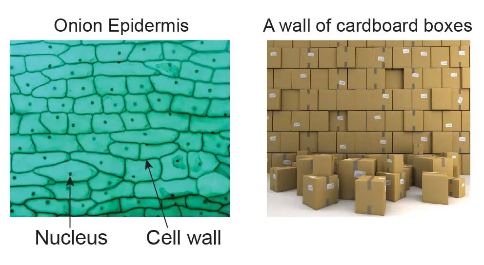
[in this figure] The illustration of the cell wall.
The cell wall acts like a cardboard box that protects the soft cell membrane and cytoplasm. Like real cardboard boxes can be piled up to build a tall wall, the plant grows by adding cells one by one as living building blocks. The weight is loaded primarily on the structural cell walls.
Cell wall also allows plants to grow to great heights. Pine trees can grow as tall as 250 ft (much taller than any animals on earth). However, the trees have no bone or skeleton system to hold them up. How do the plants support themselves? (hint: cell walls)
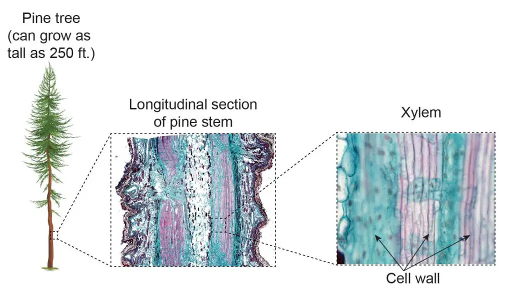
[in this figure] The longitudinal section of the pine stem.
If you look at the pine stem under a microscope, you can see the compact architecture made of many cells. Every single cell’s cell wall contributes toward the support of the entire weight of the pine tree. Dense cells in the core of the trunk can support a tree to grow very high.
Cell wall is persistent even after the plant cells die. Wood is made of the remaining cellulose fibers of cell walls after the death of matured xylem tissues of woody plants.
The discovery of “Cell”
In 1665, Robert C. Hooke (1635-1703) improved the early design of a microscope and increased the magnifications of the microscope to 50x. When Hooke viewed a thin cutting of cork, he discovered many empty spaces contained by walls, and termed them “Cells”. The term “Cells” stuck, and Hooke gained credit for discovering the building blocks of all life. These “Walls” are in fact the remaining cell walls of dead plant cells.
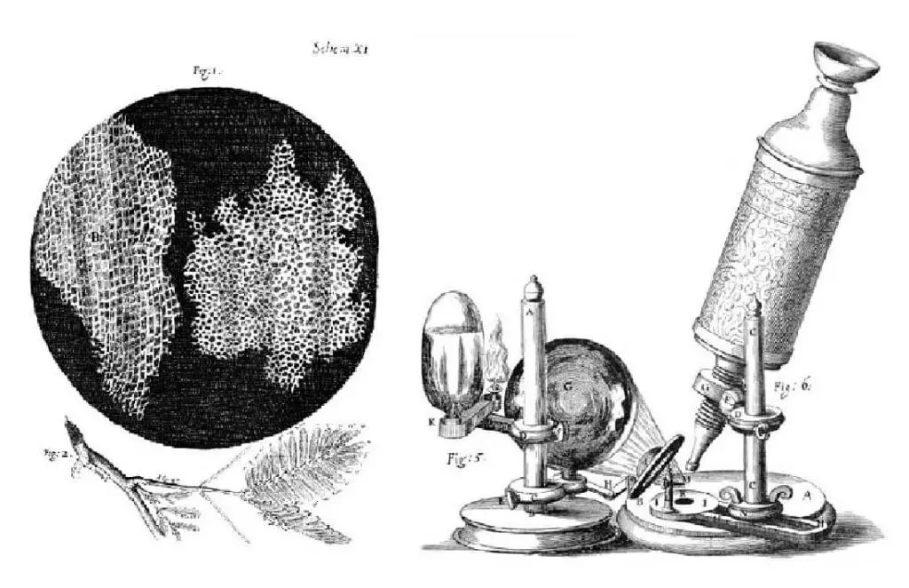
[in this figure] Left: In Hooke’s drawings, the cells look like empty chambers because he was looking at dead plant cells’ cell walls. Right: Hooke’s microscope, from an engraving in his 1665 book, Micrographia.
Photo source: Wiki
Cell walls in bacteria and fungi
Bacteria, fungi, and some protozoa also have a structure called the cell wall. They are not the same as the plant cell walls made of cellulose. These cell walls all serve the same purpose of protecting and maintaining structure, but they are very different components. In bacteria, the cell wall is composed of peptidoglycan (a polymer of sugars and amino acids). Fungi possess cell walls made of chitin (also a structural carbohydrate). Interestingly, diatoms (a group of microalgae) have cell walls composed of biogenic silica.
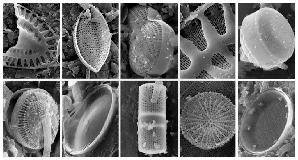
[in this figure] Silica fossil diatom frustules from Aral Sea and Baikal Lake.
The reason why using watermelon
In our previous animal model, we used banana peels to represent the softness of the cell membrane. In contrast, I would like to use watermelon rinds to highlight the stiffness of the cell wall.
Watermelon may be one of the most appropriately named fruits. It is a melon that contains 92% of water! The most popular part of the watermelon is the pink flesh. The rind, which is the green skin that keeps all that water-logged delicious fruit safe, usually ends up in the compost bin.
Today, I would like to recycle the rinds of watermelon to make my plant cell model. Here is how I do so:
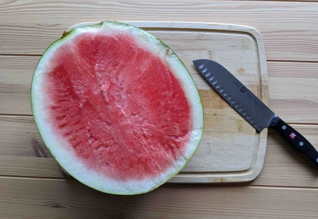
[in this figure] Cut a watermelon in half.
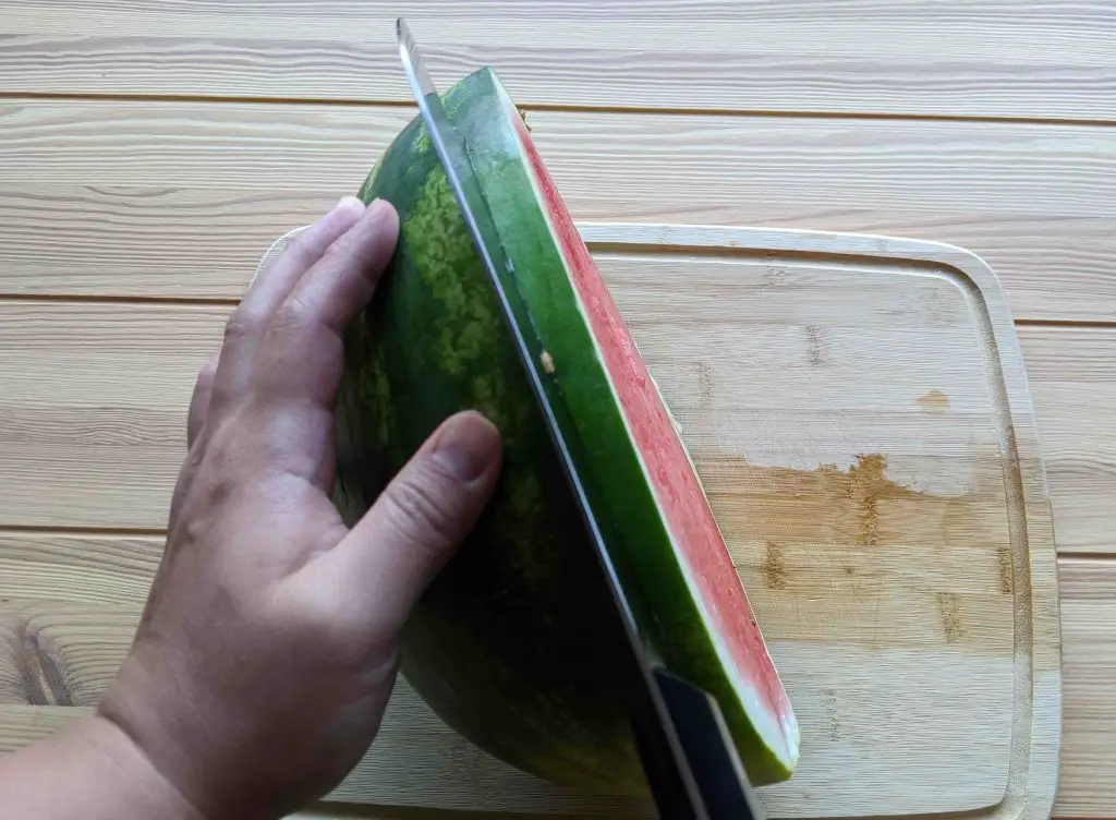
[in this figure] Carefully cut a slice of watermelon.
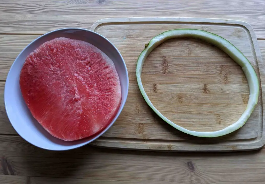
[in this figure] Cut the flesh out and leave an intact ring of rind.
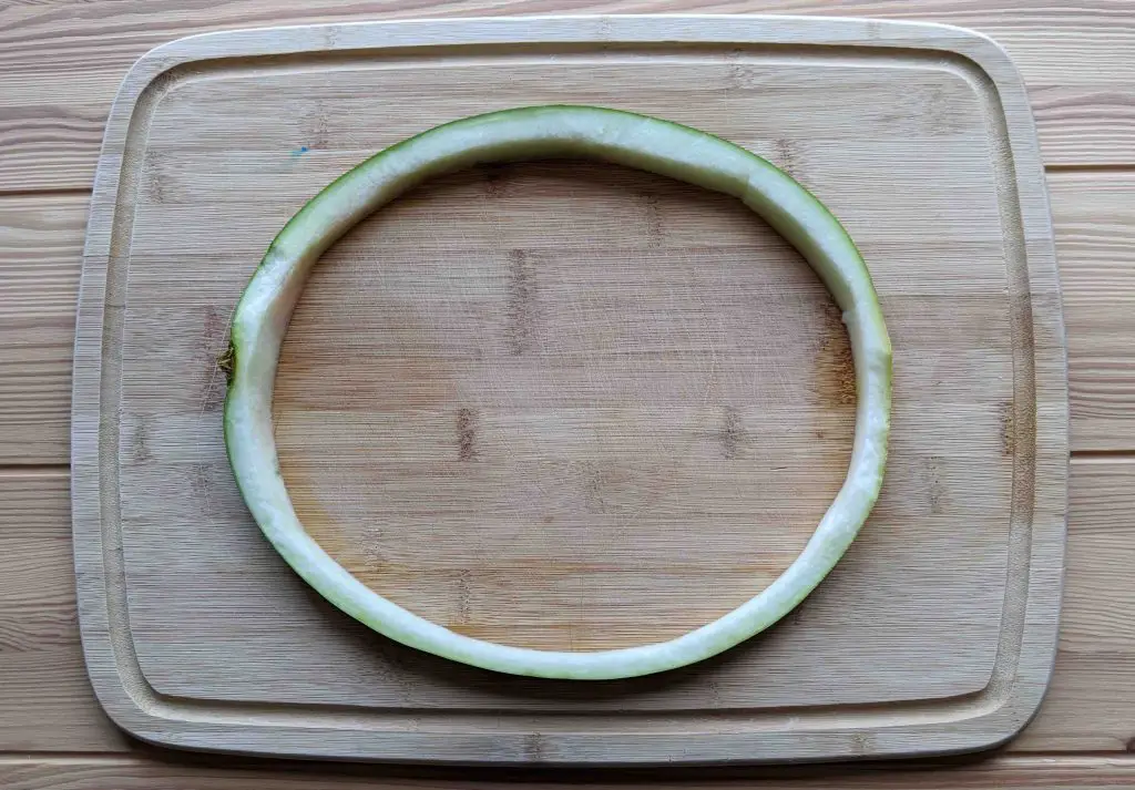
[in this figure] Use it as my cell wall.
PS. Many plant cells are closer to cubes/cuboids than spheres/ellipsoids. It is because the structural role of cell wall. In this regard, maybe we should use “Cube Watermelon” instead!🤣

[in this figure] Cube watermelons can be found in supermarkets in Japan. They grow in cubic molds to become cube shape.
Vacuole – Extra space storage
A vacuole is a membrane-bound organelle (like a bubble) that is present in all plant cells. Some animal and fungal cells also have vacuoles but they are much smaller.
Plant cell’s vacuole does not have a defined shape or size; its structure varies according to the need of the cell. Most mature plant cells have one large central vacuole that typically occupies more than 30% of the cell’s volume, and that can occupy as much as 80% of the volume for certain cell types and conditions.
The function of the vacuole is storage, defending pathogen, separating toxic waste from the rest cells, providing turgor pressure to support plant upright, and control of the opening and closure of stomata.
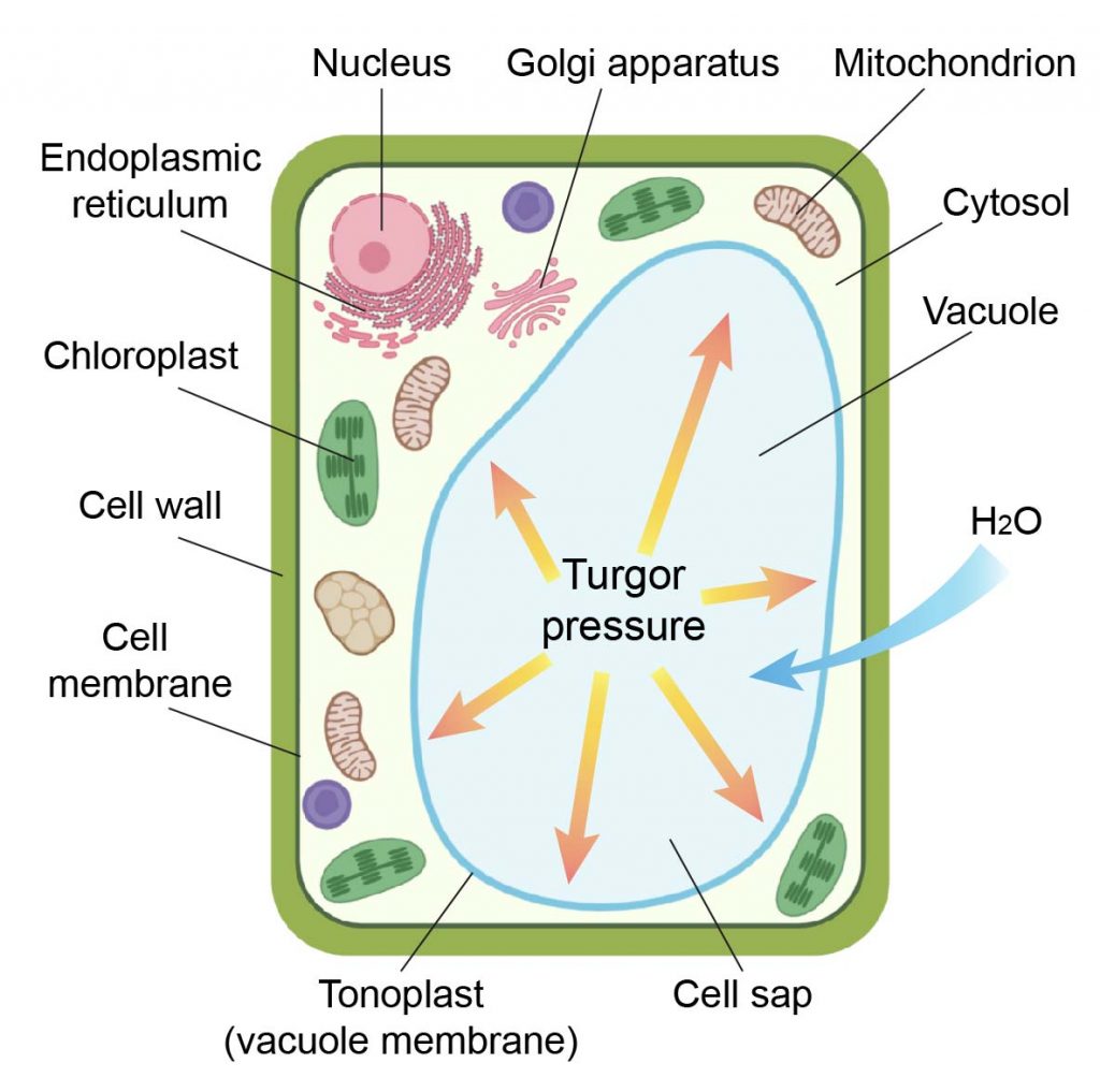
[in this figure] The anatomy of a plant cell.
Drawing of a plant cell showing a large vacuole.
The reason why using Jell-O
There is no other food item better than Jell-O to represent the water-filled, soft, jelly vacuole in my plant cell model.
Jell-O is a gelatin dessert. After mixing the gelatin powder with warm water, you allow it to cool down in a container with the shape you like. It will turn into a semi-solid, pudding-like gel.
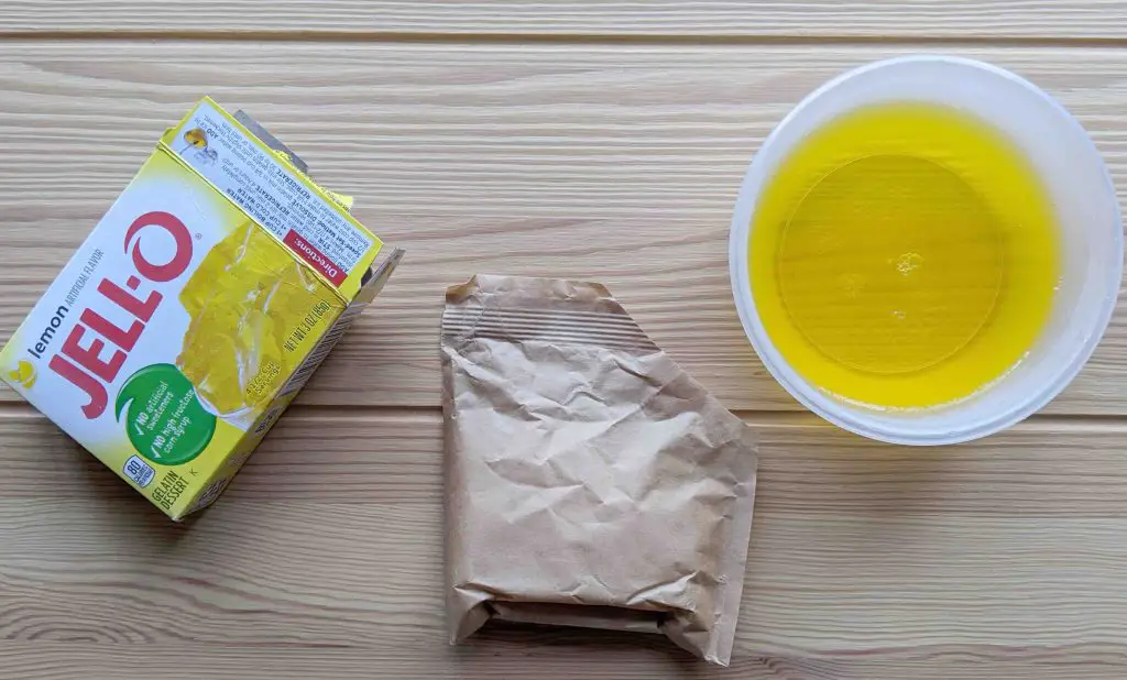
[in this figure] Mix Jell-O powder with warm water and pour it into a container.
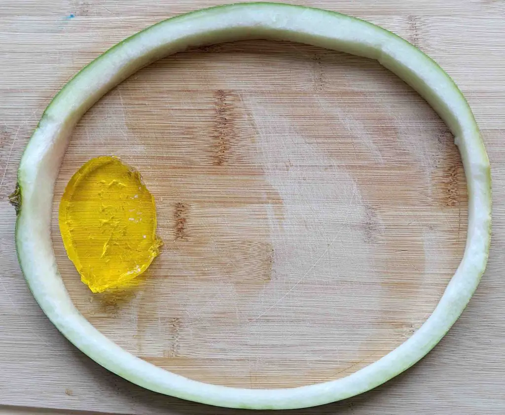
[in this figure] After gelation, scoop a piece of jelly as the vacuole in your plant cell model.
Chloroplasts – Plant cell’s solar energy panels
Chloroplasts are organelles that conduct photosynthesis and produce energy for the plant cells. Chloroplasts are only found in plant cells and some protists such as algae. Animal cells do not have chloroplasts. However, there is always an exception in biology. Check out “solar-powered sea slugs“.
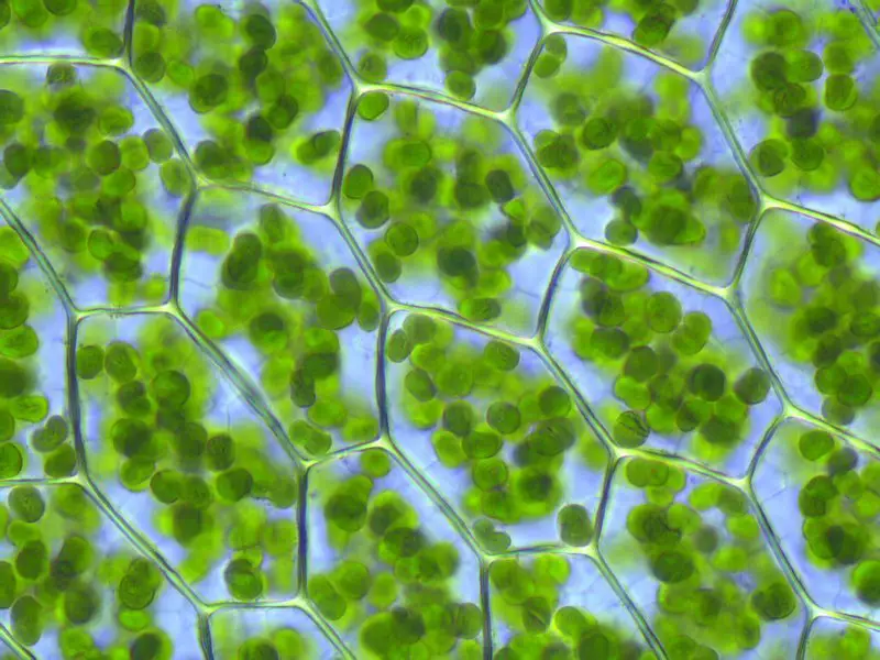
[in this figure] Chloroplasts are visible in the cells of Plagiomnium affine (a species of moss). Image credit: Kristian Peters.
Chloroplasts convert light energy of the Sun into sugars (a process called “photosynthesis”) that can be used by cells. At the same time, the reaction produces oxygen (O2) and consumes carbon dioxide (CO2). For this reason, plants are the basis of all life on Earth. They are classified as the producers of the world.
Chloroplast are motile. They can move depending on the light condition.
[in this Video] Chloroplasts circulating in Elodea (an aquatic plant) cells by cytoplasmic streaming.
The structure of chloroplast
The chloroplast stroma, enclosed by an inner membrane, is a protein-rich fluid, containing chloroplast DNA, ribosomes, starch granules, and many proteins. It is similar to the cytosol of the original cyanobacterium.
Suspended within the chloroplast stroma is the thylakoid system. Thylakoids are membranous sacks containing chlorophyll molecules and are where the light reactions of photosynthesis happen. In most vascular plants, the thylakoids are arranged in stacks called grana (singular: granum). But in certain C4 plants (like rice) and some algae, the thylakoids are free-floating.
The stacks of thylakoid sacs are connected by stroma lamellae. The lamellae act like the skeleton of the chloroplast, keeping all of the sacs a safe distance from each other and maximizing the efficiency of the organelle. If all of the thylakoids were overlapping and bunched together, there would not be an efficient way to capture the Sun’s energy.
Check this post to find out more interesting facts about chloroplast.
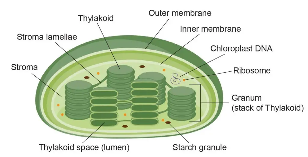
[in this figure] The structure of chloroplast.
The reason why using kale
No matter you like it or not, kale is a healthy food that contains fiber, antioxidants, calcium, vitamins C and K, iron, and a wide range of other nutrients. This green, leafy, cruciferous vegetable perfectly match the image of chloroplasts.
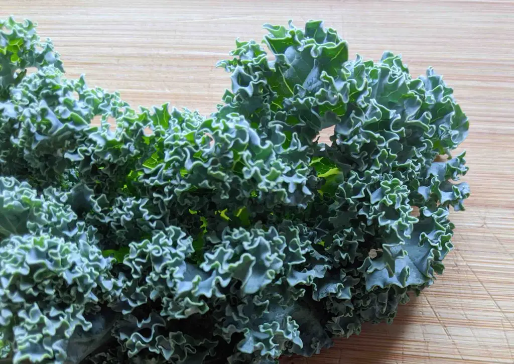
[in this figure] I like to use the fold leaves of kale to represent the thylakoid system in chloroplasts.
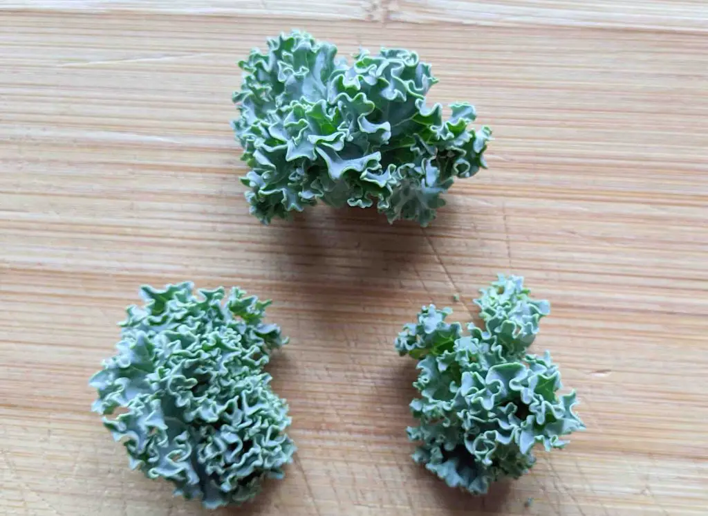
[in this figure] Prepare several small pieces of kale.
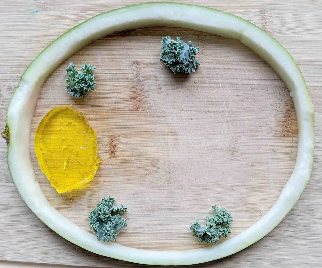
[in this figure] Decorate your plant cell model with these “chloroplasts”.
The finished “Plant Cell Model”
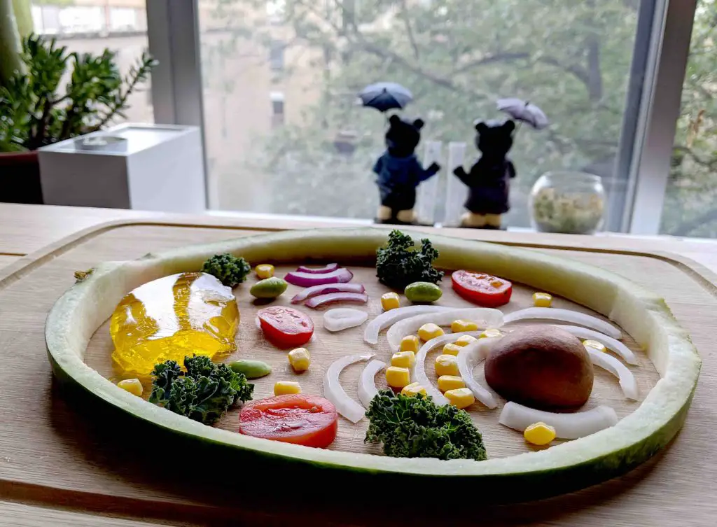
Don’t forget other organelles. Add all of them to give a final touch to your one-of-a-kind “Plant Cell Model”!
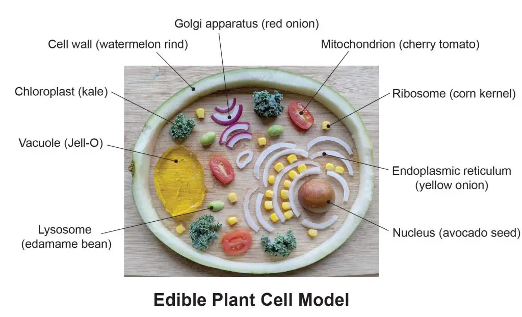
[in this figure] The nutrient content of my edible plant cell model.
Related posts
Cell Organelles and their Functions – overview of each organelle.
What is Cytoplasmic Streaming?

