
Cells are building blocks of life. Building a cell model should deepen your understanding of the cell, each organelle’s role, and how they perform their job. Some organelles are constantly present in the cell. Some organelles are temporary and only present when cells perform a particular process such as mitosis. Therefore, we have 4 blog posts series to cover the model of animal cells undergoing different processes.
- Animal Cell Model Part I – cell membrane, cytosol, nucleus, and mitochondria.
- Animal Cell Model Part II – endoplasmic reticulum, ribosome, Golgi apparatus, peroxisome, and lysosomes.
- Animal Cell Model Part III – two types of temporary organelles involving in eating behaviors, autophagosomes, and endosomes.
- Animal Cell Model Part IV – two types of temporary organelles only appearing during mitosis, centrosomes, and chromosomes.
- Plant Cell Model Part V – cell wall, vacuole, and chloroplast.
- Cell Organelles and their Functions – overview of each organelle.
In this Cell Biology on the Dining Table series Part 3, we are going to learn two types of temporary organelles involving in cells’ “eating” behavior. They are autophagosomes and endosomes!
This article covers
Autophagosome – the garbage truck
In the previous article, we left an open question, “How does the cell decide what should be recycled in the lysosomes?” Here, we are going to discover the answer by learning what autophagosome is.
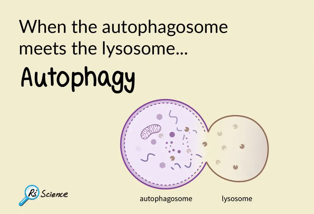
Autophagosome is a temporary organelle and only exists in the cell when the autophagy happens.
“Autophagy” (aka “self-eating”) is a biological process when cells are short of nutrient supply. When the cells sense that the environment becomes more challenging or the nutrient source is low, the cells turn on autophagy. In order to obtain the nutrient, the cells have to recycle some of their existed proteins and organelles, especially the misfunctioned ones.
The way our cells label the misfunctioned proteins is by putting “garbage tags” on bad proteins as an indication that “this guy should be recycled.” This “garbage tags” in cells is a small protein, called Ubiquitin. A group of enzymes performs the action of ubiquitination by adding Ubiquitin tags onto the bad proteins.
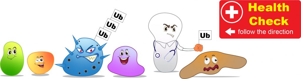
[In this figure] Cells screen for misfunctioned proteins constantly.
Once the bad proteins are founded, they will be labeled with a Ubiquitin (Ub) tag for degradation.
How does the autophagosome form?
Cells go through their inventory at the beginning of the autophagy, sort out the bad proteins, and put the “garbage tags” on them. Then, these enzymes in charge of autophagy will see these “garbage tags” and build the “garbage bags” to contain the cellular garbage. These “garbage bags” are the autophagosomes which consist of special lipid-formed vesicles.
The autophagosomes carry the cellular garbage to lysosomes. The lysosomal enzymes break down the cellular garbage into raw materials after the fusion of autophagosomes and lysosomes. After that, cells can obtain nutrients or have ingredients to build new proteins and organelles.
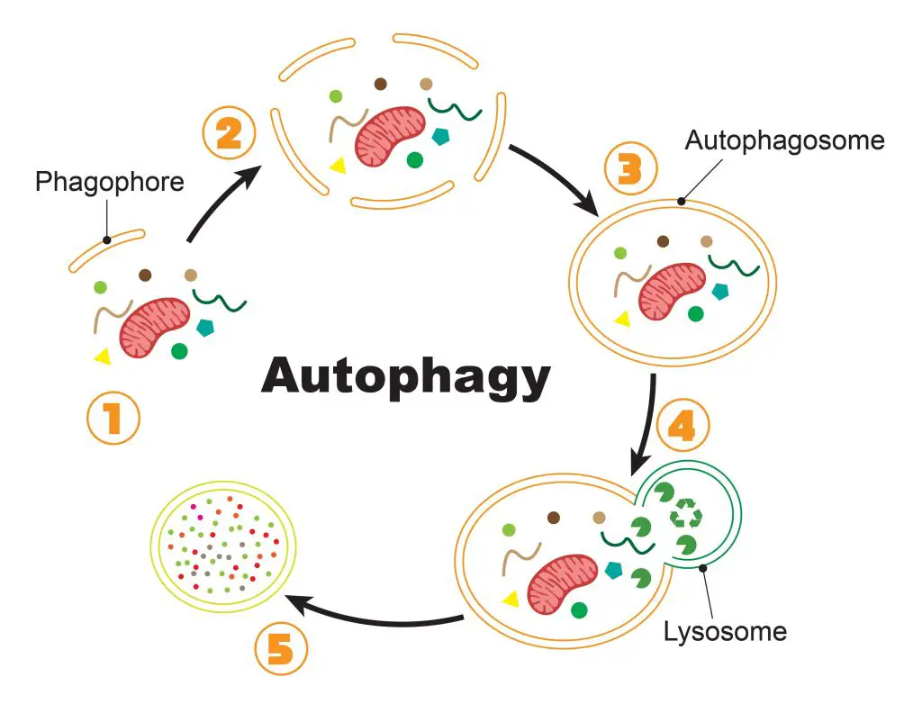
[In this figure] The process of autophagy.
(1) Once the cell initiates the autophagy, special lipid components (called phagophore) recruit close to the bad proteins/organelles tagged for recycling. (2) More and more phagophores gather around the cellular garbage. (3) Phagophores assemble into a complete vesicle, called the autophagosome. (4) Fusion with the lysosome brings in the digestive enzymes to break down the contents. (5) Bad proteins/organelles are recycled as raw materials for the cells to use.
Mitophagy
Sometimes, even entire organelles can be packed into autophagosomes for recycling. For example, the special autophagy to degrade bad mitochondria is named “mitophagy.”
In 2016, the Nobel Prize in Physiology or Medicine was award to a Japanese cell biologist, Dr. Yoshinori Ohsumi. His discoveries opened the path to understanding the fundamental importance of autophagy in our cells. Now, we already know that many diseases, including cancer and Parkinson’s disease, could be a consequence of misfunctioned autophagy.
Food model of the autophagy
In my cell mode, there is a mitochondrion (a slice of a cherry tomato) that has worked for a long time, and many parts of the electron transfer chain become dysfunctional. As a result, this mitochondrion can’t generate ATP efficiently anymore. It is time to retire and then reborn.
Here, I use small pieces of spaghetti to represent the recruited lipid components of the autophagosome.
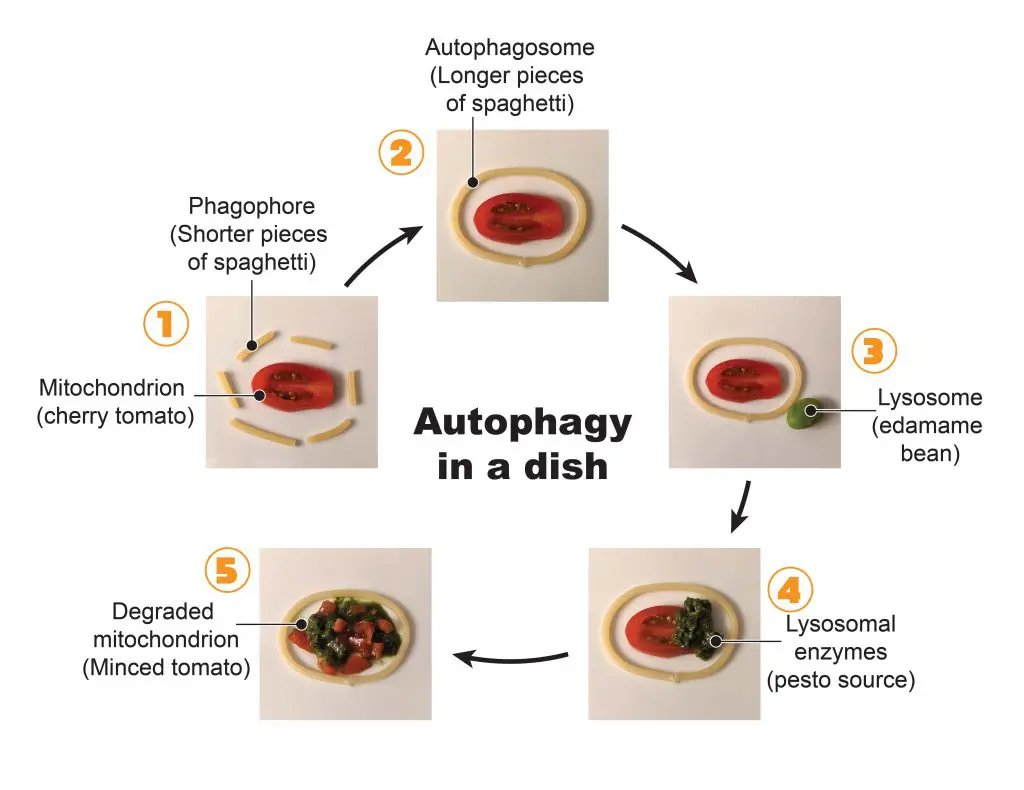
[In this figure] The process of autophagy in a dish.
(1) More spaghetti pieces are gathered around the old mitochondrion (cherry tomato) until the complete vesicle of autophagosome is formed (2). (3) Then, this autophagosome brings the mitochondrion closer to a lysosome (an edamame bean). (4) The fusion between the autophagosome and lysosome bring in the lysosomal enzymes (pesto source). (5) After a while, the mitochondrion is broken down into small pieces (minced tomato) and is further break down into nutrients for cells to use.
Endosome – takes a bite
The endosome is another membrane-bound temporary organelle inside our cell. Endosomes only form when the cell wants to engulf the stuff outside of the cell. Endosomes are formed by the invagination of the cell membrane. The molecules near or on the cell membrane’s surface could be internalized by the compartment encapsulating in the endosomes. This process is called “Endocytosis” (the opposite of “Exocytosis”).
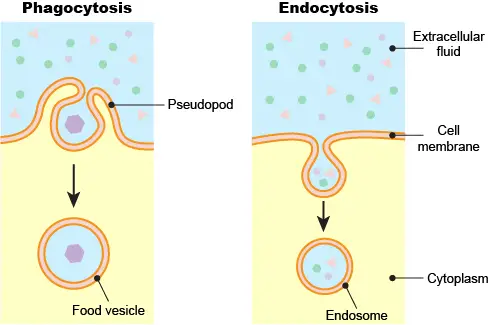
[In this figure] Phagocytosis vs. Endocytosis.
Phagocytosis is common in single-celled eukaryotic cells to engulf larger food particles. Phagocytosis requires the cell extending its pseudopods to surround the particle and bring inside the cell as a food vesicle. Endocytosis does not involve the dramatic change of cell shape like phagocytosis. Instead, endocytosis initiates a local depression of the cell membrane to create an endosome.
Endocytosis is believed to be evolved from the process of “Phagocytosis” of single-celled eukaryotic cells. A good example is the Amoeba proteus. The amoeba cell changes its cell shape by extending its pseudopods. As the amoeba moves towards its prey, its pseudopods reach out, surround, and engulf the food inside its cell membrane by forming a food vacuole. Then the digestive enzymes are released into the vacuole to break down the food to small nutrient molecules for Amoeba proteus to use.
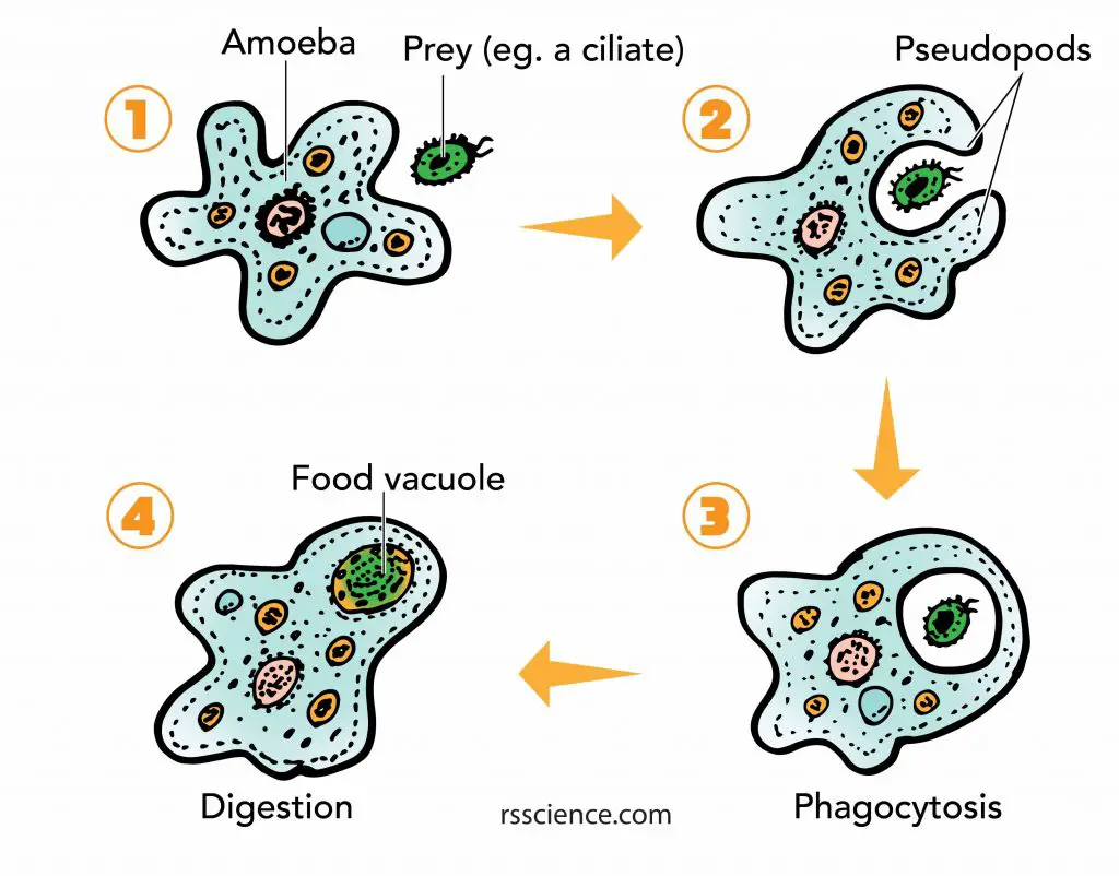
[In this figure] Amoeba phagocytosis.
The pseudopods first surround and bring the food particle close to the Amoeba. Then a part of the cell membrane opens to allow the particle to move into the cell and into a food vacuole where it is digested by enzymes.
How does the endosome form?
Our white blood cells (especially, the neutrophils and macrophages) still keep the ability to engulf bacteria as parts of our immune system. However, most other cells cannot dramatically change their cell shapes like amoebas and white blood cells. These cells retain the ability to engulf a smaller bit of external molecules by “Endocytosis”.
In many cases, endosomes are employed to recycle protein receptors and lipid molecules from the outer surface of the cell membrane. The newly formed endosomes are called early endosomes. These endosomes mature step-by-step into late endosomes. Late endosomes then merge with lysosomes to break down the recycled contents.
[In this Video] Bacterial phagocytosis by neutrophils.
Food model of endocytosis
Endosomes form by the depressing from the cell membrane; therefore, I would like to use the banana peels to demonstrate the process of Endocytosis.
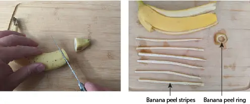
[In this figure] Banana peels represent membrane.
Cut the tip part of banana into small pieces so we can have both banana peels in rings and stripes.
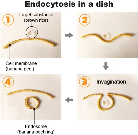
[In this figure] Food model of endocytosis.
In my endocytosis model, the cell wants to engulf some substances (brown rice) near the surface of the cell membrane (banana peel strip). The cell membrane folds in to form a cavity filled with extracellular fluid and target substance, a process known as invagination. Then, the cell membrane folds back to itself until it forms a uniformly enclosed membrane around the trapped substance. This enclosed membrane or vesicle is known as an endosome (banana peel ring).
Summary
Autophagosomes
The function of the autophagosome is to break down the misfunctioned organelles and therefore to recycle the nutrient for cells.
Endosomes
The function of the endosome is to uptake the material outside of the cell membrane and bring them inside cells.
Related posts
Animal Cell Model Part I – cell membrane, cytosol, nucleus, and mitochondria.
Plant Cell Model Part V – cell wall, vacuole, and chloroplast.
Cell Organelles and their Functions – overview of each organelle.
Pingback: How does a cell rid itself of defective or malfunctioning organelles? – Dr. Biology Questions and Answers