
The microscope is a tool to see the tiny world, but we need to prepare the “object”, mounted on the slide for a microscope to see.
In this article, we discuss several types of microscope slides and different types of mounting techniques.
This article covers
Type of slide
Common flat glass slides
The standard slides are typically made of glass, and the dimension is 1 x 3 inches (25.4 x 76.2 mm).
The coverslips are 7/8 x 7/8 inches (22 x 22 mm) in size, 0.006 inches (0.15 mm) in thickness. The coverslip is a very thin square piece of glass.
Without the coverslip in place, surface tension would cause the droplet to bunch up in a dome. The coverslip breaks this tension, flattening the sample, and allowing very close inspection with minimal focusing. The coverslip also protects the objective lens from contacting the sample drop.

[In this figure] The common flat slide
Depression or concave slides
Concave slides contain a small well, or indentation, ideal to hold a drop of water or liquid substance. They are more expensive slides.
There are also chambers or cell flasks, which can be viewed directly without preparing individual slides on the microscope stage. These are particularly useful for cultured cells.

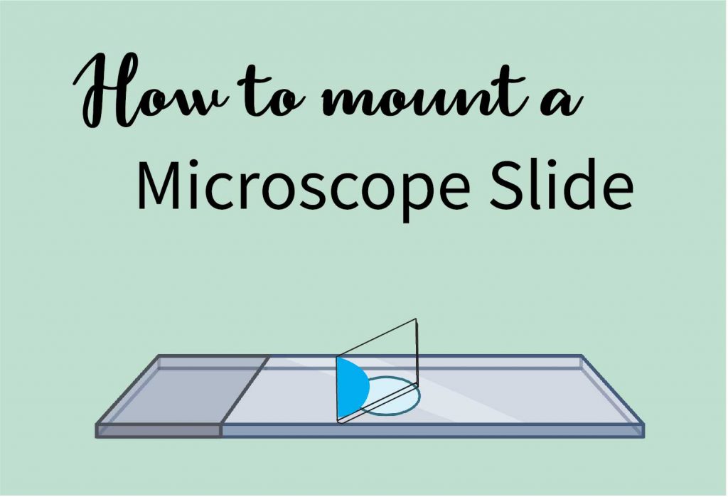
[In this figure] The concave slide
Microscope camera calibration slide
This type of slide contains a precise ruler, with 1 mm total length that is subdivided into 100 divisions, that is, each division is 0.01 mm. This precise stage micrometer is used to calibrate your microscope or microscope camera for precise measurement.
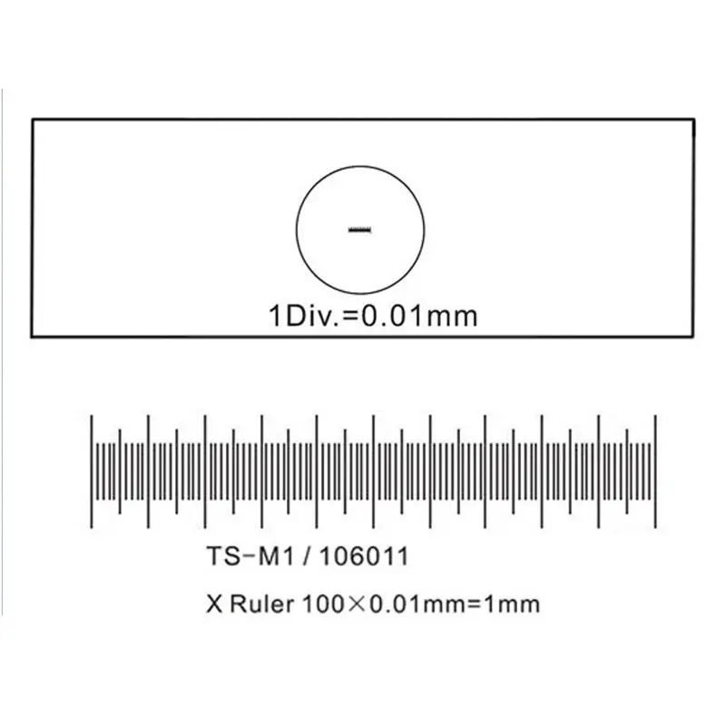
[In this figure] The microscope camera calibration slide
Type of mounts
Dry mount
The specimen is placed directly on the slide. A coverslip may be used to keep the specimen in place and to help protect the objective lens from touching the specimen directly.
Specimens suitable for dry mount
Pollen, hair, feathers, or plant materials.
Wet mount
In a wet mount, a drop of water is used to suspend the specimen between the slide and coverslip. The steps are:
- Place a sample on the slide
- Add a drop of staining solution such as Methylene Blue on the sample (optional)
- Place the edge of the coverslip over the sample and carefully lower the coverslip slowly with an angle. Allow one side of the liquid droplet touches the coverslip first. This permits air to escape from the other side. You can use the forceps or a toothpick to help you control the coverslip.
- Remove any excess solution by using a Kimwips paper to touch one side of the coverslip.
- You can also seal all the edges of the coverslip with super glue (or nail polish) for long-term storage.
- The slide is now ready for viewing.
You can see step-by-step photos in “Mount a Slide & “Look at Your Cheek Cells“.
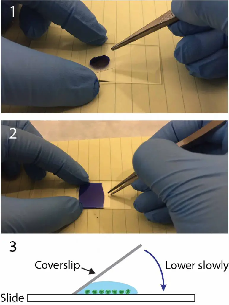
[In this figure] The wet mount technique.
Attached one side of the coverslip with an angle, lower it slowly.
Specimens suitable for wet mount
Microorganisms from the pond water, such as Protozoa, algae, paramecium, and amoeba, etc. Cells like animal and plant cells required water tension to make them extended and flat. In addition, body fluids such as saliva, blood, and urine are suitable for a wet mount.
Section Mount
Fo the section mount, a specimen needs to be cut to extremely thin pieces. Typically a microtome is needed to cut a thin cross-section of a plant stems and put it on the slide carefully. A stain can be applied directly to the specimen before covering it with a coverslip.
Specimens suitable for section mount
The plant stems, roots, and leaves. In addition, any solid that can be cut into thin pieces are good for section mount. It is very useful for a variety of specimens.
Smear
A smear is made by carefully smearing a thin layer of the specimen across a slide and should be allowed to air dry. Alternatively, a fixation like passing the slide through the flame of a Bunsen burner several times can be used, like Gram stains. Finally, a coverslip is placed.
Specimens suitable for smear
Bacteria culture, bacteria colonies and blood.
How to choose mounting media
Mounting medium (or mountant) is the medium where your sample is in while it is being imaged on the microscope. The simplest type of mounting medium is air if you don’t add anything. Liquid-based mounting media are frequently used to hold the specimens in place between the coverslip and the slide. Mounting media can also adjust the reflection index of specimens to enhance the quality of images.
Generally speaking, mounting media for whole mount slides can be categorized into water-based and organic solvent-based mounting media.
Water-based mounting media
Water-based mounting media, like pure water, phosphate-buffered saline (PBS), and 70% glycerol wet mount medium, are ideal for beginners and general usage. Water-based mounting media are easy and safe to use.
Organic solvent-based mounting media
Organic solvent-based mountant, like Canada balsam, is great for permanent-mounted slides. However, some specimens require additional dehydration and chemical treatments (with toxic solvents like toluene or xylene) before applying the mountant. Some specimens can even shrink or damaged in solvent-based mountants. For these reasons, we suggest starting with water-based mounting media with your microscopic projects.
How do professional grade specimens made?
1. The specimen is grossly cut into a suitable piece and placed in a cassette. The way you place the specimen will determine which “face” of the section you will be able to see under the microscope. Soft animal tissues may need additional processes by treating with chemicals (including formalin and alcohol) to preserve their cell structures. Plant tissues have cell walls so they may not need these steps.
2. The specimen is then embedded in a wax-like material called paraffin, and the specimen becomes a hard solid block.
3. A professional rotary microtome.
4. Carefully place the specimen block and the section blade (super sharp!)
5. By rotating the drive wheel, the blade will trim a thin section of the specimen block like the peeler in the kitchen. Each section can be as thin as 3 micrometers (1/30 the thickness of your hair!). Series of sections can be cut like a ribbon. The ribbon will then be moved to a water bath for flotation. The good sections will be selected and transferred to the glass slide.
6. Depending on the purpose of the specimen, the blank slides will be stained with specific stains, then washed, dried, and permanently mounted with coverslips. Now, the premade slides are ready.
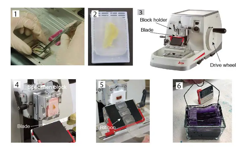
[In this figure] The steps to make professional grade specimens/slides.
Related posts
Lesson 2: Mount a Slide & “Look at Your Cheek Cells“
Lesson 4: How to Use a Microtome & “Amazing Cross-section of a Stem”