
This article covers
What is Amoeba? A quick overview
Last updated date Feb 6th, 2023
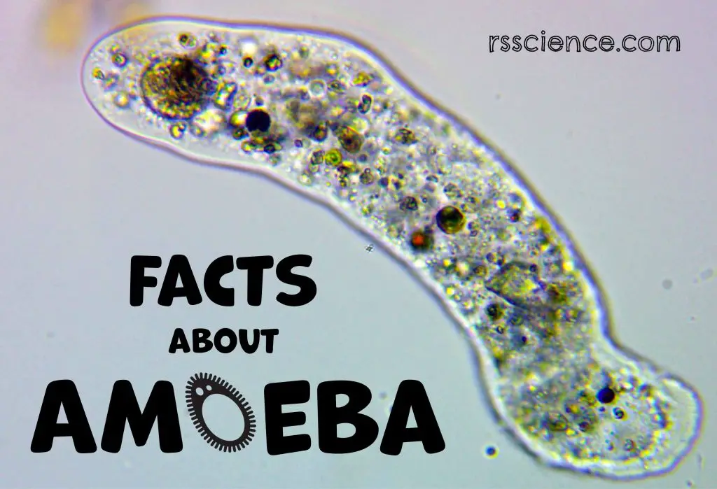
Amoeba (plural amoebas/amoebae) is a group of primitive protists. Among the big family of Amoebas, Amoeba proteus is probably the best-known member – common in classrooms and research laboratories.
Amoeba proteus is best known for the way they move, a primitive crawling manner – through extension and retraction of “false feet” (or pseudopods) over varied substrates. Amoeba proteus does not have a fixed shape – it constantly changes because it extends its pseudopods.
The ability to move by pseudopods is a common feature of the Amoeba family, although some of them look quite different from Amoeba proteus.
Classification of Amoeba – the Phylum Amoebozoa
Amoebas belong to the Kingdom of the protists (a protist is any eukaryotic organism that is not an animal, plant, or fungus). However, in terms of classification, the position of amoebas is just like their shape – consistently changing.
In the early days, when microscopy was the only way to characterize microorganisms, amoebas were classified as Phylum Sarcodina with several other species like Heliozoa. Once molecular phylogenetics (classifying a species by its genetic materials) was introduced, amoebas are now in Phylum Amoebozoa. We have, however, to keep in mind that the classification of the protists is presently much debated. Below is the current scientific classification.
Scientific classification
Kingdom: Protozoa
Phylum: Amoebozoa
Class: Tubulinea
Order: Euamoebida
Family: Amoebidae
Genus: Amoeba
Species: Amoeba proteus, Amoeba Pelomyxa, Amoeba Thecamoeba, Amoeba Vampyrella
(Reference: wiki)
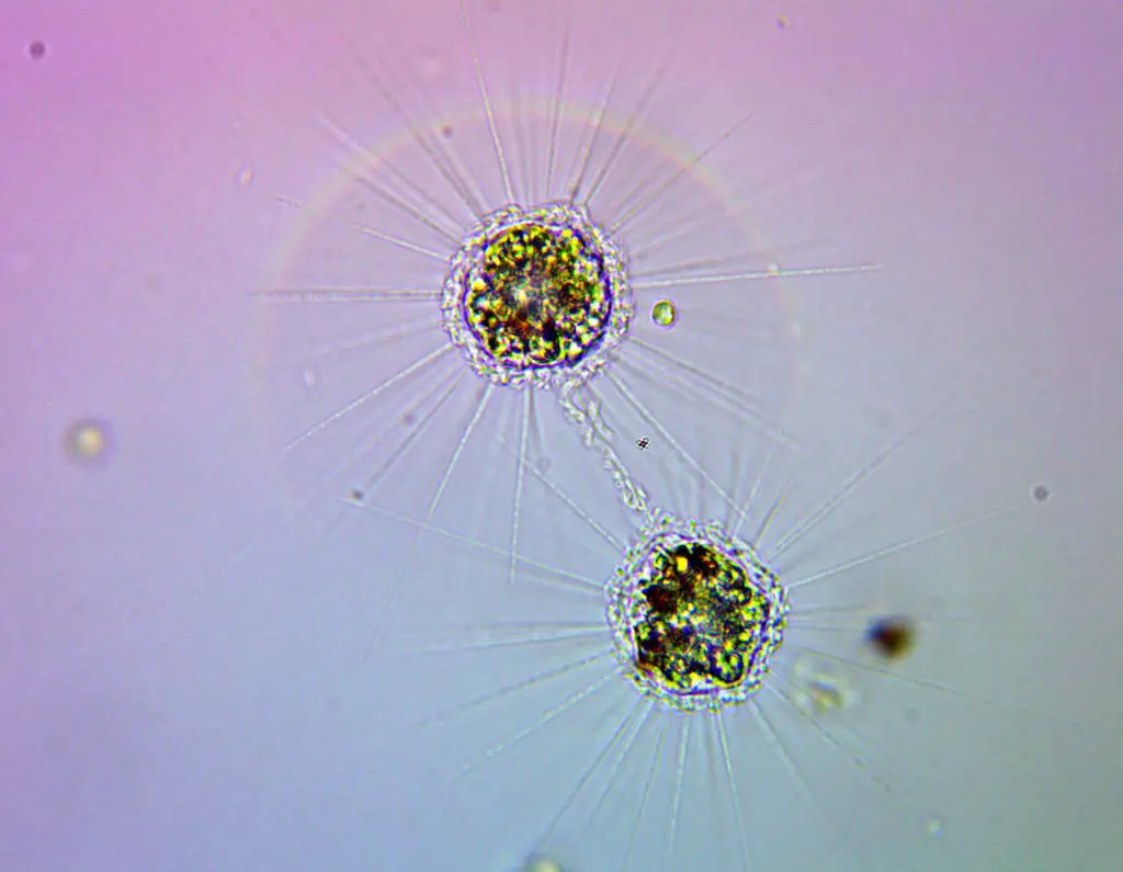
[In this image] Heliozoa is commonly known as sun-animalcules. I personally like to call it a microscopic “Uni” (sea urchin in Japanese)!
The Anatomy of Amoeba Cell
Amoeba is a single-celled organism, meaning one Amoeba consists of only one giant cell.
Amoeba belongs to eukaryotic cells, which means that their genetic material (or DNA) is well organized and enclosed within a membrane by forming a “nucleus”. In this aspect, Amoeba is closer to our human beings (also eukaryotes) than bacteria (prokaryotes).
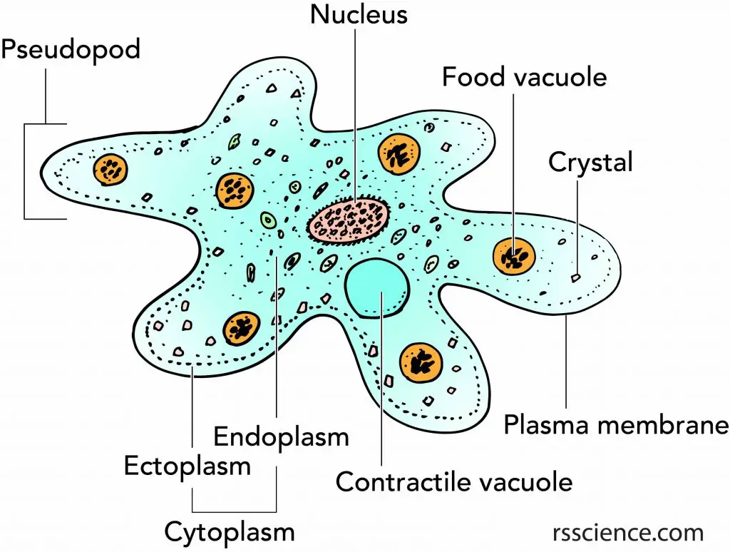
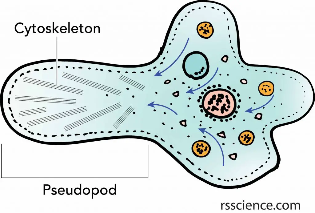
The microanatomy of Amoeba: an amoeba has a single granular nucleus containing most of the organism’s DNA.
Amoeba move and hunt by extending pseudopods.
A contractile vacuole is used to maintain osmotic equilibrium by excreting excess water from the cell.
Several food vacuoles are used to digest food particles.
The cytoplasm can be divided into two parts: a granular inner endoplasm and an outer layer of clear ectoplasm, both enclosed within a flexible plasma membrane.
Crystals are condensed wastes produced by the cell.
Nucleus – extremely large genome
Amoeba proteus is a eukaryote, meaning that its genetic materials (DNA) are enclosed in the nucleus. Scientists call the entire DNA codes in a particular organism its “genome”. Guess how big the Amoeba proteus’ genome is? Amoeba proteus has 290 billion base pairs (one base pair is equal to one DNA code), making it 100 times larger than the human genome (3 billion)!!!
One of the largest genomes belongs to a very small creature, Amoeba dubia, a cousin of Amoeba proteus, which has 670 billion base pairs! However, a large genome size does not correlate with the number of genes.
Amoeba proteus has such a large genome due to an extreme replication of the same set of genes (a classic example of polyploidy). It can have more than 500 chromosomes in a single nucleus. Human beings are diploid, and we have only two copies of the same genes (or chromosomes).
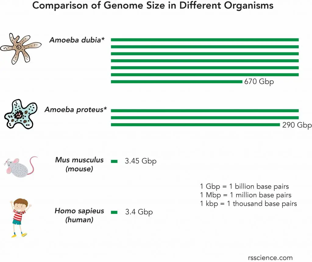
[In this figure] A comparison of genome sizes of different organisms.
Early thinking held that the genome size should be directly related to the complexity of organisms. However, this is not true. Some simpler organisms can have even larger genome sizes than the species at the higher levels of the evolution tree.
For example, Amoeba proteus and Amoeba dubia have a much bigger genome size than humans.
Note: the genome size of Amoeba dubia (also called Polychaos dubium) and Amoeba proteus were measured by the 1960s methods that analyzed the whole cell rather than single nuclei. The result could be muddled by including contributions from mitochondrial DNA, possible multiple nuclei, and anything the amoeba recently engulfed.
Pseudopods or Amoebas’ false feet
A pseudopod is a temporary arm-like projection that is developed in the direction of movement.
When the Amoeba stretches its pseudopods, the cytoskeletons (like the cells’ skeleton system) inside the cell rearrange and extrude the cell membrane to change the cell shape. Once the tips of pseudopods adhere to the substrate, the cytoplasm of the cell flow to fill the space, so the whole cell moves forward.
Under the microscope, you can see the components (including the nucleus and vacuoles) inside the Amoeba flow smoothly, like in a gel, as it moves. This form of movement by extension of cytoplasm is called “amoeboid movement”.
[In this figure] Amoeboid movement: an amoeba moves by stretching its pseudopods.
Under the plasma membrane of the pseudopods, there are organized cytoskeletons that generate the force to drive the change of the cell’s shape.
In addition to using pseudopods to move around, Amoebae also use them to engulf food particles.
Pseudopods are not exclusive to Amoeba. In fact, most eukaryotic cells can change their shapes by moving their cytoskeletons. For example, the white blood cells in our immune system can patrol and prey on invaded bacteria by pseudopods. (See video below).
Contractile vacuole – unique organelle
The third feature of Amoeba proteus is its built-in pumping system inside the cell, called “contractile vacuole”. The contractile vacuole is a water bubble within the cytoplasm of Amoeba proteus. Its function is to regulate the water content of the cell. Since Amoeba proteus is a unicellular organism, the water molecules can freely flow in or out through the semi-permeable cell membrane via osmosis.
When Amoeba proteus moves to a place with fewer ions (minerals), the environment becomes hypotonic to the cell. This means more water molecules will move into the Amoeba proteus cell to achieve a balance. When this happens, the contractile vacuoles can store extra water and help in throwing them (together with the wastes) out of the cell.
Without the contractile vacuoles, the amoeba may burst. Undoubtedly, it is a very important organelle with an essential function to the amoeba, as well as many freshwater microorganisms.
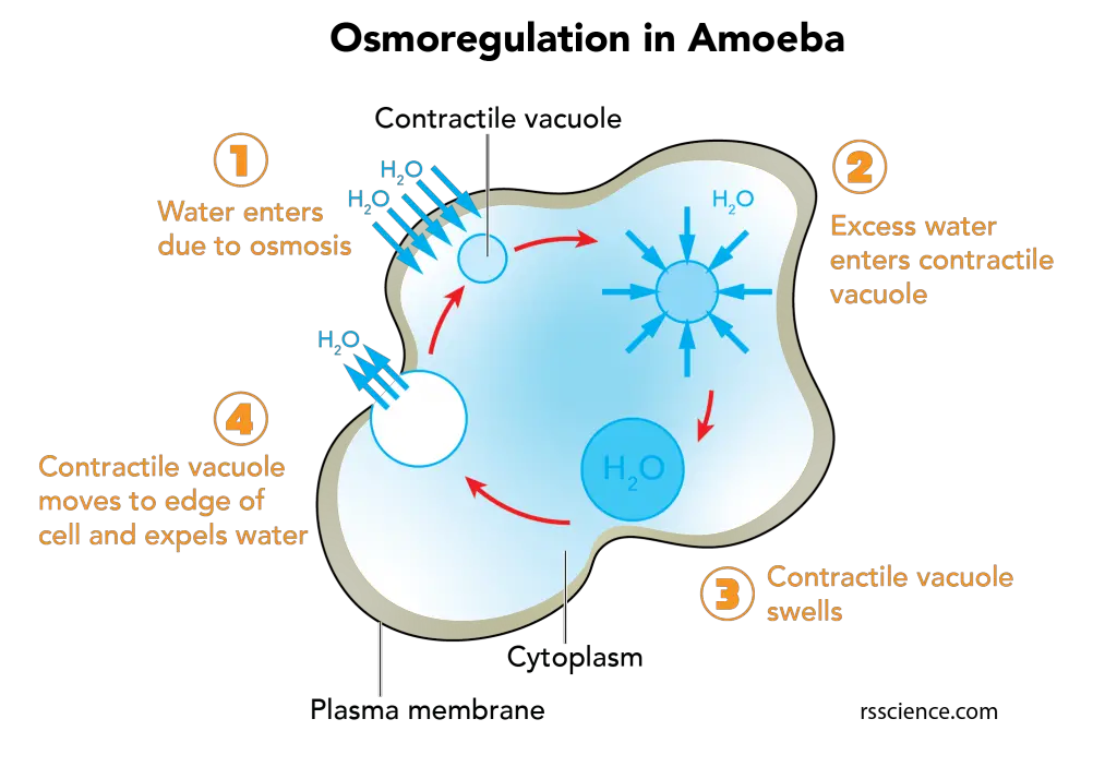
[In this figure] Osmoreulation in Amoeba.
The contractile vacuole is the key regulator of osmotic pressure in the amoeba (also in many single-celled protists). The contractile vacuole serves as a reservoir to store excess water inside the cells. Once the water reaches its limit, the contractile vacuole moves and fuses with the plasma membrane to expel water.
Many microvilli on its cell membrane
The fourth secret of Amoeba proteus is its cell membrane is not as smooth as it shows under the optical microscope. In fact, the outside face of the membrane has many microvilli attached to it (only be seen under an electron microscope). These microvilli can help Amoeba proteus attach and release from the surface of the substrate.
Crystals – shiny particles inside Amoeba proteus
Another feature that you can easily observe is the abundance of crystal-liked inclusions inside Amoeba proteus. Most crystals of Amoeba proteus are in a bi-pyramidal shape. These crystals are contained in vacuoles and composed of triuret, a nitrogen waste product. Other species of Amoebas have their crystals in different shapes, like spheres, sheets, and even croissant-shaped crystals.
Here are some examples of crystals in different species of Amoebas.
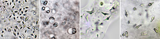
[In this figure] Crystals in different species of Amoebas.
Source: https://www.arcella.nl/inclusions/
Some large Amoebas also have glycogen bodies to store their nutrient reserve. These glycogen bodies are glossy spheroids and vary in size. Glycogen is a form of sugar stored in our body, and we store glycogen in the liver and muscle.
When the amoeba digests large amounts of diatoms, you can even see the oil droplets inside the Amoeba cell. This is because some diatoms are tiny oil producers!
Some large amoebas contain bacteria and small green algae inside their cytoplasm. These organisms have a symbiotic relationship with their host and are called “endosymbionts”. For example, green algae that live inside can provide additional energy to their host (the amoeba), making the amoeba can live in nutrient-poor environments.
How does Amoeba proteus eat?
Amoeba proteus engulfs its food through a process called “phagocytosis”. As the amoeba moves towards its food, its pseudopods reach out, surround, and engulf the food inside the cell membrane by forming a food vacuole. Digestive enzymes are then released into the vacuole to break down the food into usable nutrients.
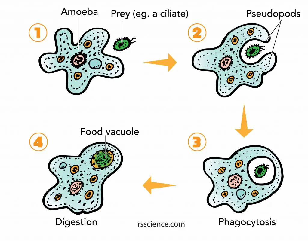
[In this figure] Amoeba phagocytosis.
The pseudopods first surround and bring the food particle close to the Amoeba. Then a part of the cell membrane opens to allow the particle to move into the cell and into a food vacuole, where it is digested by enzymes.
What is Amoeba proteus’ favorite food?
Amoeba proteus is a predator of bacteria, protozoa, and algae. It can eat almost any organic nutrients in its habitat. Paramecium is probably the most famous prey for Amoeba proteus. Check out the video of Amoeba hunting paramecia!
How big is the Amoeba proteus ?
Amoeba proteus is a large protozoan, and it can grow up to 1 mm long (average size 250-750 µm). The size ranges based on the amount of food it engulfs. It can almost be seen with the naked eye (still very difficult due to its colorless and transparent body).
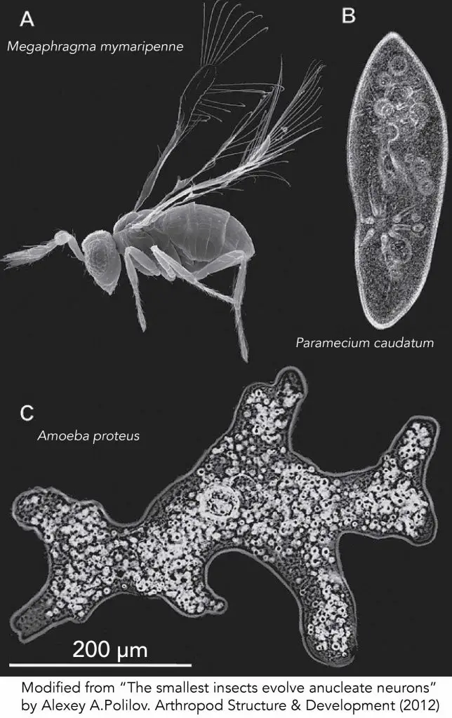
[In this figure] Size of the smallest insect and two protozoans in comparison. (A) Megaphragma mymaripenne. (B) Paramecium caudatum. (C) Amoeba proteus. Scale bar is 200 μm. Megaphragma mymaripenne, a parasitic wasp, is the smallest known flying insect.
How fast the Amoeba proteus can move?
Amoeba proteus can move at a rate of 2-5 mm per minutes.
Does Amoeba proteus have eyes?
No, Amoeba proteus doesn’t have eyes (don’t forget it is a single cell). However, Amoeba proteus can sense light and tends to move away from it. Bright light can even make all movements cease suddenly.
The scientists found that Amoeba proteus can respond to light stimulus because of reactions in its plasmagel, the gel-like cytoplasm at the tips of pseudopods. The light makes its plasmagel thicker and stiffer and, as a consequence, more difficult to move.
Other than light, Amoeba proteus can also sense several stimuli, like chemicals, toughing, temperature, and even electric fields!
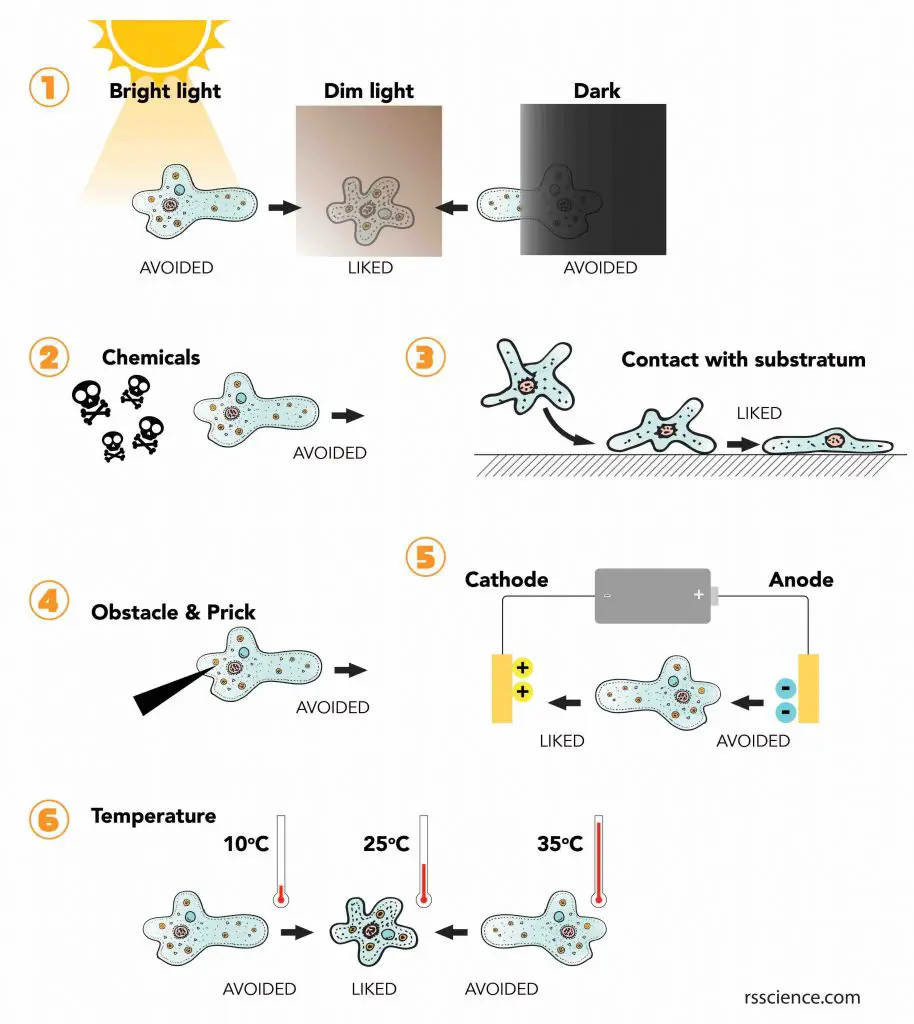
[In this figure] Even though the amoeba is just a single-celled organism, it can respond to various environmental changes.
(1) Amoeba will avoid bright light. It also does not stay in the complete dark due to the lack of food. Amoeba prefers in a dim light environment, like under the shadow of water plants or rocks.
(2) Amoeba can sense and avoid certain chemicals that are toxic.
(3) Amoeba does not like to float around. If possible, it would like to adhere to the surface of the substratum.
(4) Amoeba will avoid obstacles and sharp objects while it moves.
(5) When scientists place an amoeba in an electric field, the amoeba tends to move toward the cathode.
(6) Amoeba likes to stay at a temperature around 25oC.
How does Amoeba proteus breathe?
Because the Amoeba proteus is a single-celled organism, oxygen, and carbon dioxide can freely diffuse in and out of its cell membrane. Also, other substances (water-soluble molecules like salt) are able to transport through the membrane by osmosis.
How does Amoeba proteus reproduce?
Binary fission
Most of the time, Amoeba proteus reproduces asexually by splitting one cell into two cells, a process called “Binary Fission”. Just before it reproduces, Amoeba proteus retracts most of its pseudopods and rounds up into a ball.
After replicating its genetic material (DNA) in the nucleus, the original nucleus of the Amoeba divides to form two daughter nuclei by the process of Karyokinesis. In this process, the long DNA molecules condense into chromosomes (rod-liked shape) to facilitate the separation.
After the nucleus has divided into two, the process of Cytokinesis takes place in which the cytoplasm in the mother cell pinches in and divides into two daughter cells. This leads to the formation of the two daughter cells, having a nucleus and their own cell cytoplasm and organelles. Usually, the entire process may last anywhere from 30 minutes to an hour.
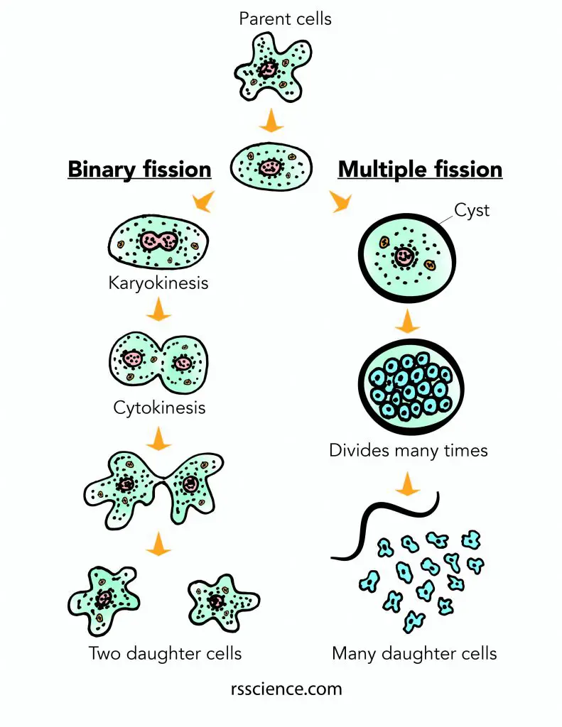
[In this figure] Two ways of amoeba reproduction: Binary fission and Multiple fission.
Most of the time, amoebas reproduce by binary fission. When the environment is turning harsh, amoebas adapt to multiple fission to increase the chance of survival.
Multiple fission
Another rare way for Amoeba to reproduce is called Encystment or Multiple Fission.
When amoeba senses the environment become unfavorable (eg. lack of nutrients, too acidic or too much bright light), it withdraws its pseudopodia and releases a protective coat (called a cyst) made of a chitin-like substance to cover its cell membrane. This cyst is able to survive in much harsher conditions. At the same time, mitosis occurs many times inside the cyst, producing more than two daughter cells. When the cyst wall ruptures (when the condition turns favorable), these daughter cells are then released to become several new amoebas.
When the environment of habitat becomes extremely unfavorable, Amoebas will reproduce through spores. This sexual reproduction can create genetic diversity and increase its chance of surviving in harsh conditions.
Amoeba under the microscope – science project
Where to look for Amoeba proteus?
Amoeba proteus resides at the bottom of clean freshwater. It feeds on decaying substances found in freshwater streams and ponds. You can use a transfer dropper to collect the bottom sediments to look for Amoeba proteus.
Amoeba proteus can also be ordered from science supply companies. It is commonly used in classrooms to illustrate the movement of pseudopods in action.
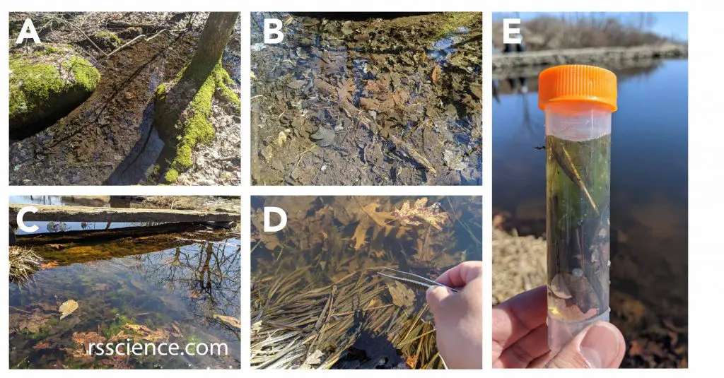
[In this figure] Where to collect the amoebas? Here are some pictures of habitats where I recently spotted Amoeba proteus.
(A-C) Amoebas like to hide in the bottom sediments (like leaves) of clear water ponds. (D-E) I used the forceps to collect some decaying leaves and water with sediments into my sample vial. I bring it home to look for amoebas and other pond lives under my microscope.
How to find Amoeba proteus under an optical microscope?
Amoebas can be directly observed under an optical microscope without additional stains. It takes patience to locate Amoebas under the microscope because they are transparent (colorless), slow-moving, and like to cover themselves under debris or bottom sediments.
-
Use a transfer pipette to get a drop of water with some bottom sediments onto a microscopic slide.
-
Gently cover the sample with a coverslip and mount it on the microscope stage for viewing. Wait 5-10 minutes to allow the microorganisms to adapt to the new environment (amoebas like to adhere to the surface of the glass).
-
Gradually increase the illumination (Amoebas are sensitive to bright light) and scan the field by low magnification (5x or 10x).
-
Looking for the tiny crystal-liked particles inside the cells of Amoebas may help you locate them. If you have phase contrast or polarized light filters, you may want to use them.
Amoebas can also be studied by dye staining to visualize cellular organelles. However, this requires the chemicals and equipment to fix and mount the dead Amoebas. If you want to know the detail, check out this link.
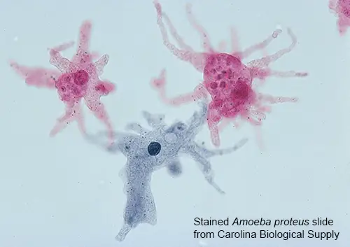
A stained Amoeba proteus slide.
What to look for under the microscope?
Direct observation of the Amoeba proteus has a significant advantage because the Amoeba proteus is still alive and actively moving when being viewed under the microscope. This allows you to see the finger-like projections (pseudopods) elongate and shorten as the Amoebas move or engulf food particles.
Some of the other organelles that are visible under the microscope include:
- Nucleus: The nucleus is found about 35 µm in diameter.
- Contractile vacuole: The size of the contractile vacuole may vary from 20 – 100 µm. Typically, it looks clear inside because it is actually a ball filled with water.
- Cytoplasm: The inner fluid containing all kinds of organelles and tiny crystals.
- Food vacuole: The food vacuoles are smaller than the nucleus. It is about 20 µm.
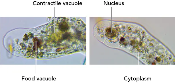
The color of the food vacuoles inside the Amoebas can also indicate the nutrient sources in the habitat. For example, I noticed that Amoebas collected in the late spring contain more green particles (could be green algae), while Amoebas from the early spring are more brownish (engrafted brown diatoms).
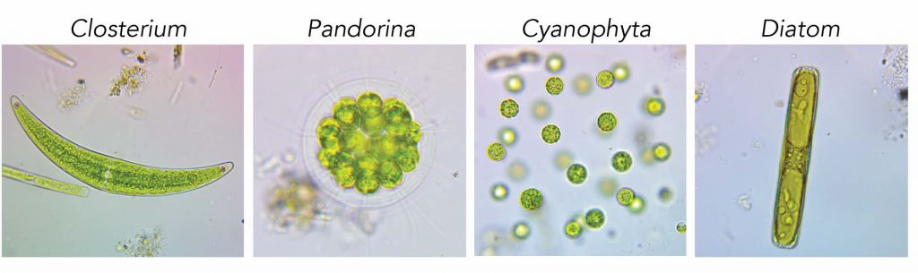
[In this figure] Examples of food sources that may affect the color of amoebas’ food vacuoles.
Sometimes, you may see Amoebas at rest and stay motionless with an oval shape.
If you have a camera or cell phone mounted on your microscope, the slow-moving Amoebas are great models to practice your microphotography and video-making skills.
Do other Amoebas also look like Amoeba proteus?
The answer is no. The family of Amoebas comprises very diverse members with over 15,000 described species. Although they all share one characteristic – moving by pseudopods, they can be totally different in shape and size.
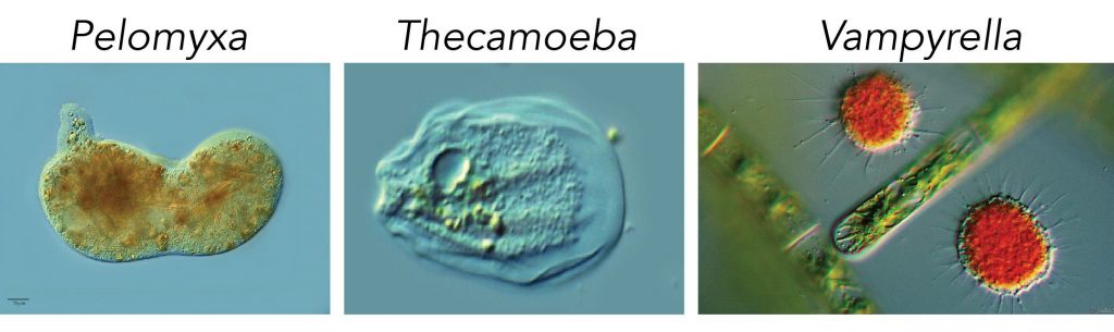
[In this figure] Cousins of Amoeba proteus
(A) Pelomyxa is a genus of giant amoeboids, usually 500-800 μm but occasionally up to 5 mm in length. (B) Thecamoeba. The body of Thecamoeba often forms a wrinkled cornucopia shape. (C) Vampyrella got its name from the way it feeds. Vampyrella sticks to its victims (usually algae), makes a large hole in the algal cell wall, and sucks the protoplast of the algae. (Credit: Pelomyxa and Thecamoeba – eol.org; Vampyrella – Sebastian Hess)
Amoebas can be divided into two major groups: naked amoeboids (subclass: Gymnamoebae) and shelled amoeboids (subclass: Testacea).
These Amoebas with soft, gel-like cell bodies, like Amoeba proteus, Pelomyxa, Thecamoeba, and Vampyrella, are all naked amoeboids.
Surprisingly, some species of Amoebas make protective shells, called “tests”, around their cells. Some shelled Amoebas make the tests entirely by themselves, and the materials could be organics, siliceous (containing silica), or calcareous (containing calcium carbonate) components produced by the Amoebas. These tests are called autogenic tests.
Some shelled Amoebas prepare their tests by collecting particles of sediment around them and gluing these mineral particles together with slime ingredients secreted from the cells. These tests are called xenogenic tests.
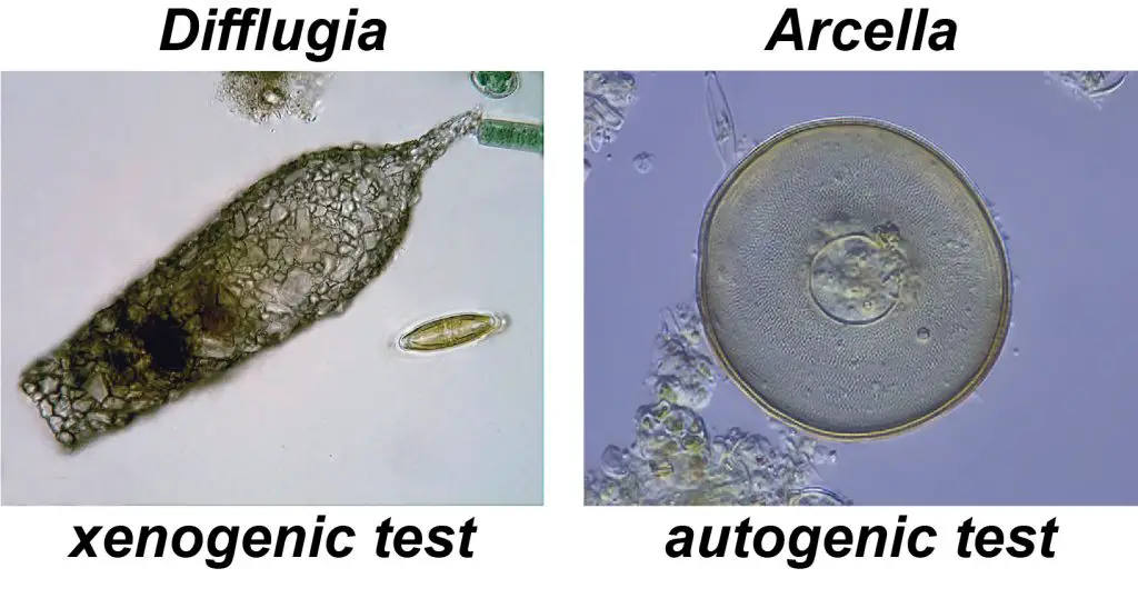
[In this figure] Left: Shell of Difflugia acuminata: the xenogenic test (about 300 µm long) made up of mineral particles glued together with secretions from within the cell. (Credit: Deuterostome on wiki); Right: The autogenic test (about 100 µm in diameter) of Arcella discoides, made up of organic plates produced by the cell. (Credit: Frank Fox on wiki)
These shelled amoebas can be collected with the same methods as naked amoebas. However, since the tests could easily break, you have to be careful when examining them under a microscope. The weight of the coverslip may crash the tests of shelled amoebas. Use the microscopic slides with a single concave or add some dots of Vaseline under the corners of the coverslip to provide more space for these creatures.
Where did the name “Amoeba” come from?
Amoeba proteus gets its name from two Greek words; “amoibe” meaning change, and “proteus” meaning Sea God. The Greek meaning describes this microbe as the Sea God Proteus that has a constantly changing shape.

[In this figure] Illustration of Proteus by Andrea Alciato from The Book of Emblems (1531)
I heard that Amoebas can eat human brains. Is it true?
Unfortunately, it is true. Although most amoebas are harmless to human beings, some rare species can be parasitic inside the human body.
Naegleria fowleri, colloquially known as the “brain-eating amoeba”, live in freshwater ponds or streams in hot geographic areas. Naegleria fowleri has two flagella (like tails) so it can swim in the water. Most of the time, Naegleria fowleri is free-living and eats bacteria. In some very rare cases, Naegleria fowleri can be inhaled through the nose and travels to the brain, causing a deadly disease called Naegleriasis. Of 154 people known to be infected in the United States from 1962–2021, only four people have survived.
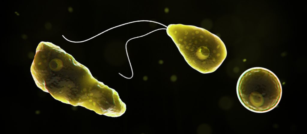
[In this figure] Naegleria fowleri (commonly referred to as the “brain-eating amoeba”) is a free-living microscopic amoeba. At a certain stage of its life circle, the Naegleria fowleri can swim by two flagella. (Source: CDC – https://www.cdc.gov/parasites/naegleria/)
Did you know?
Amoeba Proteus, Euglena, Tardigrade, and Paramecium caudatum are the most frequently studied micro life creatures in classrooms and laboratories.
References
Monaco Nature Encyclopedia – Amoeba proteus by Giorgio Venturini and Mario Beltramini
Amoeba proteus – An in-depth look at the protist, Amoeba proteus
Microbus
Microworld – world of amoeboid organisms
“Amoebas are more than just blobs” by Wim van Egmond
Wikipedia
“The nature of response to light in Amoeba proteus (Leidy)” by S. O. Mast. Published on Zeitschrift für vergleichende Physiologie volume 15, pages139–147(1931)
“Amoebae: Protists Which Move and Feed Using Pseudopodia” by David J. Patterson.
Genome size:
Genome News Network
BioNinja
Bionumbers
Integrated Taxonomic Information System
Related posts
The Biological Classification of Paramecium – Name, History, and Evolution
The Structure of Paramecium Cell
Pingback: What is an amoeba? – Dr. Biology Questions and Answers
Pingback: How does an amoeba move? – Dr. Biology Questions and Answers
Pingback: How does an amoeba obtain food? – Dr. Biology Questions and Answers
Pingback: How does an amoeba reproduce? – Dr. Biology Questions and Answers
Pingback: How big is an amoeba? – Dr. Biology Questions and Answers