
Hairstyling is a part of our everyday life, but have you ever really looked at your hair under a microscope?
In this post, I will introduce the function, structure, and finally the microscopic view of hair.
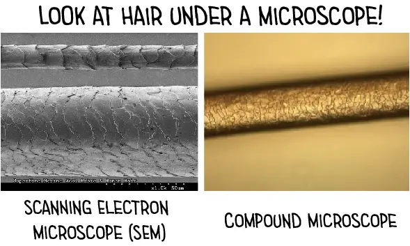
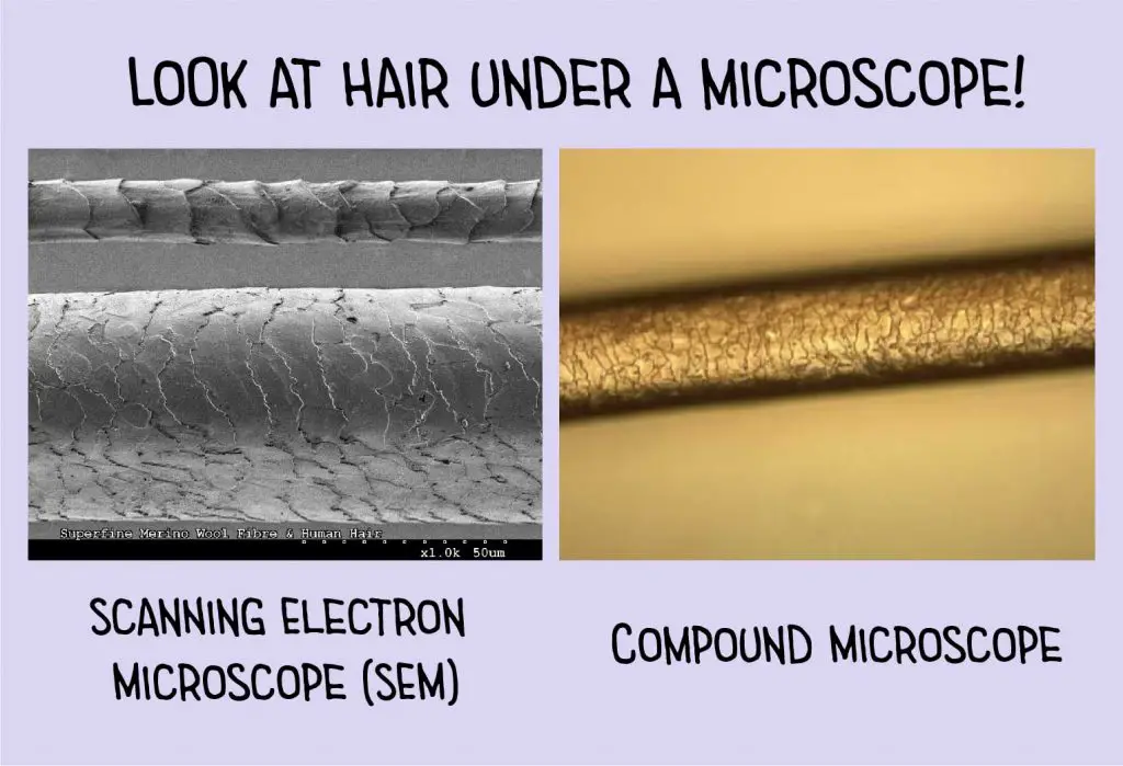
[In this figure] Hair under a scanning electron microscope (SEM) and a compound microscope.
You can see the scale (called cuticle) on the surface. The human hair is about 70 micrometers in width.
Photo source: science image
This article covers
The biology of hair
The function of hair
Hair is a characteristic attribute of mammals. It is different from fish’s scale and bird’s feathers. The fur is a thick growth of hair that covers the skin of many mammals. It consists of a combination of longer guard hair on top and shorter fleece hair (also known as underfur or down hair) beneath. The guard hair keeps moisture from reaching the skin; the underfur acts as an insulating blanket that keeps the animal warm.
Thermal insulation
The primary purpose of hair is to provide thermal insulation. Animals living in colder climates typically are covered in a thicker layer of hair (also referred to as a coat or wool). For example, the musk ox has dense guard hairs measure 30 cm (12 in) in length. Some desert mammals, such as camels, use dense fur to prevent solar heat from reaching their skin, allowing the animal to stay cool. Humans, who have little need for extra protection from the cold due to the development of clothing, are among the least hairy mammals.
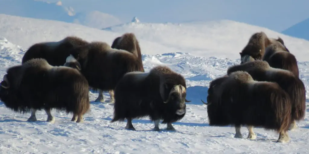
[In this figure] Alaskan muskoxen have a very dense layer of fur.
Photo credit: Bureau of land management
Waterproofing
Fur also functions as a waterproofing layer. Aquatic mammals, like beavers and otters, trap air in their fur to conserve heat by keeping the skin dry.
Protection
Hair also provides protection for the animals. Porcupines and hedgehogs have their spines, which are hollow hairs made stiff with keratin. Colors and patterns of furs can be used for camouflaging according to animals’ habitats. The bottom of the hair cells is surrounded by nerves, making hair also a sensory organ. Bristles are long hairs usually used in visual signals, such as the mane of a lion.
Basic structure of hair
A hair can be described as a long thread-liked outgrowth from the follicles of the mammal skin. A hair composes essentially of a protein, called keratin. In detail, the basic structure of hair can be divided into three anatomical regions: cuticle, cortex, and medulla.
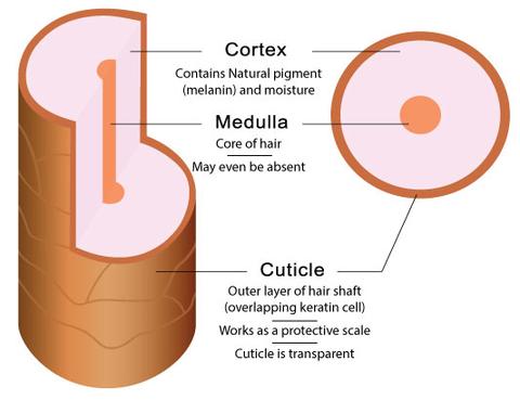
[In this figure] Hair structure: cuticle, cortex, and medulla.
Photo credit: Bigen
Cuticle
Cuticle is the outer layer of hair that works like protective scales. The cuticle is made up of overlapping transparent keratin cells. These cells are dead and cornified but preserve their keratin fibers which are resistant to chemicals and protect the cortex from chemical influences.
The condition of the cuticle layer determines the smoothness and shininess of your hair – because it can be seen and touched. Cuticle can get damaged by chemical processes, overexposure to sunlight (UV), too much heat from a dryer, abusive brushing and combing, over-chlorinated swimming pool water, etc.
The treatment of conditioners only applies additional chemical layers to make the hair look smooth and shiny. Damaged cuticle cannot repair by itself once it is damaged because of the lack of viable cells.
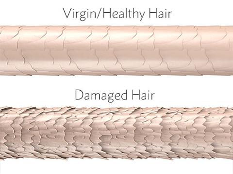
[In this figure] Cuticle on the surface determines the condition of the hair.
Photo credit: Bigen
Cortex
Cortex is the main body of hair and where moisture and natural pigment (called melanin) are held. No other part of the hair has pigments. The amount of melanin contained in cortex determines the natural color of your hair. Some hair color products, especially when lightening hair, work on melanin. Melanin is missing from gray hair.
Cortex is composed of elongated and fusiform (spindle-shaped) cells. The cortex may contain air spaces called cortical fusi, pigment granules, and large oval-shaped structures referred to as ovoid bodies.
Medulla
Medulla is the central core of hair which may or may not be present. It may be air-filled and, if so, will appear as a black or opaque structure under the transmitted light of the microscope. If the medulla is present, its structure may be continuous or fragmented. It’s not yet scientifically clear why this happens.
A schematic diagram of the microstructure of hair. Note that the cuticular scales always point from the proximal or root end of the hair to the distal or tip end of the hair.
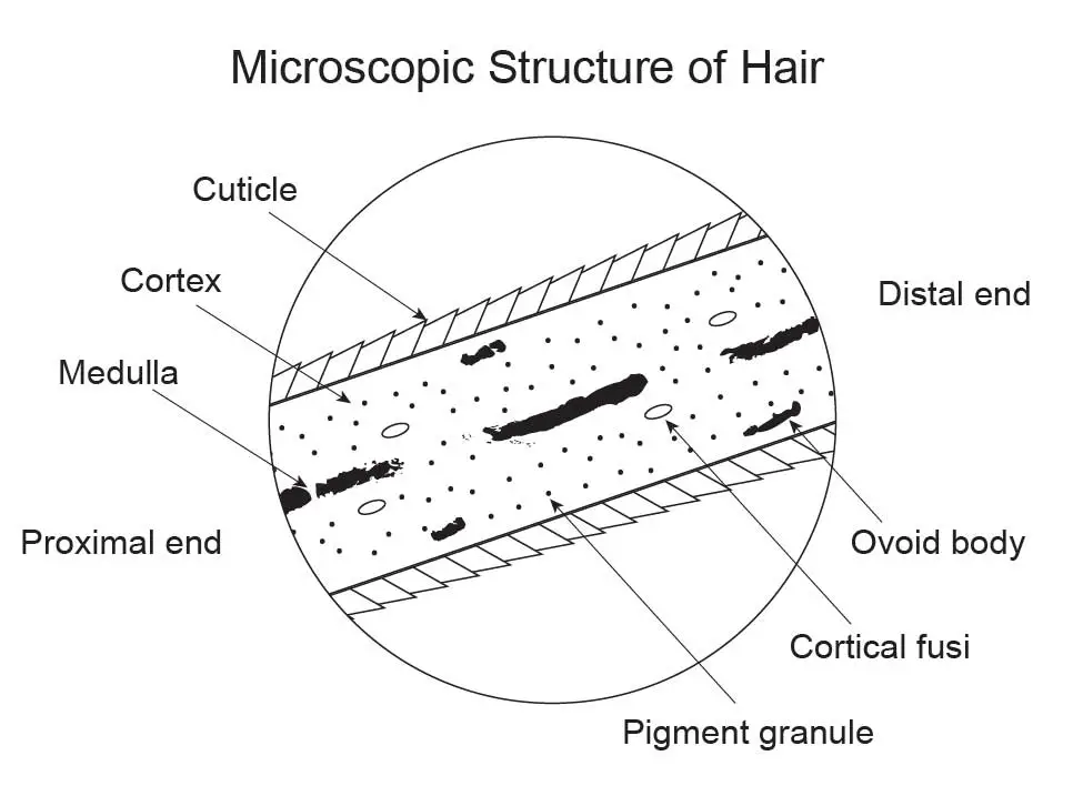
[In this figure] A schematic diagram of the microstructure of hair.
Note that the cuticular scales always point from the proximal or root end of the hair to the distal or tip end of the hair.
How a hair grows?
Our hair grows from the follicles. The hair follicle resides in the dermal layer of the skin and is made up of 20 different cell types, each with distinct functions.
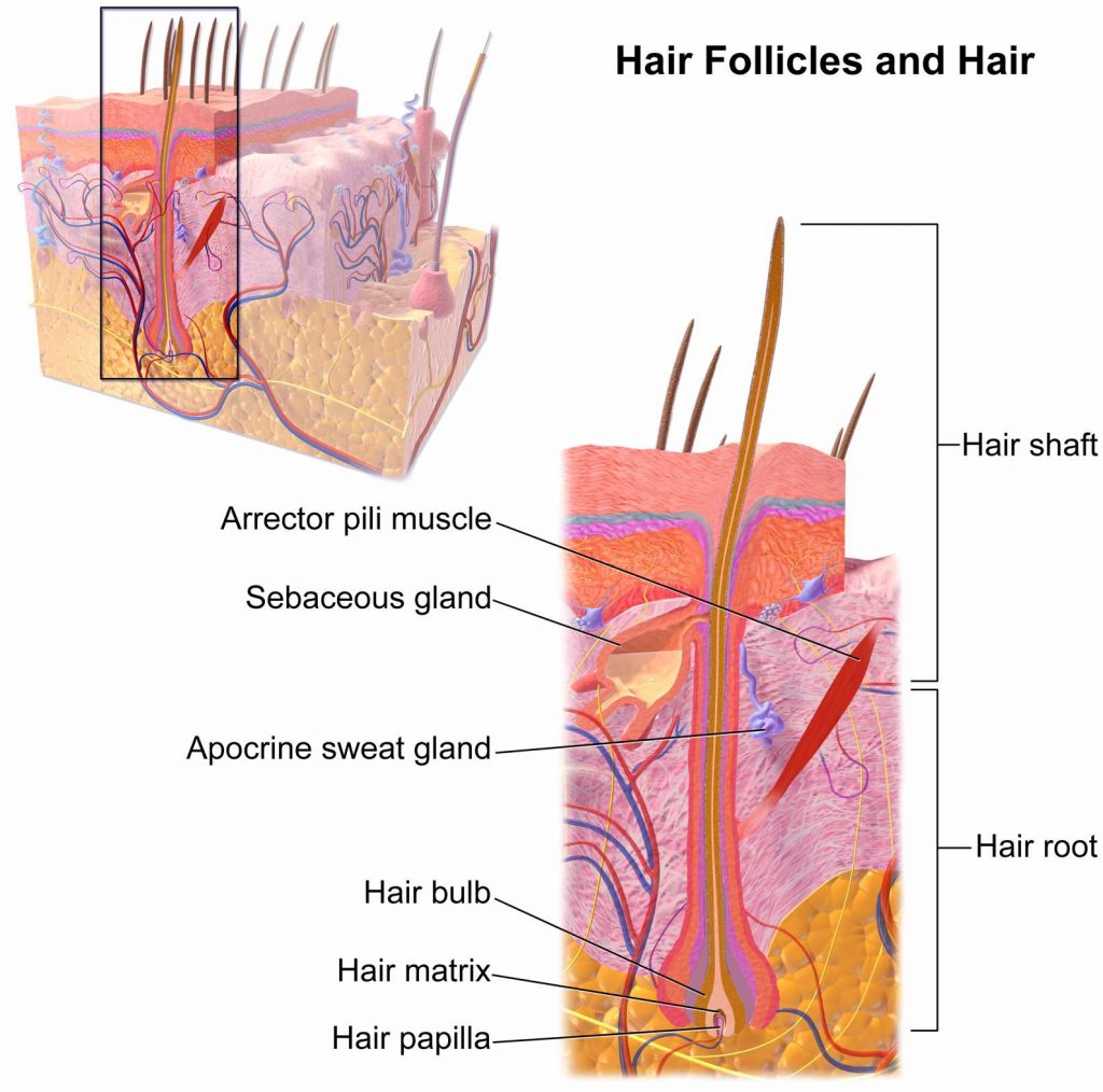
[In this figure] Structure of a hair follicle.
Photo source: wiki
We can divide hair into two parts based on its position. The part of the hair that remains under the skin inside the follicle is referred to as the hair root. The base of the hair root is called the hair bulb. The hair bulb is surrounded by blood vessels and receives nutrients for the formation of new hair cells.
As new hair cells are formed at the hair bulb, the existed hair moves up above the surface of the skin to become part of the hair shaft. At the same time, these cells also undergo a maturation process referred to as keratinization, where they lose their nucleus (dead cells) and fill with a fibrous protein, called keratin. Therefore, hair can simply be described as strands of keratinized protein.
Forensic science of hair
Trichology is the scientific study of hair. Our knowledge of trichology not only benefits medication and cosmetics, but trichology is also a valuable tool for forensic scientists to figure out the truth behind crime scenes.
The microscopic investigation of hair is a big part of forensic investigations. Just like fingerprints, hair type is unique to each individual. We have soft, medium, hard, thick, coarse, straight, curly, damaged, colored hair – with numerous combinations of all of the above. Hair also absorbs chemicals (like metals) from the surrounding environment when it grows. All these properties can be used in forensic science to identify individual persons.
Another reason that makes the hair a valuable tool for forensic investigation is its durability. Hair is more resistant to decay than most other body tissues and fluids, thus remaining intact far longer than other evidence. This durability makes hair one of the most frequently found pieces of evidence at crime scenes.
Forensic scientists perform three major types of hair analysis
The morphology under a microscope
Forensic scientists view hair under a microscope to collect evidence based on morphology. They usually study the hair’s scale pattern, its color, and the appearance of the medulla. An experienced forensic scientist can easily tell if the hair specimen is from humans or from animals. By comparing different hair samples, they also can determine if it’s from a particular person.
The chemical composition of the hair shaft
Forensic scientists can test the hair shaft for its chemical composition. For example, the chemical analysis of hair can tell if the person used a drug or had experienced nutritional deficiencies. All this evidence can provide valuable information on the body conditions of the hair’s owner.
The DNA from the cell at the root of the hair
Forensic scientists can obtain DNA from the cells at the root of the hair. These DNA specimens can provide conclusive evidence about who owns the hair.
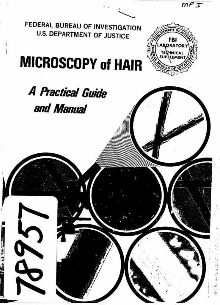
[In this figure] A practical guide and manual used in the FBI for the microscopic analysis of hair evidence. You can download it and read it on the website.
For more detail about how forensic scientists work on hair evidence in FBI, check out this link: https://archives.fbi.gov/archives/about-us/lab/forensic-science-communications/fsc/july2000/deedric1.htm
Comparison microscopy
Comparison microscopes, which give a side-by-side view of two objects (for example, hairs or fibers) is a powerful tool for forensic science.
A comparison microscope consists of a matched pair of compound microscopes with conventional specimen stages. These stages were joined by a system of lenses, prisms, and mirrors in what was termed an optical bridge. The bridge allowed the investigators to observe and compare two physically separated but optically joined objects simultaneously in a single field of view. This field of view was split by an optical hairline.
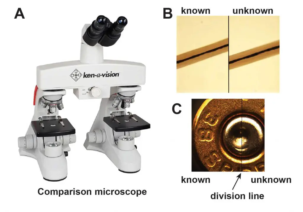
[In this figure] (A) A comparison microscope. Source: Ken-a-vision Microscopes. (B) An example of side-by-side comparison of two hair specimens in order to tell if they came from the same person. Source: kfrazierforensics. (C) A split image for firearm investigation. Source: Ludesco microscopes.
The microscopy of hair
Hair under a stereo microscope
A stereo microscope is good for the initial examination of hair before moving on to a compound microscope. Under a stereo microscope, you can easily observe basic characteristics such as color, shape, texture, and length of hair.
Below is an image to remind you of the difference between a compound microscope and a stereo microscope. We also wrote a post about “How to choose a compound microscope vs a stereo microscope.”
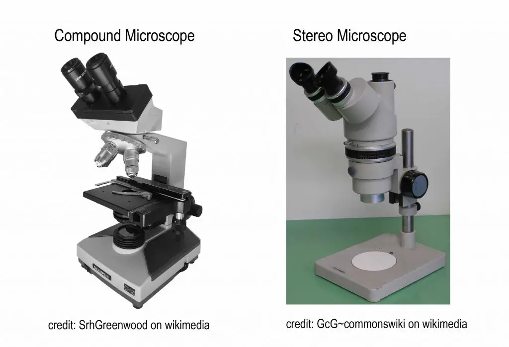
[In this figure] The difference between compound and stereo microscopes.
The materials you may need:
- Different hair specimens – try to collect different types of hair such as hair from the human body (different body area), hair from different persons, hair from animals like dog and cat, etc.
- A stereo microscope (A digital USB microscope also works)
- Tweezers
What you can look for?
Using the tweezers, gently pick and place the hair strand under a low-power stereo microscope. Observe and see if there are differences between different types of hair.
Under a stereo microscope, you should be able to see the shape of the hair (straight or twisted, etc) as well as the color of the hair strand. At higher magnification, you can also see the appearance of texture on the hair surface. When viewing different types of hair, you can also be able to differentiate them by certain characteristics, for example, the thickness between different strands.
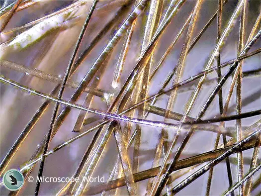
[In this figure] Human hair under a stereo microscope at 5x magnification.
Photo credit: Microscope World
Hair under a compound microscope
A compound microscope has higher power than stereo microscope to resolve the detailed structures of hair. However, the light has to pass through the hair specimen to generate a clear microscopic image. There are two main procedures that can be used to view different structures of the hair.
Method 1: Making a scale casting with clear nail polish & see “microtopography” of hair
Scale casting is a very useful technique in biology, botany, paleontology, and even forensic science. Basically, scale casting uses a molding material (like glue) to make a replicate of the surface that you want to investigate. A good scale cast can preserve the very tiny detail of the surface texture (called “microtopography”) so that you can study it under a microscope.
By applying the technique of scale casting to a hair specimen, the surface texture of hair can be well preserved. Because the characteristics of the cuticle (such as the sizes, shapes, and arrangement of scales) are the critical features of hair, the scale casting of hairs is very useful for biologists or forensic investigators to identify the origins (human or animal) of the hairs.
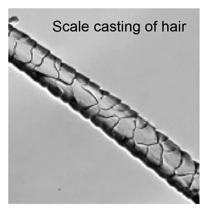
[In this figure] An example of scaled casted hair.
You can see the layer of squama-like texture on the surface of the hair.
The materials you may need:
- Nail polish (clear, no color ones are preferred)
- Microscope slides
- Tweezers or forceps
- Different hair specimens
- A compound microscope
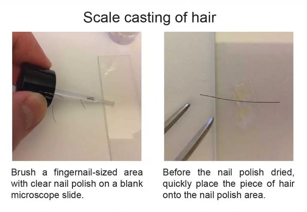
Follow these steps to make your scale casting slides:
1. Cut the hair specimen into 1-2 cm long and have them ready on hand.
2. Brush a fingernail-sized area with clear nail polish on a blank microscope slide.
Note: Latex (for molding) can be used in place of nail polish
3. Before the nail polish is dried, quickly place the piece of hair onto the nail polish area.
4. Let the slide sit undisturbed for 10 to 15 minutes allowing the nail polish to dry.
5. Once the polish is nearly dry, use forceps to pull your hair fiber out of the slide. Sometimes, hair may break in pieces during pulling off. It is ok. Try to pull as much hair as possible. It is not necessary to remove every strand of hair.
Note: the nail polish/latex is used to attach and retain the scale cast that remains on the slide once the hair strands are pulled off.
6. The clear replica reflecting the texture of the hair will be left on the microscope slide. Your slide is ready for viewing now!
Note: Although the nail polish is transparent, the texture of the surface scale of hair is preserved. Under a microscope, you can tell the ups and downs that represent the texture of hair with the contrast of shadow. Lower light intensity could give you higher contrast and better images.
What you can look for?
The scale casting technique is ideal to view the outer layer of hair that contains the scales (not for the internal structure of the hair strand). Once the hair strands are pulled off, the shapes of scales (or called the “microtopography”) remain attached to the slide and can be viewed using lower power objectives.
Examine the slide using the low-power objective of a microscope. Look for impressions of individual scales. Compare different types of hairs and differentiate the characteristics of the scales.
Biologists study the casts of the hair collected from animals for determining scale patterns. The arrangement and shape of hair scales can vary greatly from species to species and are often very distinctive. Scientists usually classify scales into two major categories:
- Coronal — Completely encircling the entire width of the hair shaft
- Imbricate — Short, wide, and not encircling the hair shaft
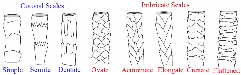
[In this figure] Examples of different cuticle scales of mammalian hair.
photo credit: MicrolabNW Photomicrography
Try to collect as many different types of hair specimens (from humans and animals) as possible. Well-made casting slides can be stored in slide boxes (away from dust) for a long time. You can establish your own library of hair collections.
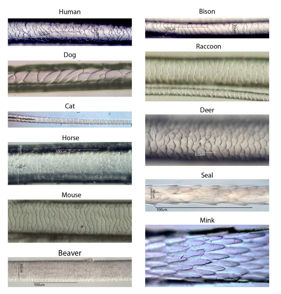
[In this figure] A collection of hair casting from the different mammals.
Please note that the magnification of each image is different. Also, for each animal, hair from different types (guard or fleece) and body locations can be very different. For more detail, check out this website: MicrolabNW Photomicrograph Gallery.
Method 2: Making a whole mount slide of hair to look at internal features (medulla)
In order to look at the internal structures of hair such as medulla, a whole mount slide is ideal.
The materials you may need:
- Microscope slides
- Mounting media (it could be water or 70% glycerol wet mount medium)
- Coverslips
- Tweezers or forceps
- Different hair specimens
- A compound microscope
Follow these steps to make your whole mount slides:
1. Cut the hair specimen into 1 cm long (smaller than the size of your coverslips).
2. Obtain a clean microscope slide and place a drop of mounting medium or water on it. You can learn “how to choose mounting media“
3. Place several strands of hair on the drop of mountant or water.
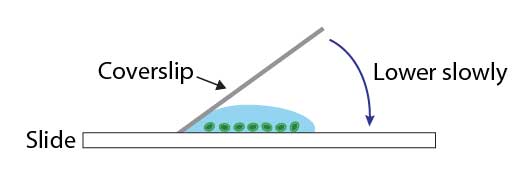
4. Lower the coverslip slowly with an angle. Allow one side of the liquid droplet to touch the coverslip first. This permits air to escape from the other side. You can use the forceps to help you control the coverslips.
5. Remove any excess solution by using tissue paper to touch one side of the coverslip.
6. Optional: You can also seal all of the edges of the coverslip with super glue (or nail polish) to prevent the movement of the coverslip and for long-term storage.
What you can look for?
Examine the whole mount slides first under the low-power objectives of a microscope. Once you find the right focus, switch to the high-power objectives.
Thick hair or hair with dark color may need stronger light intensity to illuminate its internal structure. Examine several sources of hair (human vs. animal, or hair from different body areas) and look for different internal features such as granules or air spaces. Draw the features of the hair you observed or take some pictures. You can also measure the width of your hair if you have a microscopic ruler.
Identifying hair by the appearance of the medulla
The medulla is a central core of cells that may be present in the hair. If it is filled with air, it appears as a black or opaque structure under transmitted light.
A whole mount allows the study of the appearance of the medulla; however, a medulla is not always present in a hair (especially, for human hair). When medullae are present, they often show distinctive variations between species. The appearance of a medulla is classified as continuous (unbroken), intermittent (regular intervals), or fragmented (irregular intervals).
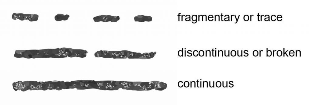
[In this figure] Diagram of the three basic medullary types.
The difference in medulla between humans and animals
Identifying whether the hair is from a human or animal is the first step in forensic hair analysis. In human hairs, the medullae are generally amorphous in appearance, whereas in animal hairs, their structures are frequently very regular and well defined. Human hairs usually contain a thin (less than ⅓ of the hair’s diameter), fragmented or absent medulla region. Animal hairs usually have thick medullae (more than half of the hair’s diameter) with regular patterns.
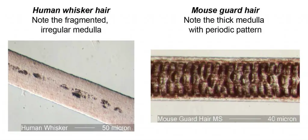
[In this figure] The typical difference of medulla between human and animal hair.
The appearance of human hair medulla looks fragmented and irregular, while that of mouse hair medulla contains the periodic pattern.
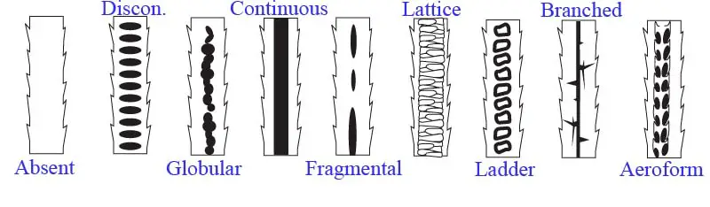
[In this figure] Examples of different medullary patterns of mammalian hair.
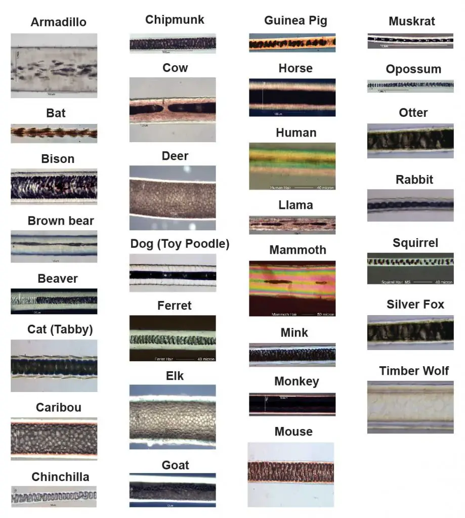
[In this figure] A collection of hair medullary patterns from different mammals.
The most interesting specimen is the hair of a frozen woolly mammoth discovered from Siberian ice. Please note that the magnification of each image is different. Also, for each animal, hair from different types (guard or fleece) and body locations can be very different. For more detail, check out this website: MicrolabNW Photomicrograph Gallery.
Advanced microscopy of hair biology
Like other fields of biological research, trichology is also benefited from the advance of microscopic technology. For example, a scanning electron microscope (SEM) can reveal the detailed surface texture of hairs (similar to the scale casting, but with a much higher resolution).
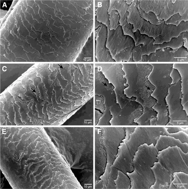
[In this figure] Scanning electron microscopy analysis of the effect of heat and enzymatic hydrolysates treatments on hairs.
A, B –Control; C, D – After the treatment with the enzymatic hydrolysates and straightener at 180°; E, F- After the treatment with the enzymatic hydrolysates without heat. Source: BMC Biotechnology.
The combination of fluorescent microscope and molecular biology provides a great tool for biologists to study the growth of hair, especially the stem cells that resided inside the hair follicles. This knowledge may improve our success in treating hair loss due to aging or diseases.
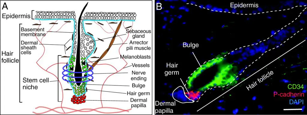
[In this figure] (A) The anatomy of the hair follicle. (B) A fluorescent image of a hair follicle showing two types of stem cells. The colors are labeled by antibodies that can recognize specific protein markers on the stem cells. Source: Development.
Conclusion
Turn your students into a CSI team and let them solve a “crime” using hair analysis. The forensic mystery will both engage and intrigue them while they learn science concepts.
Teaching basic physiological science concepts is interesting from a forensics point of view. By incorporating a problem-solving approach to science education, teachers engage their students in exciting and innovative ways. Forensic labs also provide “real-world” applications of science and math.
References
“Under the Microscope: Get Forensic with Hair Analysis”
“History of Hair and Fiber Analysis”
“Comparison Microscopy”
“Identification of Human and Animal Hair”
“Hairs, Fibers, Crime, and Evidence” by Douglas W. Deedrick. Forensic science communications, July 2000 – Volume 2 – Number 3.
“MicrolabNW Photomicrograph Gallery”
Related posts
Lesson 5: How to Do Scale Casting with Clear Nail Polish & See “Microtopography”
How to Choose the Right Microscope (Compound Microscope vs. Stereo Microscope)