
Paramecium is an incredible creature that is absolutely worthwhile to spend several posts talking about it. Therefore, we will have 4 series of paramecium.
Part I. The Biological Classification of Paramecium – Name, History, and Evolution
Part II. The Structure of Paramecium cell
Part III. Paramecium Reproduction, Physiology, and Behaviors
Part IV. The Natural Habitation and Cultivation of Paramecium – Find Paramecium for Your Microscopic Project
This article covers
What is paramecium? A quick overview of paramecium
Paramecium (pair-ah-me-see-um; plural, Paramecia) is a unicellular (single-celled) living organism with a shape resembling a slipper. Paramecium is naturally found in aquatic habitats. You need a microscope to see the paramecia because they are only 50 to 300 µm (micrometers) in length.
Their sizes vary from species to species. P. caudatum is among the largest protozoan and can grow up to 200-300 micrometers. It is barely visible with the naked eye. Another common paramecium, called P. aurelia, is smaller (50-150 micrometers). P. caudatum is more elongated and P. aurelia is more ovoid in shape.
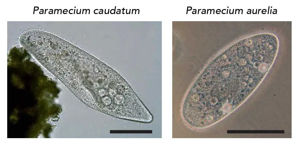
[In this figure] The appearance of P. caudatum and P aurelia.
P. caudatum is elongated, rounded anteriorly, and pointed posteriorly. P. aurelia is more ovoid in shape. Both scale bars are 100 micrometers. Although P. aurelia is smaller than P. caudatum, P. aurelia grows faster.
Photo credit: P. caudatum (Deuterostome), P. aurelia (Barfooz on Wiki)
The organelles of paramecium
Although paramecium is small and has only one cell, it can do everything that a living creature can do: Paramecium can swim, digest food, and reproduce. Paramecium’s cell contains several complex organelles performing specific functions to make its survival possible.
Paramecium constantly moves by beating rows of microscopic hairs, called cilia (singular cilium), that work like miniature oars. This is why paramecium is commonly studied as a white rat of ciliates. Certain paramecia are also easily cultured in labs and serve as useful model organisms.
Paramecium collects foods via its mouth, called oral groove. The food materials enter the cell body and then are digested in food vacuoles. Paramecia eat other microorganisms such as bacteria, yeasts, and algae.
Paramecium has a protective skin layer consisted of pellicle and ectoplasm. Trichocysts are special organelles situated in the ectoplasm which can discharge long spikes for protection from their predator.
The most unusual characteristic of paramecia is their nuclei. They have two types of nuclei, micronucleus, and macronucleus. The macronucleus controls non-reproductive cell functions, expressing the genes needed for daily functions, such as movement and feeding. The micronucleus is the germline nucleus, containing the genetic material that passed along from one generation to the next generation.
Paramecium has two star-shaped contractile vacuoles. The contractile vacuoles regulate the quantity of water (osmosis) inside of a cell. They expel water out of the cell by contracting and they prevent the cell from absorbing too much water.
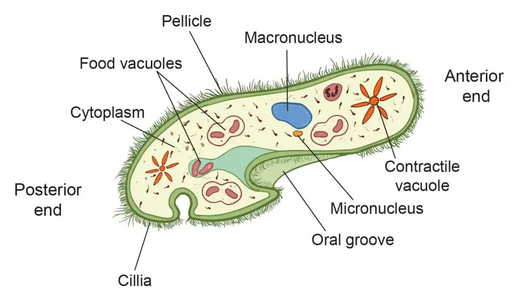
[In this figure] The organelles of a paramecium.
Paramecium reproduction
Paramecia can reproduce either asexually or sexually, depending on their environmental conditions. When nutrients are plentiful, the population of paramecia increases rapidly by asexual reproduction, called binary fission. One paramecium cell divides into two daughter cells with identical genetic information.
Under unfavorable conditions, such as starvation, paramecia switch to sexual reproduction. Odd types of paramecia can mate with even type of paramecia. Each parent paramecium provides half of the genetic information to give rise to a new paramecium. When the starvation lasts longer, paramecia can also undergo self-fertilization.
Paramecium is the most frequently studied microorganisms in classrooms and science fairs. Paramecia can be found in freshwater habitation and are easy to observe under a light microscope. Common paramecia are also easily cultured in the laboratory without the need for special equipment. Hay infusion (boiled hay in water) is an ideal medium for culturing paramecia.
Where did the name “Paramecium” come from?
Paramecium, also called “slipper animalcule”, was among the first ciliates to be seen by microscopists, in the late 17th century. The Dutch pioneer of microbiology, Antonie van Leeuwenhoek (1632-1723), clearly described this tiny creature during his establishment of microbiology as a scientific discipline.
Later in 1718, the French mathematics teacher and microscopist Louis Joblot (1645-1723) published an illustration of a microscopic “fish”, which he discovered in an infusion of oak bark in water. Joblot gave this creature the name “Slipper” based on its shape.
The name “Paramecium” came from the Greek “paramēkēs”, meaning “Animalcules which have no visible limbs or tails, and are of an irregularly oblong figure.”
Animalcule (“little animal” from Latin) is an older term for a microscopic animal or protozoan, which was first used by Antonie van Leeuwenhoek to refer to the microorganisms he observed in rainwater.
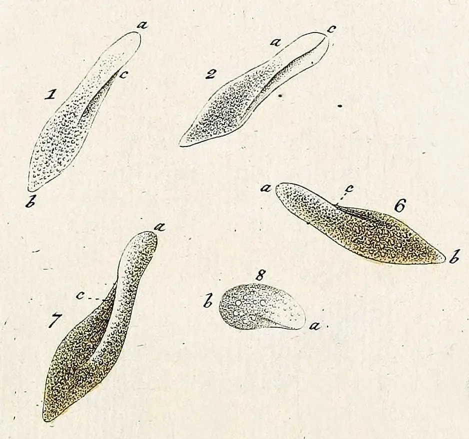
[In this figure] “Slipper animalcule” illustrated by Otto Müller, 1773

[In this figure] Left: A portrait of Antonie van Leeuwenhoek (1632–1723) by Jan Verkolje. Middle: The original design of Leeuwenhoek’s microscopes. Right: A replica of Leeuwenhoek’s microscope (photo credit: Jeroen Rouwkema and wiki).
Paramecium classification
The general term “Paramecium” refers to the organisms within the genus Paramecium. “Genus” is a level of biological classification which refers to a closely related group of organisms that share similar characteristics. Under the genus of Paramecium, there are currently about 30 species. The most two common species are P. aurelia and P. caudatum. “P.” is the common abbreviation for “Paramecium”.
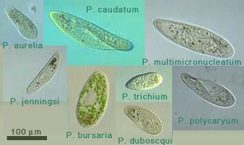
[In this figure] A photo collection of several Paramecium species. This image is from Protist Information Server, a Japanese image database for protists and microorganisms.
Morphology classification
The methods of classifying paramecia have changed over the years. The early methods were based on the morphology through visual observation under the microscope. At that time, all paramecia were described as either P. aurelia or P. bursaria.
Molecular classification
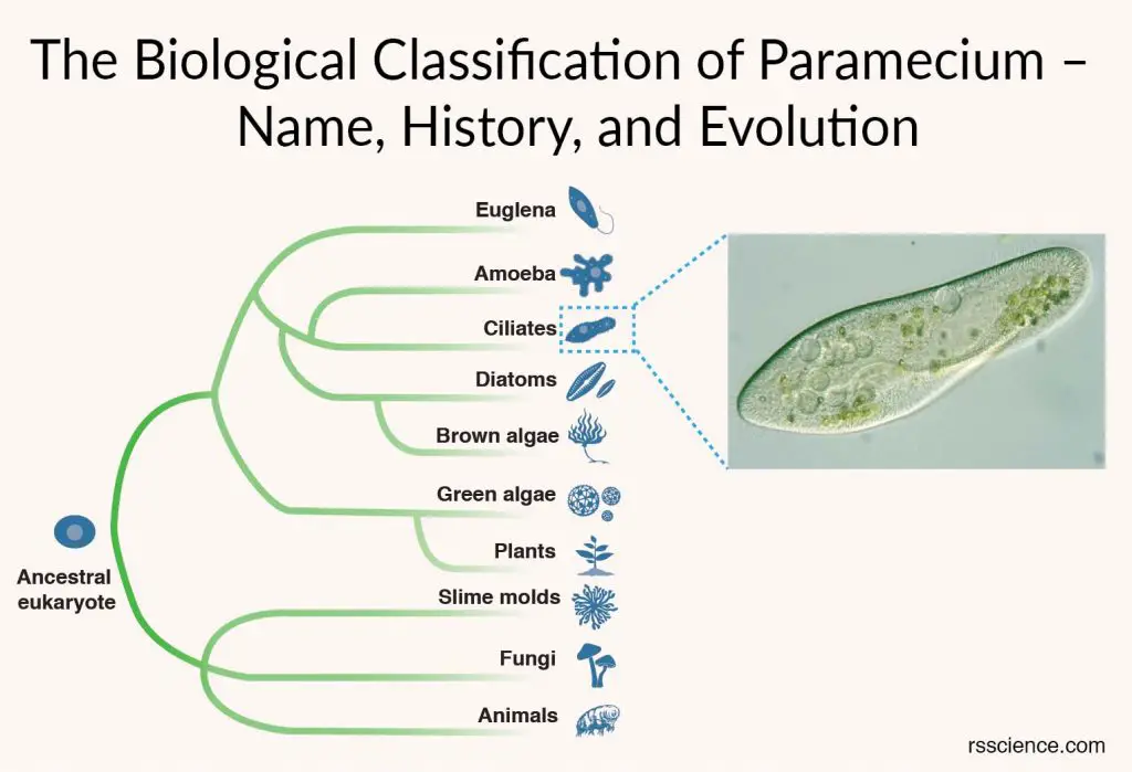
More recently, DNA sequencing became a powerful tool for biological classification. This allows us to read the organisms’ genetic information and develop a family tree, known as a phylogenetic tree, which represents evolutionary relationships. This shift from the morphology to the genetic evolution has a huge impact on the understanding of relationships within the Paramecium genus and species diversity.
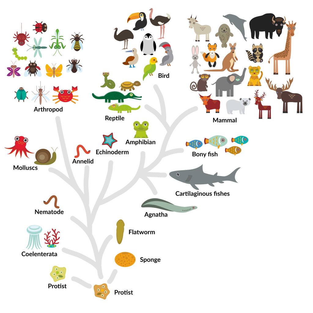
[In this figure] An example of phylogenetic tree or life tree showing the relationships between organisms during the evolution.
Based on DNA sequencing, scientists are able to classify sibling species that look alike with no distinguishing characteristics, but they differ in biochemical and genetic aspects and cannot mate with one another. With this powerful tool, new species of Paramecium continue to be described even today.
For example, a 2015 research study described three new species, which are morphologically difficult to distinguish, found in Germany, Hungary and Brazil. However, their DNAs indicate that they are separate species. If we look closely at the freshwater pool around us, we may still find undefined species.
Paramecium taxonomy – the position in the history of life
In biology, Taxonomy (from Ancient Greek taxis, meaning ‘arrangement’, and -nomia, meaning ‘method’) is the science of naming, defining, and classifying groups of biological organisms on the basis of shared characteristics.
In the Taxonomic Hierarchy, Paramecium can be classified into the following position based on their certain characteristics.
Kingdom: Chromista
Phylum: Ciliophora
Class: Oligohymenophorea
Order: Peniculida
Family: Parameciidae
Genus: Paramecium
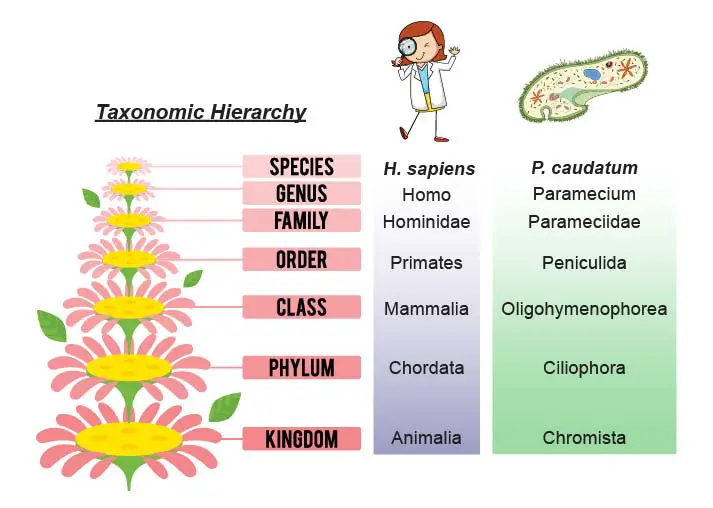
[In this figure] Taxonomy is the science of defining and naming groups of biological organisms on the basis of shared characteristics. The ranking system we used here (also currently used on Wikipedia) is the newest system proposed by Ruggiero et al. in 2015. This system has 7 kingdoms (Bacteria, Archaea, Protozoa, Chromista, Plantae, Fungi, and Animalia).
The old classification of paramecium – Protista kingdom
The system of taxonomic hierarchy is still under debate and evaluation, especially for Protists. Protists are a diverse collection of eukaryotic organisms. They are primarily microscopic and unicellular. In older literature, you may see Paramecium was grouped in the kingdom of Protista (based on five-kingdom scheme proposed by Robert Whittaker in 1969).
A protist could be any eukaryotic organism that is not classified as an animal, plant, or fungus. In other words, the kingdom of Protista was like a temporary folder for these organisms that scientists don’t know how to classify. As a result, you will find both animal-like (called Protozoa, including Amoeba, Paramecium, etc.) and plant-like (called Protophyta, including green algae, Diatom, etc.) organisms were all grouped together.
The current classification of paramecium – Chromista kingdom
In this post, we (as well as Wikipedia) use the 7-kingdom system (Bacteria, Archaea, Protozoa, Chromista, Plantae, Fungi, and Animalia) proposed by Ruggiero et al. in 2015. In this system, green algae are classified into Plantae (together with land plants) for their chloroplasts containing pigment molecules, called chlorophyll a and b.
The rest of the Protista split into Protozoa and Chromista. Animal-like protists that don’t have chlorophyll pigments belong to Protozoa. Organisms containing chlorophyll c are grouped together in the kingdom of Chromista. Paramecium is classified in Chromista for this reason. Other members of Chromista include marine algae, potato blight, dinoflagellates, brain parasite (Toxoplasma), and malarial parasite (Plasmodium).
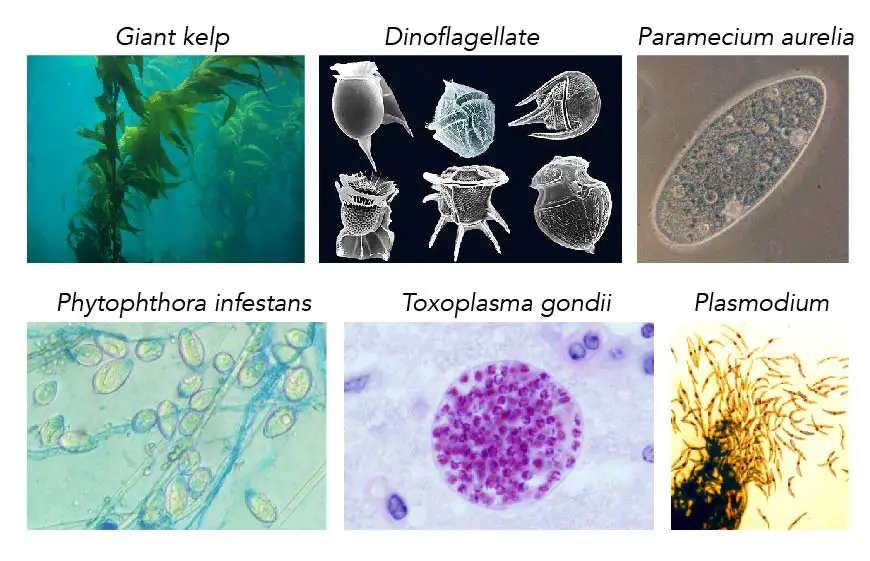
[In this figure] The Kingdom of Chromista.
Organisms grouped in Chromista due to the similar features of pigment molecules, called chlorophyll c. The members are quite diverse in shape and size. For example, marine algae-like giant kelp can grow up to 45 meters (150 feet). Single-celled dinoflagellate (5-2000 µm) has a hard shell and a flagellum to swim. Phytophthora infestans is a mold-like microorganism that causes tomato disease known as potato blight. Toxoplasma gondii, one of the world’s most common parasites, can infect the brain and cause Toxoplasmosis. Infection usually occurs by eating undercooked contaminated meat and causes flu-like symptoms. Plasmodium is also a parasite that causes malaria in humans. Mosquitos carry Plasmodium to infect patients’ red blood cells.
Photo credit: giant kelp (NOAA); dinoflagellate (fickleandfreckled on flickr); Paramecium aurelia (Barfooz on Wiki); Phytophthora infestans (Sanjai K Dwivedi); Toxoplasma gondii (Jitinder P. Dubey from USDA); Plasmodium (NIAID).
The classification may change, like Euglena
There are still organisms that cannot be properly classified using the current taxonomic system. For example, Euglena is a single-celled organism that can swim freely with a tail (flagellum). However, Euglena has chloroplasts and provide energy for itself like plants. Currently, Euglena is marked as “unclassifiable” at the level of Kingdom. Therefore, don’t be surprised if the classification of Paramecium changes in the future.
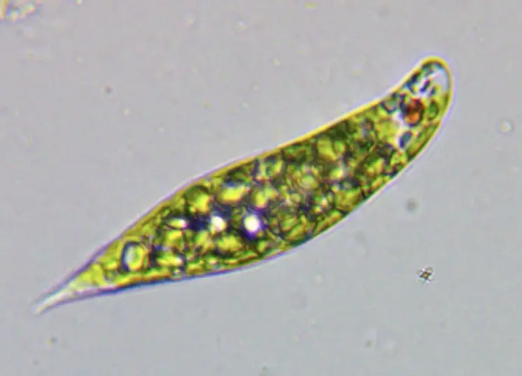
[In this figure] Euglena.
Photo credit: RZ Lin
Paramecium, a great model to study ciliates
Paramecium belongs to the Phylum of Ciliophora and is a typical model of ciliates. Paramecium’s whole body is covered with small hair-like filaments called the cilia. The movement of cilia drags the food closer to the oral groove (functions as paramecium’s mouth). We will discuss the structure and function of cilia in detail in the near future post.
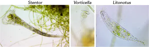
Other well-known ciliates include Stentor, Lacrymaria, Litonotus, Dileptus, Vorticella, and Coleps. They are common pond water living organisms. We have a post about how to find and observe pond life under the microscope.
[In this figure] The Phylum of Ciliophora.
Cilia are the shared characteristic of all ciliates. Here are some commonly found ciliates in pond water specimens: Stentor, Vorticella, and Litonotus.
Related posts
The Structure of Paramecium Cell
Paramecium Reproduction, Physiology, and Behaviors
Pingback: Where does Paramecium live? – Dr. Biology Questions and Answers
Pingback: What is the size of the Paramecium? – Dr. Biology Questions and Answers