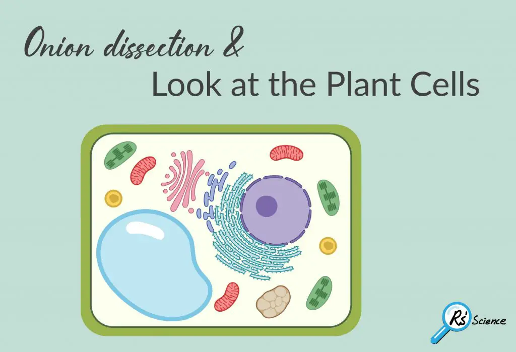
The goal of this lesson is to look at the plant cells. An onion is very easy to get and it is also easy to collect cells from. Therefore, let’s look at onion cells under a microscope! Below are steps showing how to do it.
This article covers
Onion skin slide preparation
The material you need
- Blank microscope slides and coverslips
- Forceps
- Eosin Y staining solution
- Dissecting knife
- Petri dish
- Onion
Preparing onion cells slide for a microscope
- Peel the brown skin away from the outside of the onion. Take one layer of the onion flesh and carefully cut out a piece.
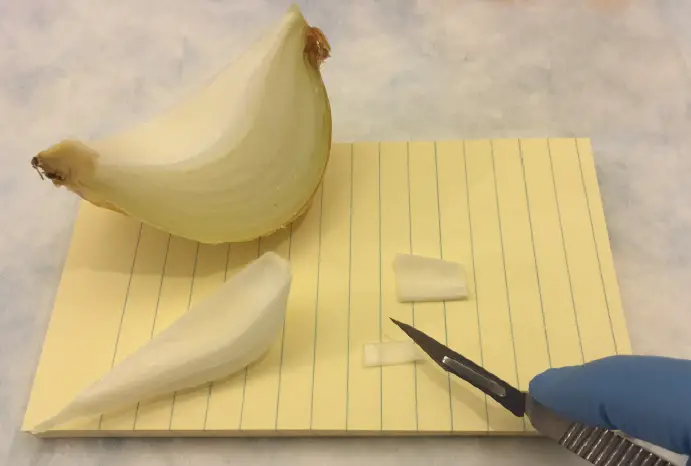
- On the inside of this piece is a thin sheet of the membrane. Use tweezers or dissection needle to peel the membrane away.

- Place the specimen in a small dish of stain (Eosin Y) and leave it for about 3 minutes.

- Use tweezers to transfer the specimen out of the dish. Rinse it well in a dish containing clean water.
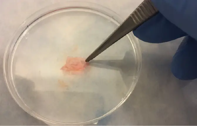
- Make a wet mount and look at your stained section under the microscope. See if you can find onion cell under a microscope.

What will you see?
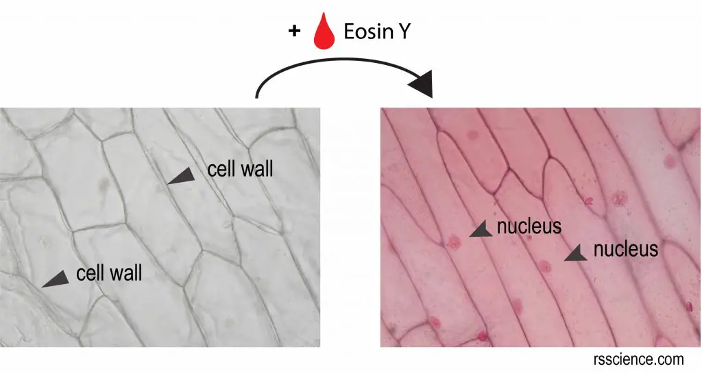
[In this figure] Microscopic view of onion skin.
Without stains, you can only see the cell walls of onion cells. With the staining of Eosin Y, now you can see a nucleus inside an onion cell.
What do your onion cells look like under the microscope? Please share it with us below!
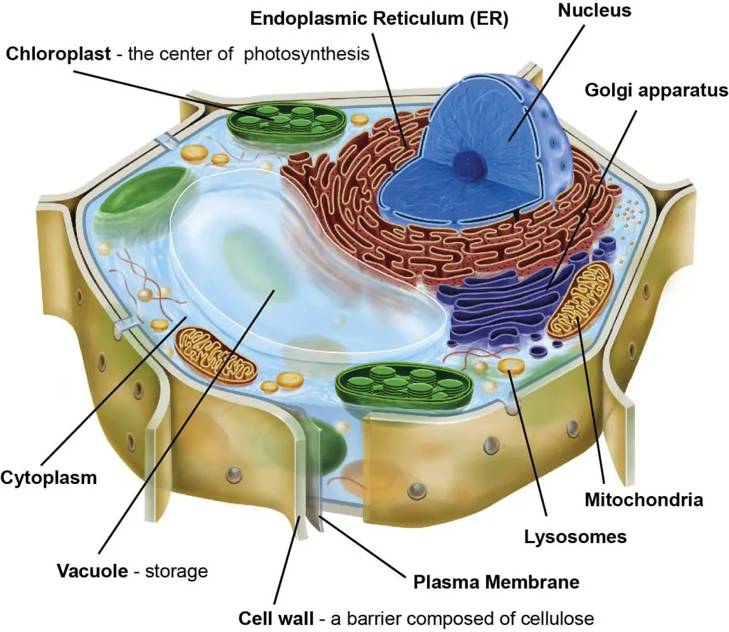
Anatomy of the plant cell
Structurally, plant and animal cells are very similar because they are both eukaryotic cells. However, plant cells have three additional structures including chloroplasts, the cell wall, and vacuoles.
Chloroplasts
Chloroplasts are the center of photosynthesis, where the green pigment chlorophyll captures energy from sunlight and stores it in the energy storage molecules, like sugar.
Cell wall
A cell wall is a structural layer surrounding plant cells (also in bacteria and fungi), just outside the cell membrane. It is tough and rigid providing the cell with both structural support and protection.
Vacuoles
Vacuoles are like storage rooms for the cells.
Related posts
Lesson 4: How to Use a Microtome & “Amazing Cross-section of a Stem”

