
Electron microscopy is a valuable tool to obtain high-resolution images. The way that an electron microscope works is to accelerate a focused stream of electrons in a vacuum towards the specimen. Interactions between the electron beams and the sample create an image, which is similar to how optical microscopes use light to capture images.
This article covers
1. Do not mix up SEM and TEM images
There are two types of electron microscope: scanning electron microscope (SEM) and transmission electron microscope (TEM). SEM and TEM differ in how they work and what types of images they are able to capture.
Can you tell SEM and TEM images below? It is not difficult once you know the difference between these two types of electron microscopes.
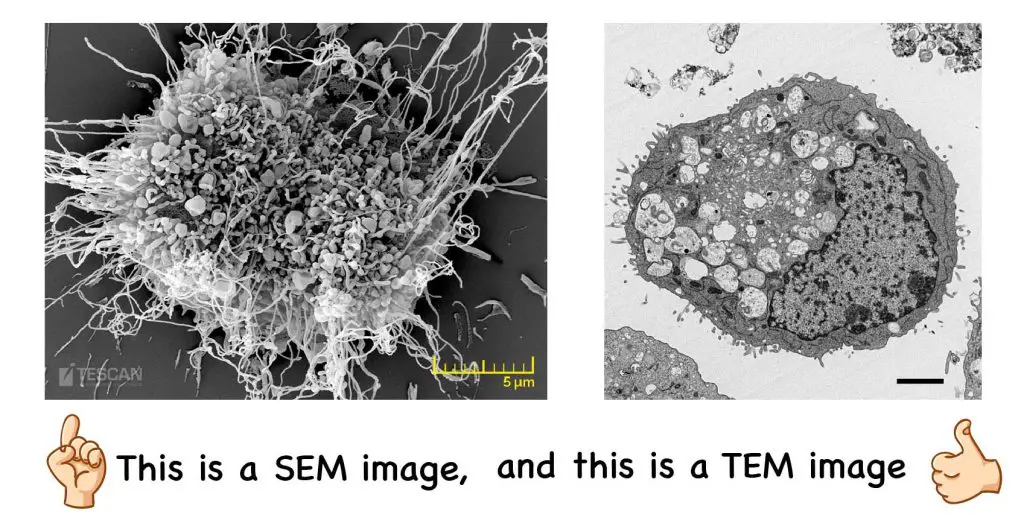
[In this figure] The examples of SEM and TEM images.
The left image was taken by a SEM. In contrast, the right image was taken by a TEM.
What is SEM?
SEM stands for Scanning Electron Microscope. An SEM is a type of electron microscope that uses a fine beam of focused electrons to scan a sample’s surface. SEM images give insight into a sample’s surface topography; therefore it creates 3-D images.
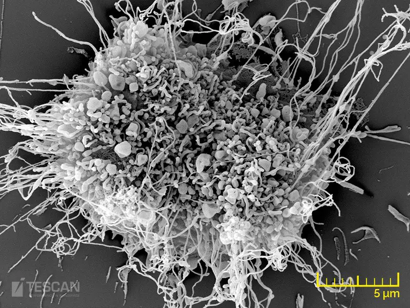
[In this figure] SEM image of a fibroblast.
A fibroblast is a type of cell that synthesizes the extracellular matrix and collagen for connective tissues. SEM image allowed us to see all kinds of cellular protrusions and extracellular matrix on the surface of the cell. (Credit: tescan.com)
How does a scanning electron microscope work?
To obtain a high-resolution image, an electron source (also known as an electron gun) emits a stream of high-energy electrons towards a sample. The electron beam can be focused using electromagnetic lenses. When the focused stream reaches the sample, it scans its surface. The interaction between the electron beam and the sample creates secondary electrons, backscattered electrons, and X-rays. These interactions are captured to create a magnified image.
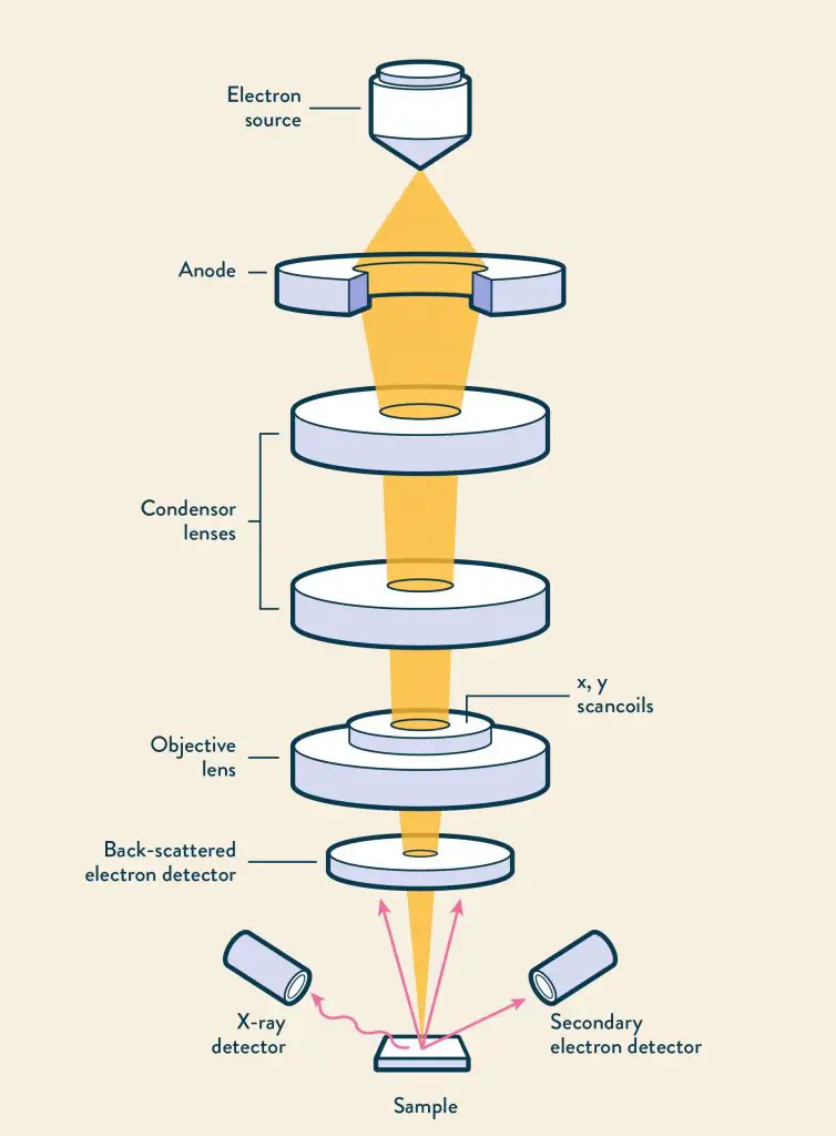
[In this figure] The scanning electron microscope principle.
Photo credit: https://www.technologynetworks.com/analysis/articles/sem-vs-tem-331262
What is TEM?
TEM stands for Transmission Electron Microscope. A TEM is a type of electron microscope that uses a broad beam of electrons to create an image of an internal structure of the sample. A beam of electrons is transmitted through a sample to create an image. Samples must be incredibly thin, often less than 150 nm thick, to allow electrons to pass through them. Therefore, TEM creates 2-D images. Below is an example of a TEM image.
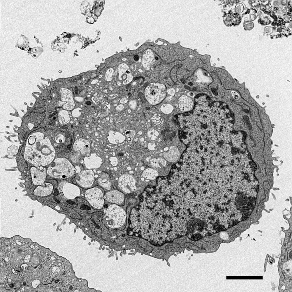
[In this figure] TEM image of pancreatic ductal adenocarcinoma (PDAC) cancer cell.
PDAC is one of the deadliest cancers in Western countries. Scientists learned from this TEM image that abnormal mitochondria contribute to the growth of cancer cells. (Credit: High Resolution Electron Microscopy Facility at MD Anderson Cancer Center)
How does a transmission electron microscope work?
An electron source sends a beam of electrons through an ultrathin sample. The electron passing through the samples then reaches the detector. The detector can be a fluorescent screen or a charge-coupled device (CCD) camera.
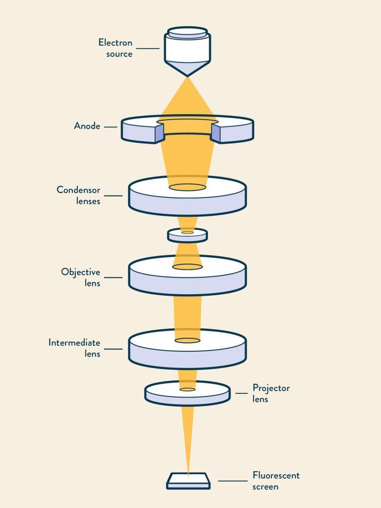
[In this figure] The transmission electron microscope principle.
Photo credit: https://www.technologynetworks.com/analysis/articles/sem-vs-tem-331262
SEM vs. TEM
SEM and TEM are both valuable tools in the biological, physical, and chemical sciences. The major difference is that SEM creates 3-D images of a specimen’s surface topography and TEM sees through the internal structures of the ultrathin sample. TEMs have an incredible magnification potential of 10-50 million times. On the other hand, SEM has the potential to magnify an image up to 2 million times.
2. Can we see colors under electron microscopes?
The answer is No.
The reason is pretty basic: color is a property of light (i.e., photons), and since electron microscopes use an electron beam to image a specimen, there is no color information recorded. The area where electrons pass through the specimen appears white, and the area where electrons do not pass through appears black.
So, what you are looking at the image produced by an electron microscope is basically the “contrast”. This is the reason why the image is black and white.
The color image of an electron microscope below on the right is actually colored after the image is produced.
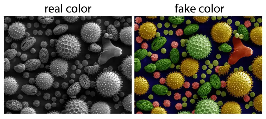
[In this figure] Image of pollen grains.
The image on the left was taken on an SEM shows the characteristic depth of the field of SEM micrographs. Pollen from a variety of common plants: sunflower (Helianthus annuus), morning glory Ipomoea purpurea, hollyhock (Sildalcea malviflora), lily (Lilium auratum), primrose (Oenothera fruticosa) and castor bean (Ricinus communis).
Photo credit: Wiki.
The image on the right was colored by a computer, not a real image directly from an electron microscope.
Another example of a real image from an electron microscope and colored later (right).
[In this figure] Hydrothermal worm marine organism imaged on a Quanta SEM.
The left image is the real color, and the color on the right image was added by the computer.
Photo credit: Thermo Fisher.
3. Not real electron microscope images
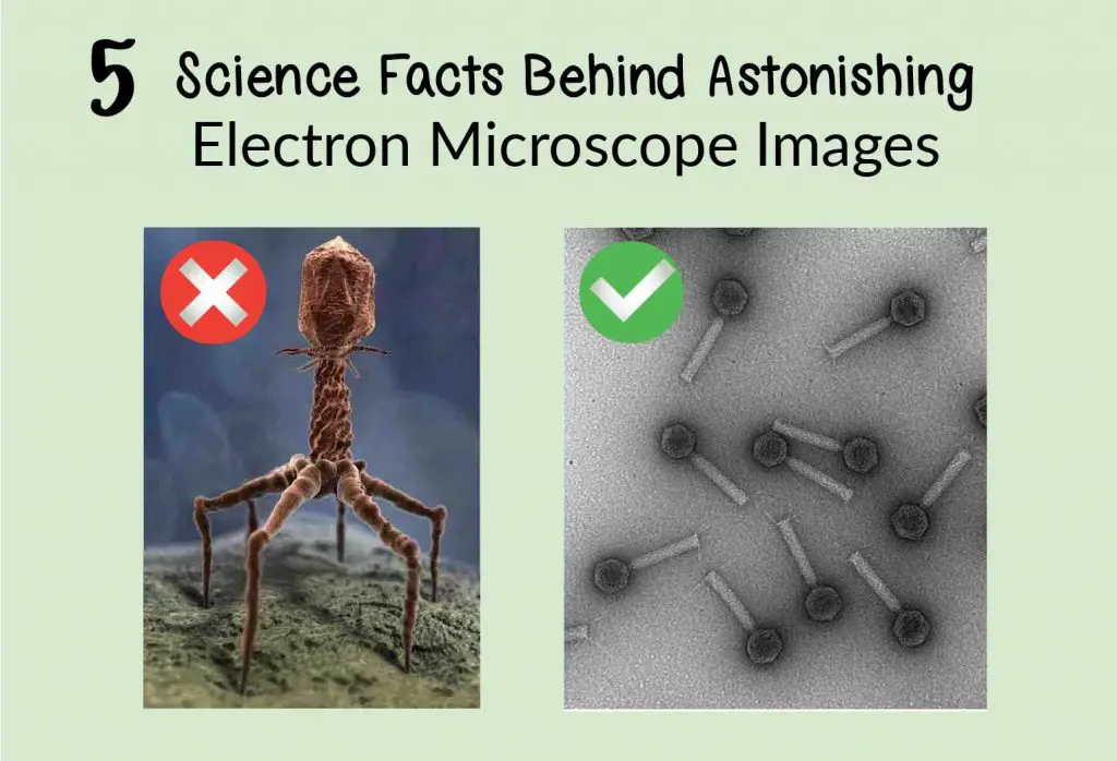
We often found very cool images online or Pinterest, like the virus images below on the left. Do you know if it is taken by an electron microscope?
The answer is No.
The image below on the left is most likely computer-simulated. Electron microscopes do not have a depth of field on this scale, and the model has perspective distortion which implies the “lens” would be in an unusual position for a typical electron microscope.
The image below on the right is the real image taken by a transmission electron microscope. You can see the scale bar (100 nm) below with a magnification 150,000x. In addition, the EM images are black and white. Therefore, the right image is the real image via an electron microscope.
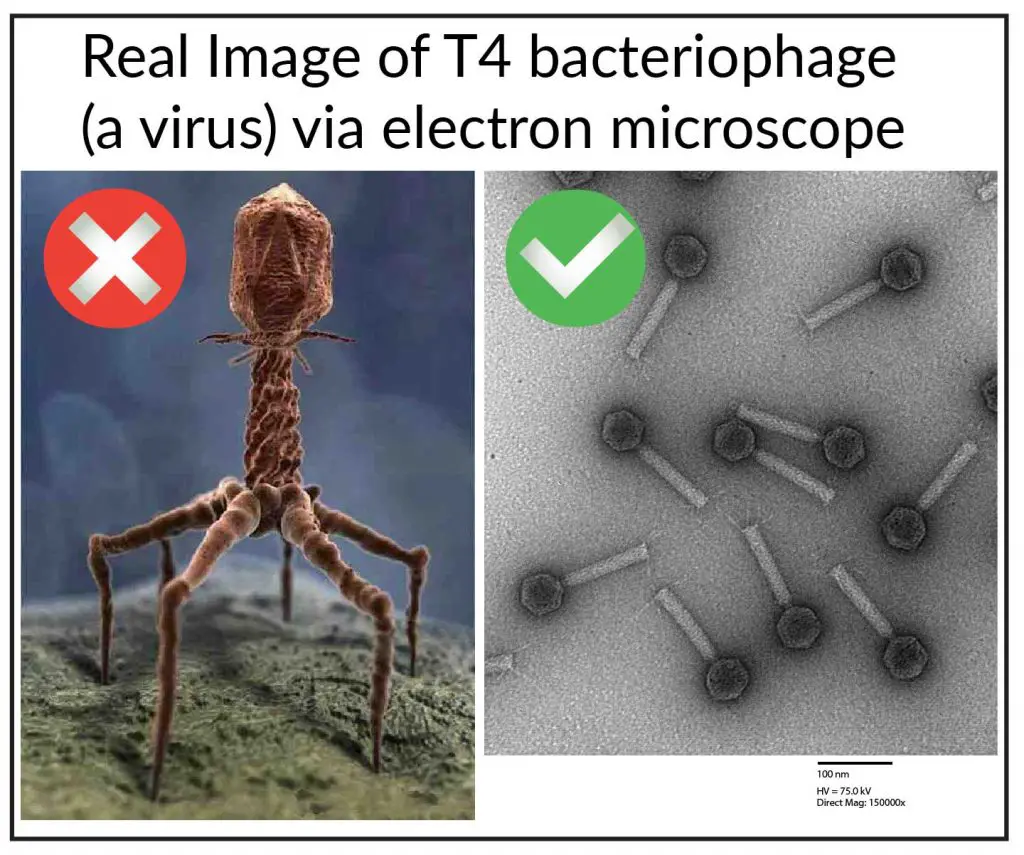
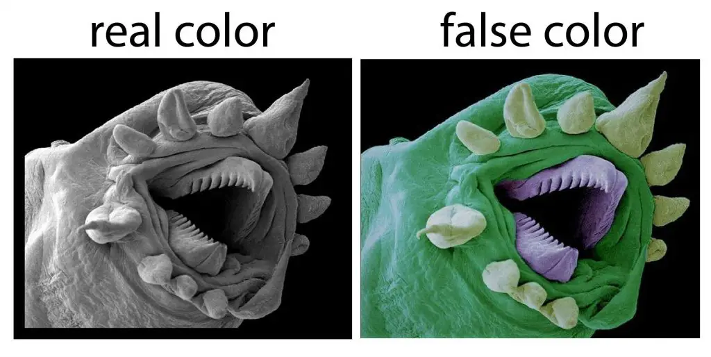
[In this figure] Which one is the real image from an electron microscope?
The left image is likely a model simulated by a computer. The right image was taken on the Hitachi H-7000 TEM by Carina Buttner, Laboratory of Dr. Alan Davidson.
4. Wrong organisms of electron microscope images
There are websites saying that the image below are “Chalk under an electron microscope”.
This is not ‘what chalk looks like’. The image below is an organism called Coccolithophore cells whose calcium carbonate ‘coccoliths’, or shells, accumulate to form chalk.
Here is the research paper as a reference.
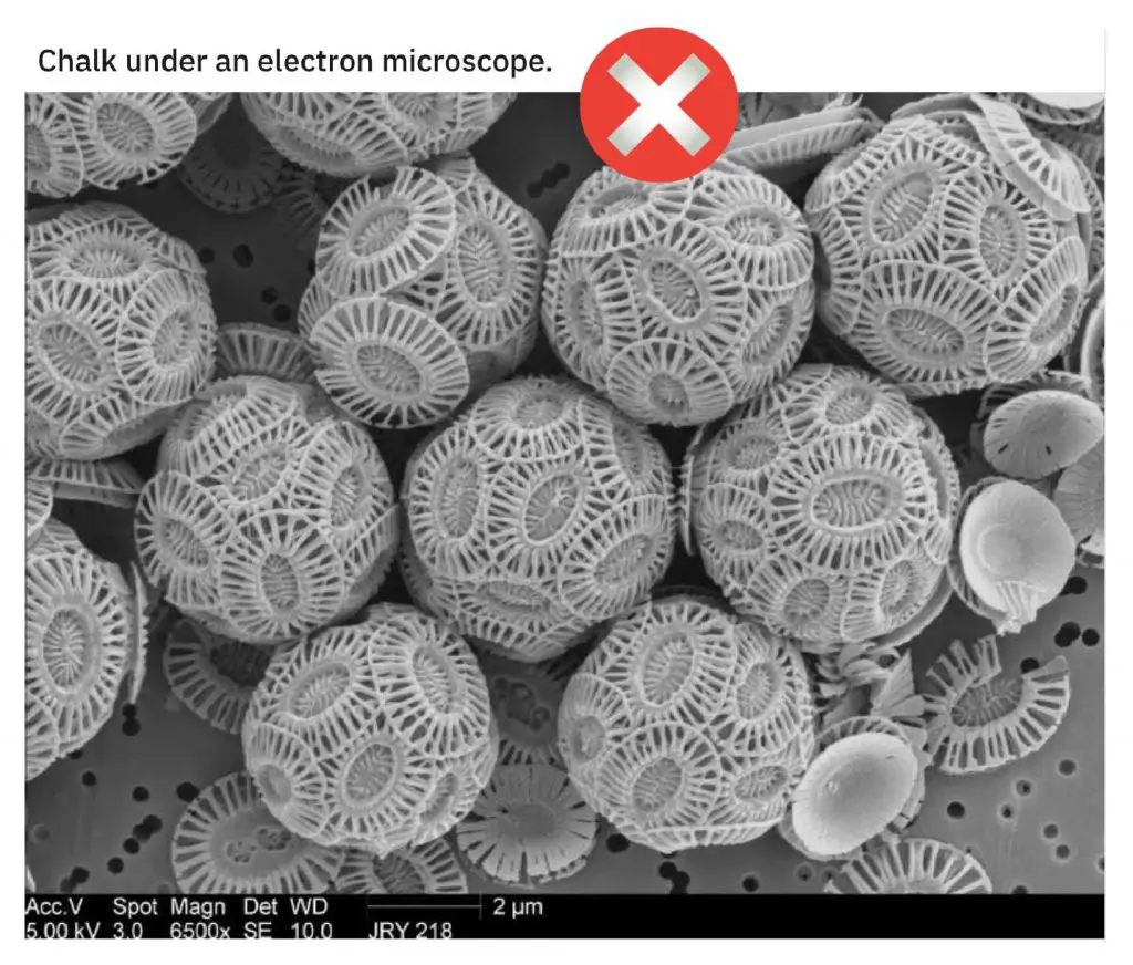
[In this figure] This is not “Chalk under an electron microscope”.
This is Coccolithophore cells covered with calcium carbonate scales.
5. Not an electron microscope image
Sometimes you saw horrible images like one below. It is thought that is is an electron microscope image, but actually it is not. It is image by a technique called confocal.
This photo won 4th place in the 2017 Nikon small world photomicrography competition. These eye-looking things are not eyes. They’re suckers for attaching to the intestinal lining.
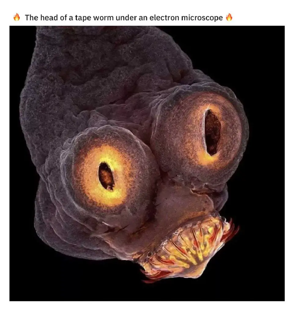
[In this figure] This is imaged by confocal, not electron microscopy.