Our previous article quickly covered the biology, classification, and characteristics of planaria (nickname, “cross-eyed worms”). I also briefly talk about their remarkable regenerative capability. However, there are still so many interesting scientific findings of regeneration in planarians that I don’t have space in the previous article. This is why I want to dedicate a follow-up article about this topic.
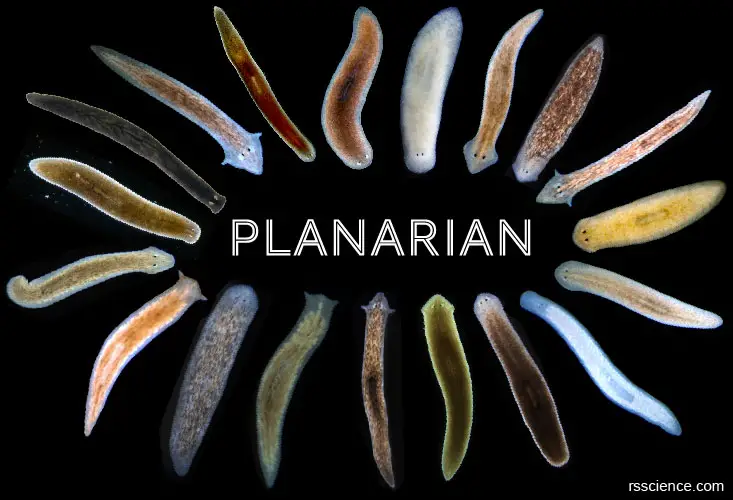
[In this image] Planaria are free-living flatworms in the phylum Platyhelminthes.
They come in different sizes, shapes, and colors. They are tiny (3 to 15 mm in length) but not simple; in fact, they have several organ systems, including digestive, nervous, reproduction, and excretory systems, just like the human bodies. Planaria can move smoothly across a surface with many cilia on their belly. If you want to learn more about the basics of the planarian, see “Planarian – Biology, Classification, Characteristics, and Regeneration”.
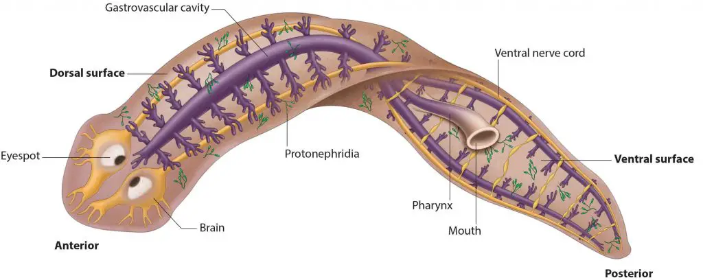
[In this image] The anatomy of a planarian shows its digestive (purple), excretory (green), and nervous (yellow) systems.
Photo source: cuttingclass
This article covers
These flatworms can regrow a body from a fragment. How do they do it and could we?
How flatworms can regrow a body from a fragment?
This is exactly the question that many scientists are asking and putting huge effort into investigations.
More than 100 years ago, scientists figured out that some planarians could regenerate parts of their bodies. Planaria are remarkable in that they can regenerate an entire worm from just a tiny fragment of the original. For example, a planarian that is cut into three pieces (head, body, tail) can regenerate into three separate individuals.
In the laboratory, scientists utilize planaria as a model organism to investigate the mechanism of regeneration. The ultimate goal is regenerative medicine. If we can figure out the mechanisms and copy the planarian’s regenerative powers to human patients, he/she may cure themselves by replacing damaged organs or lost limbs.
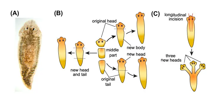
[In this image] Planarian’s Regeneration: (A) The most frequently used planarian in high school and first-year college laboratories is the brownish Girardia tigrina. (B) A planarian cut into three pieces (head, body, tail) can regenerate into three separate individuals. (C) Two incisions on a planarian’s head can regenerate into a three-headed planarian.
Does a planarian feel pain?
Because of their simple nervous system, planarians do not feel pain when cut, only pressure.
The process of planarian’s whole-body regeneration
A fragment of planarian can regrow into a new animal in about two weeks. As you can see in the image below, a planarian cut into five pieces become five new planaria. The head piece regrows a body and a tail. The tail piece gains a new head and a body. The most amazing part is the body fragments which have neither a head nor a tail, but can easily regenerate everything a new animal needs in just a few days!
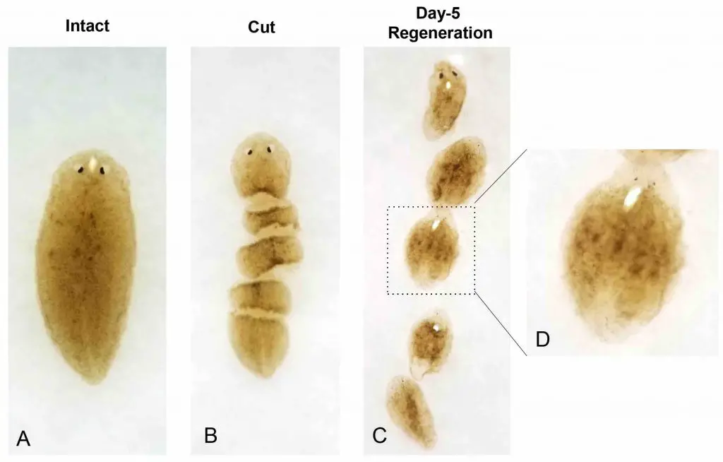
[In this image] Scientists demonstrated that an intact planarian (A) was cut into five pieces (B). After 5 days, these fragments were in the process of regeneration (C, D). Planarians can regenerate all missing tissue within one week of amputation, including a brain, eyespots, and pharynx (feeding tube).
Photo source: HKU Med
[In this video] See how the planaria regenerate from fragments.
Two mysteries – What are the materials and blueprints for planarian to regenerate
Scientists use planarians as model organisms to study the remarkable process of regeneration. They are seeking for understanding these two fundamental questions:
(1) What does a planarian use to regenerate its missing or damaged body parts?
Does a planarian have a reservation of stem cells (as materials) to regrow organs?
Our (human) body can not do that. For example, damaged brain tissue can not repair because neuronal cells stop growing in adults and there are very few stem cells in the brain.
(2) How does a planarian know how to rebuild the body the right way ?
How does a planarian know to grow only one head instead of two? Does it have many copies of blueprints saved in each cell?
If we transplant stem cells without giving them the right instruction, these stem cells grow into a tumor (called teratoma) that contains a mess of several types of tissues.
Let’s see what our scientists know about these two mysteries so far:
Superpower (1) – Planarian has adult stem cells everywhere
Planaria can do this remarkable feat because roughly 25~40% of their body is made up of adult stem cells, which are able to regenerate all tissues the worm may need to replace.
These adult stem cells are called neoblasts, which can collectively differentiate into any of 40 to 50 different cell types present in a planarian. Neoblasts locate throughout the body, except for the anterior tip of the head and the pharynx.
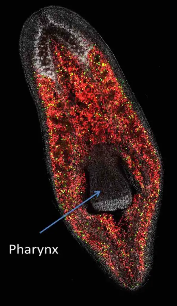
[In this image] The distribution of neoblasts in a planarian. The green ones are dividing, and the red ones are not dividing.
Photo source: Janet Rossant, eLife, 2014
Individual neoblasts are considered pluripotent, meaning they can make most, but not all, cell types. However, a collection of neoblasts can make any cell type, making them a collectively totipotent cell population.
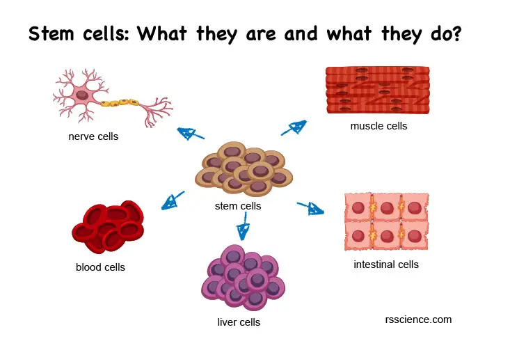
[In this image] Cells are the basic units of our body. Each cell type has its specialized functions. “Stem cells” mean these cells have the ability to become other cell types.
When a planarian is cut, its neoblasts multiply to make more stem cells. These stem cells then differentiate into cells needed to replace the missing body parts. These regeneration abilities are far beyond those of the human body. However, understanding how planarians can regenerate lost body parts may provide clues for improving wound healing in humans.
Another remarkable feature of planarian neoblasts is their high mitotic activity; in other words, they are almost constantly dividing. The frequent division of neoblasts results in a fast turnover of all planarian tissues. As a result, planarians are likely to lack any long-lived cells and can turn over all the cells in their bodies in just a few weeks, keeping all their organs “young.” Planarians can also quickly increase and decrease body size by changing their overall number of cells, typically in response to food availability.
Human doesn’t have that powerful adult stem cells
Now, we turn around to look at the situation in our bodies. For humans and other mammalians, totipotent and pluripotent stem cells only exist shortly in the early embryo stage. We do have “adult” stem cells, but these cells have very limited potentials. For example, our bone marrow stores hematopoietic stem cells (HSCs). These HSCs can become all kinds of blood cells (red blood cells, white blood cells, and platelets), but not others. Bone marrow transplantation transfers these HSCs from a healthy donor to rebuild the blood system in a patient.
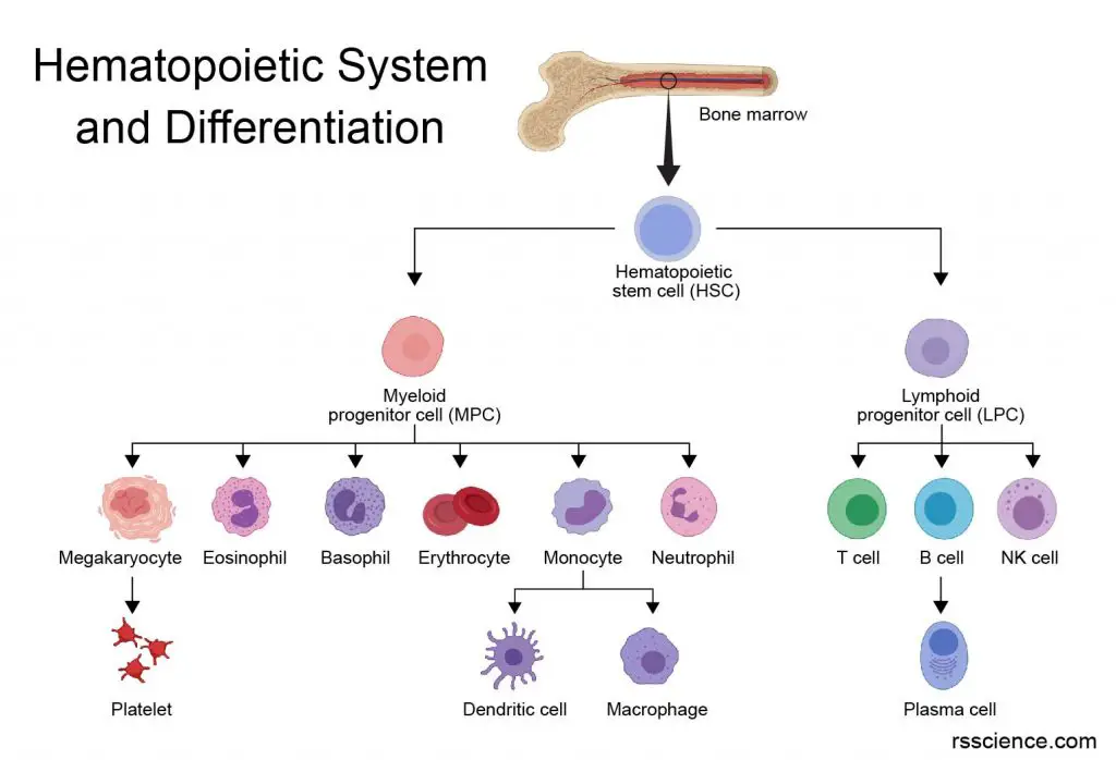
[In this image] Hematopoietic stem cells (HSCs) are the root of the entire blood system.
Another example is the intestinal stem cells (ISCs). The ISCs reside in the epithelial layer of our small intestine and they replicate and replace the dying cells frequently. Fortunately, our ISCs are active adult stem cells that can maintain a high turnover rate. However, most of the cells in adult humans are not replaced as frequently. Therefore, damaged tissues and organs can not be repaired as well. For example, we have to use the same neuronal cells (brain) and cardiomyocytes (heart) throughout our life because these cell types cease cell division after birth. This is why the recovery after a stroke or heart attack is usually bad.
Induced pluripotent stem cells (iPSCs) bring us hope!
Induced pluripotent stem cells (iPSC) are among the most exciting discoveries in biomedical science in the past 20 years. In 2006, Professor Shinya Yamanaka of Kyoto University, Japan, discovered a new way that allows us to manufacture our own pluripotent stem cells. This technique is called “induced pluripotent stem cells or iPSCs”. He found that four genes – Oct3/4, Sox2, c-Myc, and Klf4 (we now called them “Yamanaka factors”) can reverse the cell fate.
These four genes working together can teach adult cells to “forget” their differentiation state and to be reprogrammed to an embryonic-like state. These embryonic-like cells are what we called “Induced pluripotent stem cells or iPSCs”. These cells, once given the right environment, can grow to any type of cells we need. Now, we may have unlimited cell sources to replenish the old and damaged cells in our bodies.
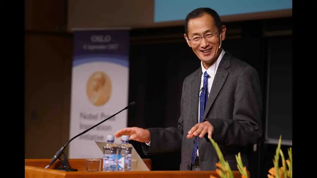
[In this image] Professor Shinya Yamanaka received The Nobel Prize in Physiology or Medicine 2012 for the discovery that mature cells can be reprogrammed to become pluripotent.
Photo credit: nobelprize.org
Superpower (2) – A genetic blueprint for rebuilding the planarian body
A piece cut from a planarian can even form a new planarian. This new planarian regrows a head and a tail in the appropriate places. How is this possible? How does a small fragment know where should be a head or a tail?
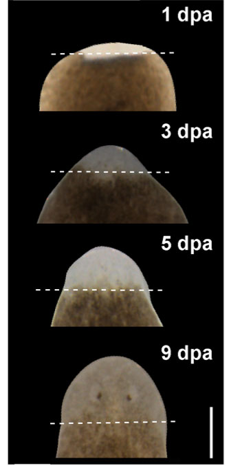
[In this image] The cut piece decided where to regrow the head within the first-day post-amputation (dpa). On day 9, the head had regrown to show two eyespots.
Photo source: HHMI/BioInteractive
We know that the planarian’s powerful stem cells can provide functional cells for its new organs. However, many different activities, including cell migration, cell division, gene regulation, even cell death, must coordinate across thousands of cells, so that a regenerating planarian always ends up looking like a “normal” planarian. Disrupting this coordination, the new worms could end up wildly misshapen – with tiny shrunken heads or even multiple heads!
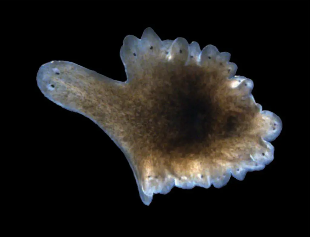
[In this image] An example of a misshapen planarian.
Image from Sánchez Alvarado Lab.
Several genes map the body plan of planarian
Scientists found that several genes and their protein products can coordinate and instruct the regeneration of planarian. See the image below as an example. Gene notum produces the “anterior factors” that mark the pole where should regrow into the new head. On the opposite side, gene wnt1 encodes the “posterior factors” that guide the formation of a new tail. There are several pairs of these genes that control the polarity of planarian. Polarity is a property of organisms that have distinct ends, for example, distinct heads and tails, which determine the axis of the body patterning. In fact, the same genes are also found in our bodies.
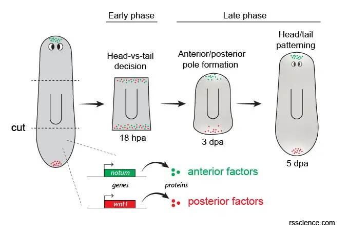
[In this image] Genes and their protein products control the regeneration of the head/tail in a Planarian.
In this study, two genes, notum and wnt1, encode anterior and posterior factors, respectively. In uninjured animals, notum is expressed at the anterior pole (head), while wnt1 is expressed at the posterior pole (tail). During regeneration, both genes are induced at all wounds in a dispersed “salt and pepper” pattern in the early phase. Then, notum clusters at the anterior-most tip and wnt1 shifts to the posterior-most tip of the fragment. As regeneration continues, notum and wnt1 expression domains elongate until the head/tail pattern is restored. hpa: hours post-amputation. dpa: days post-amputation.
The detail of this study can be found here
Two-headed planarian while its genes were messed up
You may ask what will happen if these genes go wrong. You can see the image below – it regenerated into a two-headed planarian!

[In this image] Two planarians from the same species.
The one on the left is a typical individual for this species. The one on the right regenerated from a cut planarian treated with RNAi.
Photo source: HHMI/BioInteractive
In this study, scientists used RNA interference (RNAi) to block the action of posterior factors. RNAi is a technique that uses small pieces of RNA to turn off the function of specific genes. Because of RNAi, this planarian could no longer produce an important protein for maintaining polarity. As a result, the planarian’s stem cells incorrectly formed two heads instead of one head and one tail.
[In this video] See how scientists find the key genes that control planarian regeneration.
Bioelectricity controls planarian regeneration
Genes are powerful regulators of planarian’s regeneration but not the whole picture. In fact, environmental cues also play an important role. Scientists are especially interested in how bioelectrical signaling controls the process of regeneration.
For example, the planarian which losses the function of H, K-ATPase (a protein channel that transports ions across the cell membrane), fails to regenerate properly.
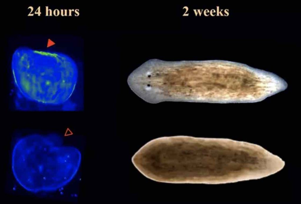
[In this image] Top: Normal regeneration of a planarian. Bottom: While the H, K-ATPase activity was blocked by a drug, the trunk fragment regenerated without a head and failed to reshape their old tissues.
Photo source: Beane lab
External electric field changes planarian’s regeneration as well. If you place the (-) anode near the head wound, the animal will regrow normally. However, if you reverse the direction of the electric field, low-intensity currents cause double-headed worms, whereas high-intensity currents cause reversed-polarity worms.

[In this image] Electric field changes planarian’s regeneration.
Modified from Lobo D. et. al., PLoS Comput Biol. 2012
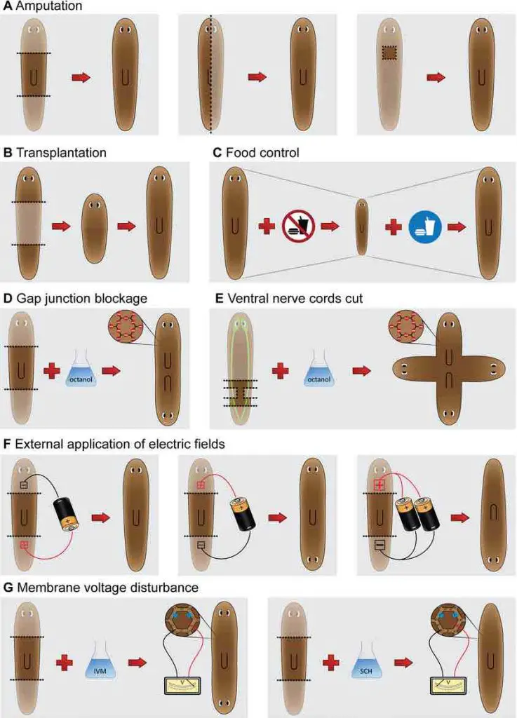
[In this image] Scientists did a variety of experiments, including changes in electric fields, food supplies, and membrane voltages, to understand the regulation of Planarian’s regeneration. If you are interested to learn more, see the original article.
What can we learn from a planarian?
In nature, there are many animals with remarkable healing abilities like the Wolverine in X-men. For example, a gecko can regrow its lost tail, and a salamander can regenerate its entire arm. However, no one can regenerate more remarkably than planaria.
Humans have very limited regeneration ability. Our liver can regenerate from 1/3 of the original size. However, other major organs, such as the brain, heart, and lung, can not regenerate at all.
When we get older, we lose our reserve pool of stem cells. The ability to repair wounds declined, which is a major cause of many aging and metabolic-related diseases, including heart attack, diabetes, and neurodegenerative diseases. If we can replenish the old and damaged cells in our body, we may fundamentally treat many diseases. This concept is called “Regenerative Medicine,” obtaining these powerful stem cells for replenishment will be the key to success.
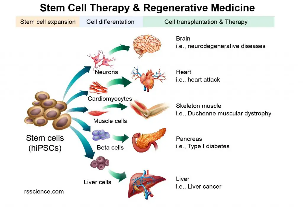
[In this image] The concept of Stem Cell Therapy and Regenerative Medicine.
Although we have a powerful technique, iPSCs, induced stem cells from mature cells, we are still learning how to make a functional organ out of a dish of iPSCs. This is why scientists are so passionate about planaria. If we can figure out how the planarian’s regeneration is determined, this will provide strategies for regenerative medicine’s goal, replacing lost organs or limbs in human patients.
The future of regenerative medicine is promising! As we know more and more about our genes (Human Genome Project), scientists found that we share many of the same regenerative genes with planaria. It is possible that our ability to regenerate is just locked. At the same time, our capability to control gene functions has greatly advanced in the past decades. Amazing techniques like RNAi, CRISPR gene editing, and modified mRNA enable us to turn on/off or replace any given gene in our cells. These tools may help us to unlock our ability to regenerate. I optimistically believe the era of Regenerative medicine is not too far.
References
“Go ahead, grow a head! A planarian’s guide to anterior regeneration”
“Model systems for regeneration: Planarians”
“Tissue Regeneration in Animals”

